Antibodies


1 Introduction 2 Recombinant antibody technology 3 REAfinity™ Antibodies: recombinant antibodies for flow cytometry
1 Introduction
Antibodies are arguably one of the most important components of a molecular biologist’s toolbox. They have revolutionized biological research and are now making their impact felt in the clinic as effective therapeutic agents. Following an introduction to antibody structure and types as well as recombinant antibody technology, this chapter details the properties of REAfinity™ Recombinant Antibodies and their use in flow cytometry.
 MACS Handbook:
1.1 Antibody structure
Antibodies are characterized by their unique Y shape. Each antibody consists of four polypeptides – two heavy chains and two light chains. According to their functional characteristics, antibodies can be subdivided into variable and constant regions. The variable region, made up of the N-terminal ends of the heavy and light chains, contains the antigen binding site. The constant region, which consists of the rest of the molecules, determines the immune response or the mechanism used to destroy antigens (fig. 1).

 View details
Each antibody can be proteolytically cleaved into three fragments – two Fab regions and an Fc region. Fab, short for fragment antigen binding, includes the variable ends of an antibody. The Fc fragment, short for fragment crystallizable, determines the antibody biological effector functions, such as antibody-dependent cellular cytotoxicity (ADCC) and complement-dependent cytotoxicity (CDC). In ADCC, the Fc region of an antibody binds to Fc receptors (FcγRs) on the surface of leukocytes, resulting in phagocytosis or lysis of the targeted cells. In CDC, the antibodies trigger the complement cascade at the cell surface leading to cell death.
1.2 Antibody isotype classifications
Antibodies are divided into five major isotypes, based on their structure, biological properties and ability to deal with antigens. Table 1 outlines the isotypes and their properties, distribution, and immune functions. (Janeway, E. A. et al. (2001) The distribution and functions of immunoglobulin isotypes. Immunobiol. Immune Syst. Health Dis. 5th Ed.)
Most assay antibodies belong to the IgG subclass which is subdivided into four isotypes: IgG1, IgG2, IgG3, and IgG4 in humans, and IgG1, IgG2a, IgG2b, and IgG3 in mice. IgG isoforms exert different levels of effector functions – the level of ADCC effector function is high for human IgG1 and IgG3, low for IgG2 and IgG4, whereas the level of CDC effector function is high for human IgG1 and IgG3, low for IgG2, and null for IgG4.
Table 1: Antibody isotypes
| Isotype | Subclasses | Structure | Major immune function | Distribution |
| IgA | IgA1 and IgA2 | Monomer, dimer | Neutralization | Extracellular fluid (monomeric IgA), secretions across epithelia like breast milk (dimeric IgA) |
| IgD | Monomer | Function not clear | Mostly bound to B cells | |
| IgE | Monomer | Sensitization of mast cells | Mast cells beneath epithelial surfaces (specifically of the respiratory tract, gastro-intestinal tract and skin) | |
| IgG | IgG 1–4 (in humans) | Monomer | Neutralization, opsonization, complement system activation, sensitization for killing by NK cells | Plasma, extracellular fluid |
| IgM | Pentamer | Complement system activation | Plasma, extravascular spaces |
1.3 Primary-secondary antibody classification
Antibodies that bind to specific antigens of interest help biologists visualize a variety of biological entities – from proteins on a Western blot membrane to cellular and subcellular structures.
Primary antibodies bind directly to the antigen. Depending on the type of experiment, primary antibodies are used alone or in combination with a secondary antibody. For example, in flow cytometry, primary antibodies conjugated to fluorochromes are almost always used alone, whereas in microscopy, a primary-secondary antibody combination is used to increase the signal.
Secondary antibodies help detect, sort, or purify antigens of interest by binding to the primary antibody. They are raised against the species and isotype of the primary antibody. For example, for a primary antibody raised in rabbit, an anti-rabbit secondary antibody raised in a host other than rabbit would suffice. Primary antibodies are often IgG isotypes and therefore will need an anti-IgG secondary antibody. Secondary antibodies are often conjugated to enzymes, dyes, or fluorescent proteins. The choice here depends on the type of experiment; for instance, in immunofluorescence experiments, secondary antibodies conjugated to fluorescent proteins or dyes are the right choice.
A two-antibody system – the primary-secondary system – is often employed for i) stronger dye localization to an antigen, ii) multicolor labeling within a specimen and across experiments using one primary antibody and multiple fluorescent secondary antibodies, and iii) economical reasons.
1.4 Polyclonal vs. monoclonal antibodies
Polyclonal antibodies (pAbs) are a heterogeneous mixture of antibodies, produced by various B lymphocyte clones. They mostly consist of a mixture of IgG subtypes and recognize multiple epitopes of an antigen. Polyclonal antibody serum can be obtained within 4–8 weeks by injecting the host animal with the antigen of interest followed by observation of antibody response and collection of antisera. Their production is inexpensive compared to monoclonal antibodies. (PMID:15953834)
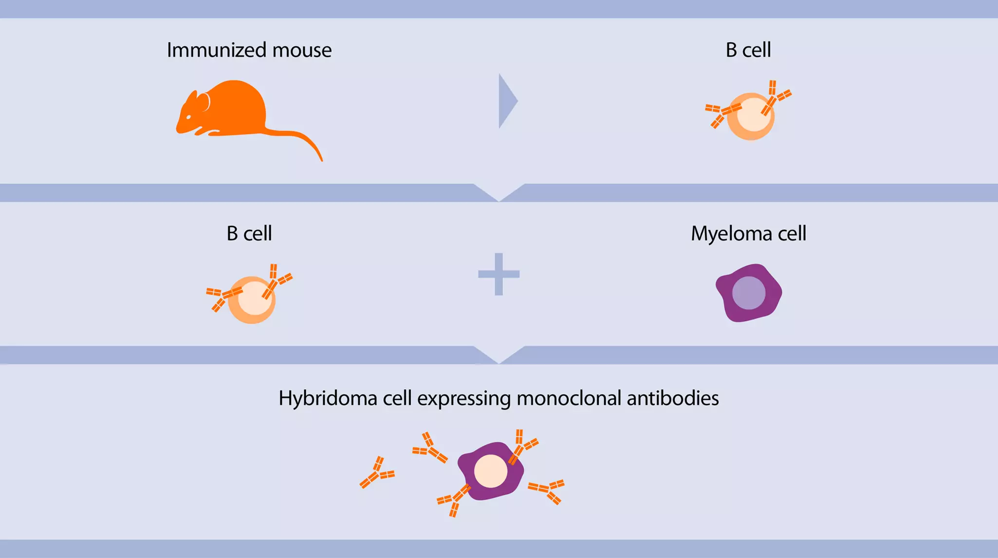
Monoclonal antibodies (mAbs) are produced by a single clone of B lymphocytes and recognize only a particular epitope on the antigen. They consist of only one antibody subtype. The mAbs are produced by hybridomas, i.e., B cells immortalized by fusion with myeloma cells (fig. 2). (PMID: 15728446)
Compared to pAbs, mAb production is more complex, expensive and time-consuming (3–6 months). (PMID: 15953834)
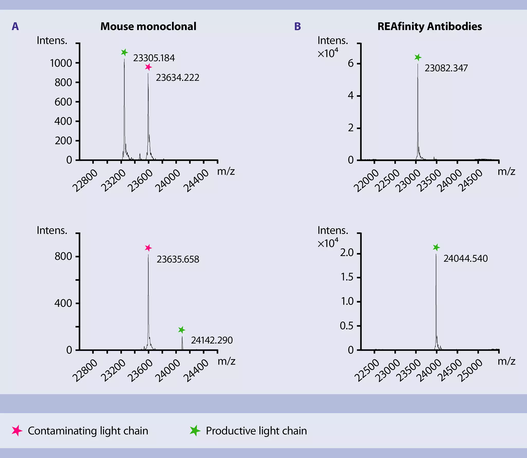
Hybridoma-derived mAbs are often contaminated with antibody impurities from two sources: myeloma derived–Ig light chains and serum-derived IgGs. (PMID: 29485921) These impurities lower the specificity of the antibody resulting in irreproducible results (fig. 3).

1.5 The reproducibility crisis in life science research: Antibodies are a major culprit
Irreproducibility of results is a major challenge faced by the scientific community today. Several factors contribute to this complex problem. According to the Second Annual State of Translational Research 2014 Survey Report conducted by the Sigma-Aldrich Corporation on the issue of reproducibility, antibodies were one of the major product classes contributing to the issue (around 36%) and second only to animal models (around 41%). In 2014 another survey conducted by the American Society for Cell Biology, the ASCB member survey on reproducibility, 50% of respondents named reagents including antibodies, sera, and plasmids to be the reason for the failure to replicate experiments.
Antibody-related reproducibility can be improved by adhering to the following guidelines by all parties involved – vendors, users, and journals:
- Application-specific validation of antibodies
- Transparent and clear reporting of reagent use
- Selling and usage of consistent-quality antibodies
One solution is to use antibodies produced by recombinant antibody technology. (PMID: 25652980) Recombinant antibodies exhibit higher purity, lot-to-lot consistency, and definability because of standardized cell culture systems and access of DNA sequence, when compared to hybridoma-derived monoclonal antibodies.
1.6 Antibody validation
All our antibodies are rigorously tested and validated before release. In 2020, we began providing validation data directly on our product pages. There, we provide extended validation data highlighting details of antibody performance, specificity, and fixation compatibility. We do this in order to make it even easier for our customers to choose the antibodies that best match their needs, and to decrease the validation efforts required on the researcher side. Our validation is based on three pillars: reproducibility, specificity, and sensitivity, and all antibodies for which any of these datasets are already available will be indicated with the extended validation stamp. Some details follow below:
Antibody reproducibility and consistency
- Recombinant engineering ensure high lot-to-lot consistency, whilst mass spectrometry analysis is used to confirm purity of antibody products
- All antibodies are additionally tested for lot-to-lot consistency at two stages: during antibody raw material production, and in the fluorochrome-conjugation process (including purification steps to remove unconjugated fluorochromes and antibodies)
Antibody specificity
- Epitope competition assay compare the epitope specificity of REAfinity Clones with other known clones in the market
- Knockout validation via targeted genome editing involves the target gene being knocked out in a suitable cell line using site-specific nucleases; The knockout is confirmed by sequencing of the target locus, and specific binding is confirmed if no antibody binding to the knockout cells can be detected
- RNAi knockdown involves the target antigen being knocked down using RNA interference or RNAi. The translation of the target RNA is inhibited by transfecting cells with small non-coding RNA oligonucleotides. A comparison between the transfected cells with the control cells reveals the specificity of the tested antibody to its antigen
Antibody sensitivity
- Functional testing of every product prior to release: All conjugated antibodies, including multiple conjugates of the same clone, are tested on primary samples, and in multicolor panels wherever possible
- Antibodies are routinely tested on cells derived from tissues using enzymatic treatment to allow for validation of sensitivity to epitopes that have undergone enzymatic processing
- The performance of our conjugated antibodies are compared to those from other vendors to ensure similar or better performance in flow cytometry applications
- Each lot is validated by a series of dilutions to make sure the product is working within the expected antibody titer range
- Our antibodies are also tested for their stability during fixation to verify that the epitope of an antigen can still be detected after the fixation process
With well over 10,000 antibodies in our portfolio, this is an ongoing project, and we are continuously extending the availability of data that demonstrates our rigorous production, quality control, and validation standards. We hope this inspires customers with the confidence to trust their precious research with our high-quality products, as well as help them minimize their own validation efforts.
Validation is of course not solely the responsibility of the supplier because antibody validation methods and antibody validation protocols are highly dependent on the intended application and experimental setup. For this reason, we recommend thoroughly researching the most suited validation methods for your application(s). Several articles have been published providing antibody validation guidelines, as well as publication guidelines. A selection is given below:
- Uhlen M., et al. (2016) A proposal for validation of antibodies. Nature methods, 13(10), 823–827 (PubMed ID: 27595404)
- STAR methods from CellPress (https://www.cell.com/star-authors-guide)
- Lucas, F., et al. (2020) MiSet RFC Standards: Defining a Universal Minimum Set of Standards Required for Reproducibility and Rigor in Research Flow Cytometry Experiments. Cytometry. Part A: the journal of the International Society for Analytical Cytology, 97(2), 148–155 (PubMed ID: 31769204)
- Roncador, G., et al. (2016) The European antibody network's practical guide to finding and validating suitable antibodies for research. mAbs, 8(1), 27–36. (PubMed ID: 26418356)
2 Recombinant antibody technology
Recombinant antibodies (rAb) are monoclonal antibodies generated in vitro and animal-free using rDNA technology. Recombinant antibodies overcome many of the limitations of conventional mAbs (see table 2). (PMID: 26482034)

 Related videos:
2.1 Recombinant antibodies: production methods
The production workflow of recombinant antibodies consists of the following steps:
- Determination of the sequence of the desired antibody genes
- Gene synthesis and cloning into expression vectors
- Expression of antibody fragments in mammalian or bacterial cells
- Purification and analysis of the antibody
For recombinant antibody production, stable cell lines such as CHO and HEK293 are often used.
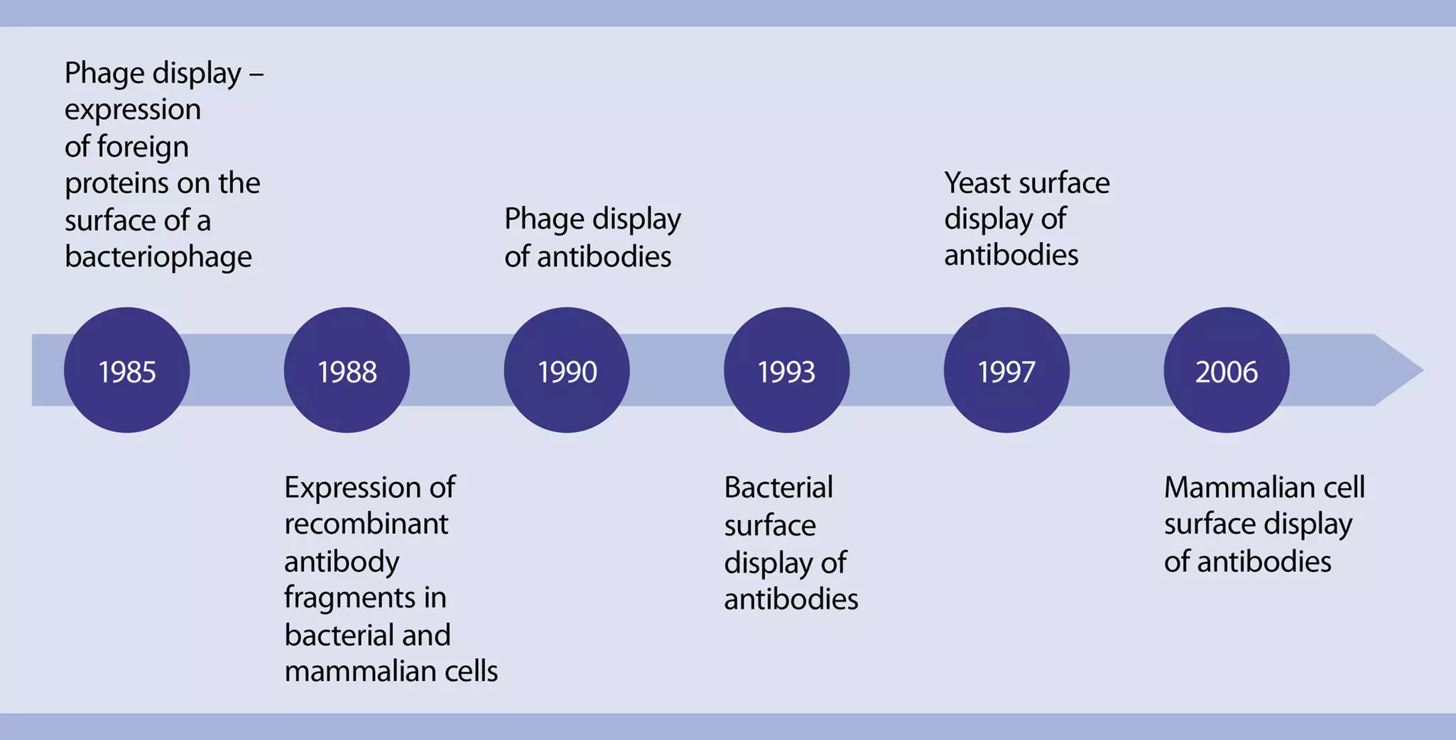
Antibody fragments for recombinant antibody production can be obtained from existing monoclonal antibodies or can be selected from libraries of genes (encoding slightly different antibody proteins) according to the affinity of the antibodies for binding to target antigens. The library-based recombinant antibody production methods are divided into in vitro display platforms, such as phage display and ribosome display, and in vivo display platforms, such as bacterial, yeast, and mammalian cell-surface display. Figure 4 below outlines the early milestones in the production of recombinant antibodies. (PMID: 15953834, 4001944, 3140379, 3045807, 3210233, 2247164, 8248129, 9181578, 16763048)
Phage display is the most common method which involves the following steps:
- Library construction: The gene fragments encoding antibody heavy and light chains are cloned into phage vectors resulting in a phage library displaying antibodies with varied antigen binding sites.
- Antibody selection by panning: Panning is a process by which antibodies are selected by incubating the phage library with the target antigen immobilized on a solid phase. Unbound phage particles are washed away, and the bound phages are eluted and amplified in E. coli.
- Cloning and selection: The resulting antibody DNAs are cloned into expression vectors and expressed in E. coli.
- Screening and affinity maturation: To improve antibody affinity, sequence diversity is introduced into the selected antibody molecules by methods like error-prone PCR or site-directed mutagenesis. Antibodies are evaluated for improved affinity and the selected clones are expressed and purified.
3 REAfinity™ Antibodies: recombinant antibodies for flow cytometry
REAfinity™ Recombinant Antibodies are a new generation of recombinantly generated antibodies specifically designed for flow cytometry applications. They provide superior lot-to-lot consistency and purity, as compared to mouse or rat monoclonal antibodies. The following structural features of the REAfinity Antibodies makes them the superior antibody choice for flow cytometry experiments (fig. 5).
- Universal human IgG1 isotype reduces the complexity of experiment planning by eliminating the need to include multiple isotype controls during flow cytometry analysis.
- Mutated Fc region eliminates background signals by abolishing any binding of the recombinantly engineered antibodies to FcγRs.

 View details
 Related PDFs:
 Related products:
3.1 REAfinity Recombinant Antibodies: production workflow
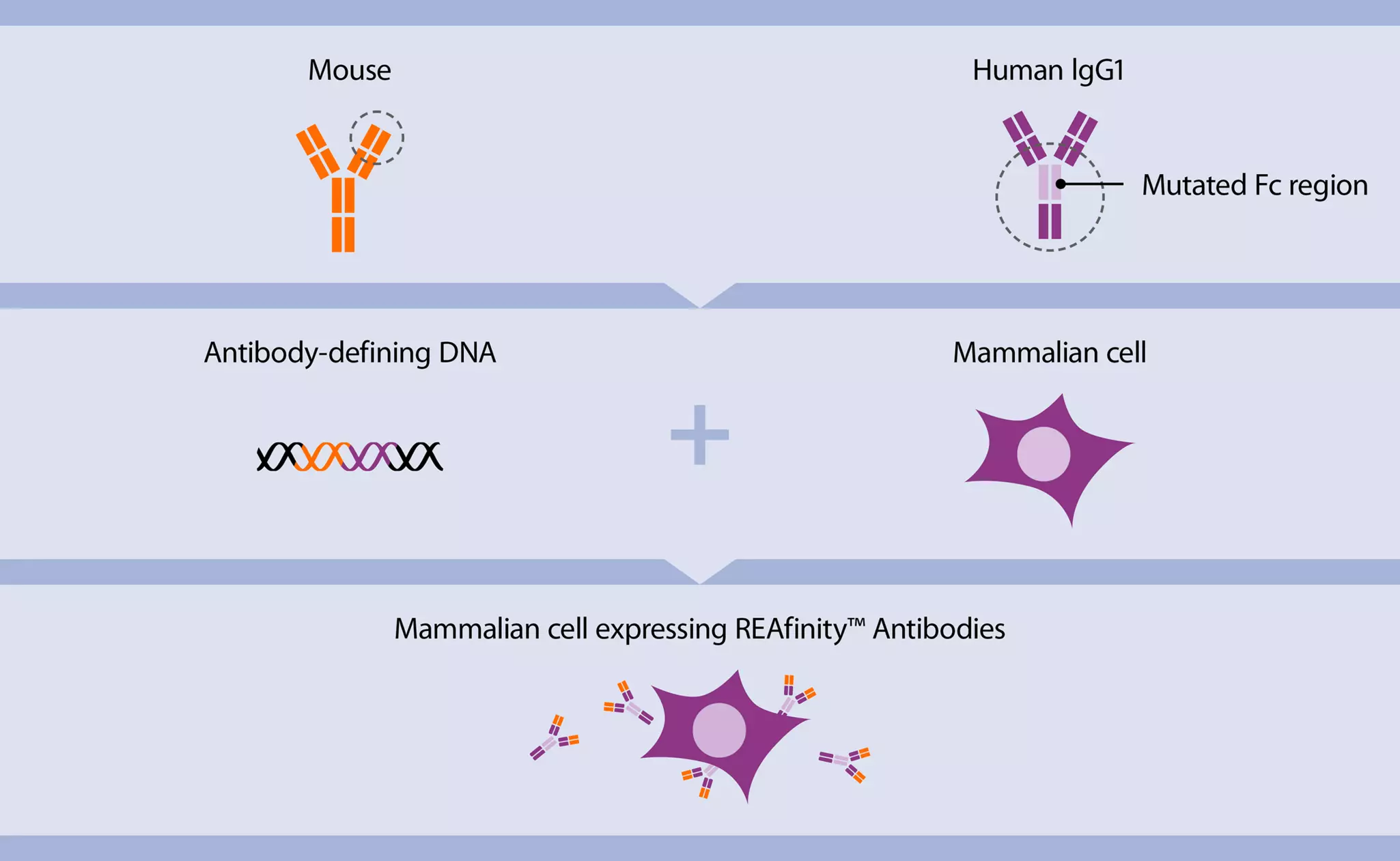
REAfinity Antibodies are generated by cloning the antigen binding region from a traditional hybridoma-derived monoclonal antibody (mouse IgG) with the human IgG1 Fc region. The Fc region is mutated to abolish binding of the recombinant antibodies to Fcγ receptors. This chimeric DNA is expressed in a mammalian cell line cultured under standardized conditions. To ensure lot-to-lot consistency, the same cell line is used to produce all REAfinity Antibodies (fig. 6).
Advantages of this production method include:
- No light chain impurities because of a highly defined chimeric IgG sequence
- No cell-derived impurities as the cells are programmed to solely express the defined antibody
- No culture media–derived impurities as the mammalian cells are cultivated in standardized and defined culture conditions
3.2 REAfinity Recombinant Antibodies: unique features and advantages
3.2.1 High lot-to-lot consistency – reproducible results
Hybridoma-derived monoclonal antibodies often contain contaminating light chains and serum-derived antibodies. The presence of these impurities and a non-standardized production method result in lot-to-lot variability and the lack of reproducibility of experiments.
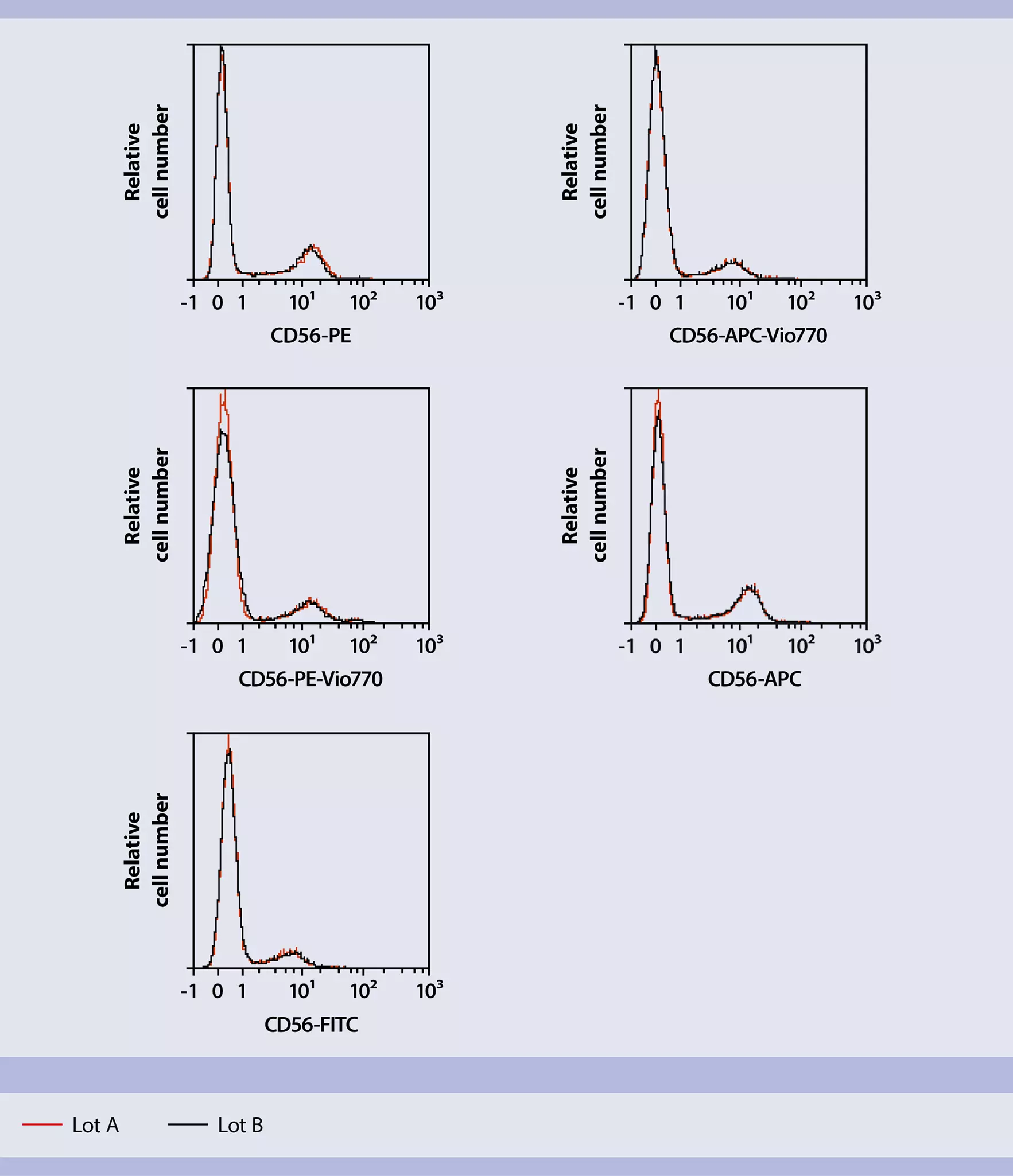
REAfinity Antibodies are derived from a defined DNA sequence, which encodes only one type of heavy and light chain, ensuring high antibody purity (fig. 7). They are produced in mammalian cells under biologically defined, standardized culture conditions, resulting in high lot-to-lot consistency and purity as compared to mouse or rat monoclonal antibodies. Moreover, every manufactured lot undergoes thorough analytical, biochemical, and cell-based analysis (fig. 8).
3.2.2 No Fcγ receptor binding – reduced background signal
High background signals from antibodies often compromise the quality of flow cytometry results. One of the reasons for high background signals is non-specific binding of antibodies to Fcγ receptors on leukocytes. To avoid this type of non-specific binding, Fc blocking reagents are used during antibody staining. However, the blocking procedure is time-consuming and costly.
REAfinity Antibodies feature a mutated human IgG1 Fc region which abolishes their binding to Fcγ receptors (fig. 9). This allows for background-free analysis and eliminates the need for additional blocking steps such as using an FcR blocking reagent (fig. 10). Therefore, staining protocols are much faster with REAfinity Antibodies (table 3).
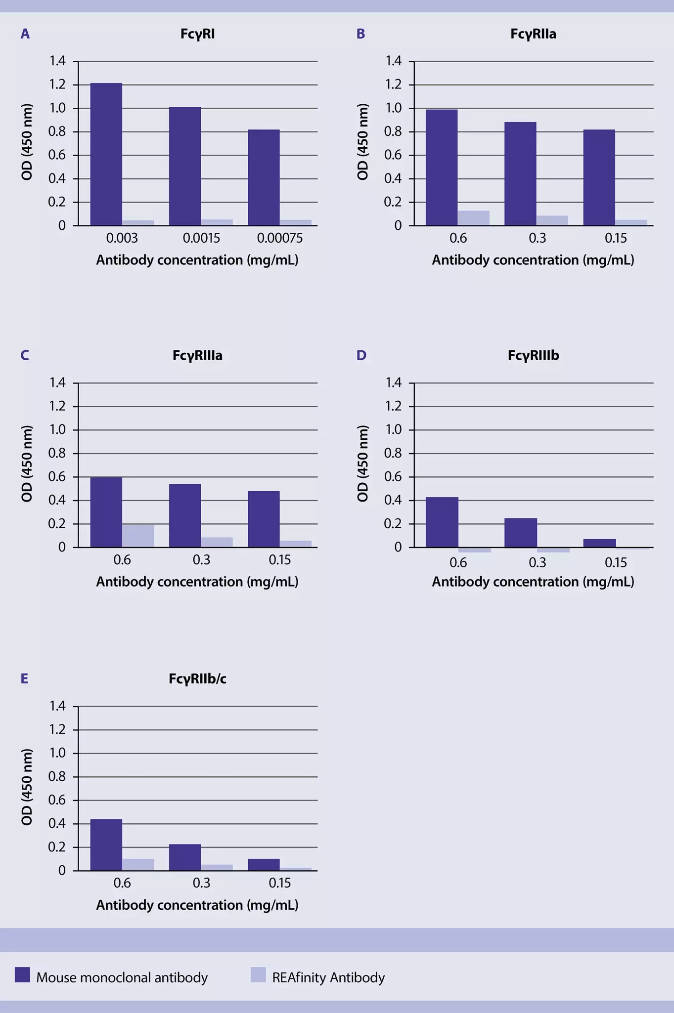

Table 3: Staining protocols for immunophenotyping of human and mouse surface markers
| Steps involved | Duration of each step | |
| Hybridoma-derived antibodies | REAfinity Antibodies | |
| Prepare 106 cells | - | - |
| Add blocking reagent to cells and incubate at 4° C or RT | 10–15 min | - |
| Add primary antibodies and incubate at 4° C or RT | 10–15 min | 10 min |
| Wash cells | 1–2× 10 min | 10 min |
| Resuspend and analyze cells | - | - |
| Total staining procedure | 30–65 min | 20 min |
3.2.3 One universal isotype – easy experiment planning
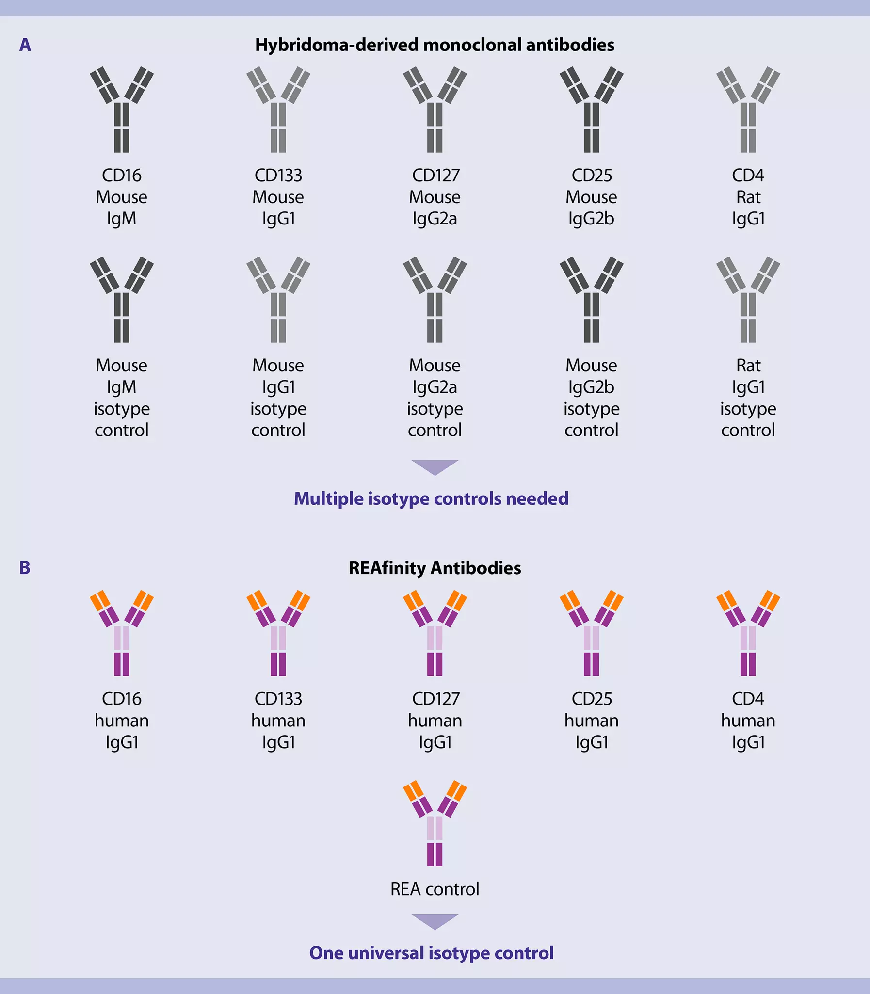
All REAfinity Antibodies, i.e., REA clones, have the same human IgG1 isotype, eliminating any need to include multiple isotype controls during flow cytometry analysis (fig. 11). This offers the possibility of working with only one type of isotype control, reducing the complexity of experiment planning and saving time.
In contrast, hybridoma-derived monoclonal antibodies are composed of antibody isotypes derived from different species and therefore require multiple isotype controls. This complicates not only the management of reagent inventory but also panel design when setting up experiments.
Besides these unique features, all REAfinity Antibodies undergo strict production and quality control processes. For each specificity, premium antibody screening technology is applied to identify the clone with the highest binding affinity and specificity. Only the top candidate is selected to become a recombinantly engineered antibody (REA) clone.
3.3 REAfinity Recombinant Antibodies: optimized for flow cytometry applications
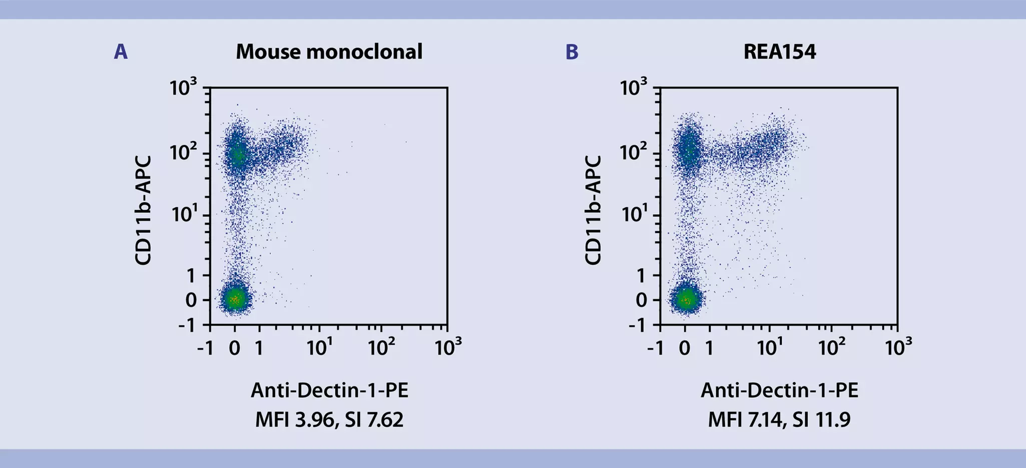
REAfinity Antibodies are designed with flow cytometry in mind. Their high purity, lack of background signal, and a standardized fluorochrome conjugation process ensure a superior performance in flow cytometry experiments in comparison to hybridoma-derived monoclonal antibodies (figs. 12–14).


3.4 Multicolor flow cytometry with REAfinity Recombinant Antibodies
REAfinity Antibody clones are available for hundreds of specificities and are conjugated to a variety of fluorophores to address multicolor flow cytometry needs. Current options include traditional dyes, like FITC, PE, APC, and newer versions like Vio® and VioBright™ Dyes (fig. 15). REAfinity Antibodies conjugated to Vio Dyes are powerful and reliable tools for multicolor cytometry analysis, resulting in highly sensitive, reproducible, and rapid cell profiling.

MACS Handbook:
Related content:
Related products:
You have previously chosen International as your location.
What do you want to do?