3D graphic showing regulatory T cells
Logo

Transforming gamma delta (γδ) T cell research: efficient workflow and key solutions in cell therapy

Isolating and expanding gamma delta (γδ) T cells can be challenging due to their low frequency in the blood. Their cell-killing ability can be affected by the method of isolation and expansion. We offer efficient solutions for the isolation, activation, gene transfer, and γδ T cell expansion. Combined, our instruments, reagents, and software work in harmony to ensure consistent and reliable results. Discover how to jumpstart your γδ T cell immunotherapy research with ease!

Content overview

Content

  • Workflow overview
  • 1. γδ T cell isolation
  • 2. γδ T cell activation and expansion
  • 3. Gene delivery in γδ T cell transduction
  • 4. γδ T cell characterization using flow cytometry analysis
  • Targeted γδ T cell discovery with spatial imaging
  • Functional profiling of γδ T cells

A translational workflow to accelerate gamma delta (γδ) T cell research in cell therapy

Gamma delta (γδ) T cells are a unique subset of T lymphocytes characterized by their distinct T cell receptor (TCR), which comprises of one gamma (γ) chain and one delta (δ) chain, differing from the more common alpha-beta (αβ) T cells. γδ T cells recognize conserved non-peptide antigens of stressed cells and tumor cells.1

In the peripheral blood, γδ T cells represent 1–10% of total T cells and the main population is characterized as Vγ9Vδ2+ T cells.2

These cells have gained interest within the allogeneic cell therapy field due to their HLA-unrestricted antigen recognition and their natural capacity to recognize and kill tumor cells.3

Working with γδ T cells can be tricky. That's why we offer convenient and efficient solutions for all process steps, with instruments, reagents, and software designed to ensure consistency. Begin your γδ T cell research effortlessly!

Illustration on Miltenyi Biotec's workflow solutions for every step of gamma delta γδ T cell research.
Gamma delta (γδ) T cell workflow at a glance.

Gamma delta (γδ) T cell workflow at a glance

Our workflow is based on Miltenyi Biotec's expertise in γδ T cell research to standardize a procedure that seamlessly adapts to translational applications.
This robust process includes cell isolation, activation, transduction, expansion, and product analysis, using tools specifically designed for efficiency and reproducibility.

Generate gamma delta (γδ) T cells in streamlined steps

Miltenyi Biotec's expertise in γδ T cell research has been leveraged in our workflow to standardize procedures that can be seamlessly adapted to translational applications. This robust process encompasses cell isolation, activation, transduction, expansion, and product analysis, employing tools specifically designed for efficiency and reproducibility.

1. Gamma delta (γδ) T cell isolation

γδ T cell isolation from PBMCs

γδ T cells can be positively isolated from PBMCs with our Anti-TCRγ/δ MicroBead Kit, human or by the REAlease® Anti-TCR γ/δ MicroBead Kit, human which offers MicroBead- and label-free target cells with very high purity.

TCRγ/δ T Cell Isolation Kit, human offers the possibility to isolate untouched γδ T cells from PBMCs by depleting magnetically labeled non-target cells.

Depletion of αβ T cells from PBMCs can be facilitated with our research-grade TCRα/β T Cell Depletion Kit, human. When transitioning to clinical studies, our CliniMACS® TCRα/β Kit provides the solution for the depletion of unwanted TCRα/β+ T cells in combination with the CliniMACS Plus System.

Specific γδ T cell subpopulations can be isolated via magnetic separation using fluorochrome antibodies such as TCR Vγ9 Antibody, anti-human, REAfinity® (APC) combined with our anti-fluorochrome MicroBeads (Anti-APC MicroBeads).
Additionally, Vδ1 T cells can be flow-sorted using our TCR Vδ1 Antibody, anti-human, FITC, REAlease®.

Dot plot illustration showing the efficient isolation of gamma delta (γδ) T cells with TCR γ/δ+ T Cell Isolation Kit, human.
Isolation of γδ T cells with the TCR γ/δ+ T Cell Isolation Kit, human.

Isolation of γδ T cells with the TCR γ/δ+ T Cell Isolation Kit, human.

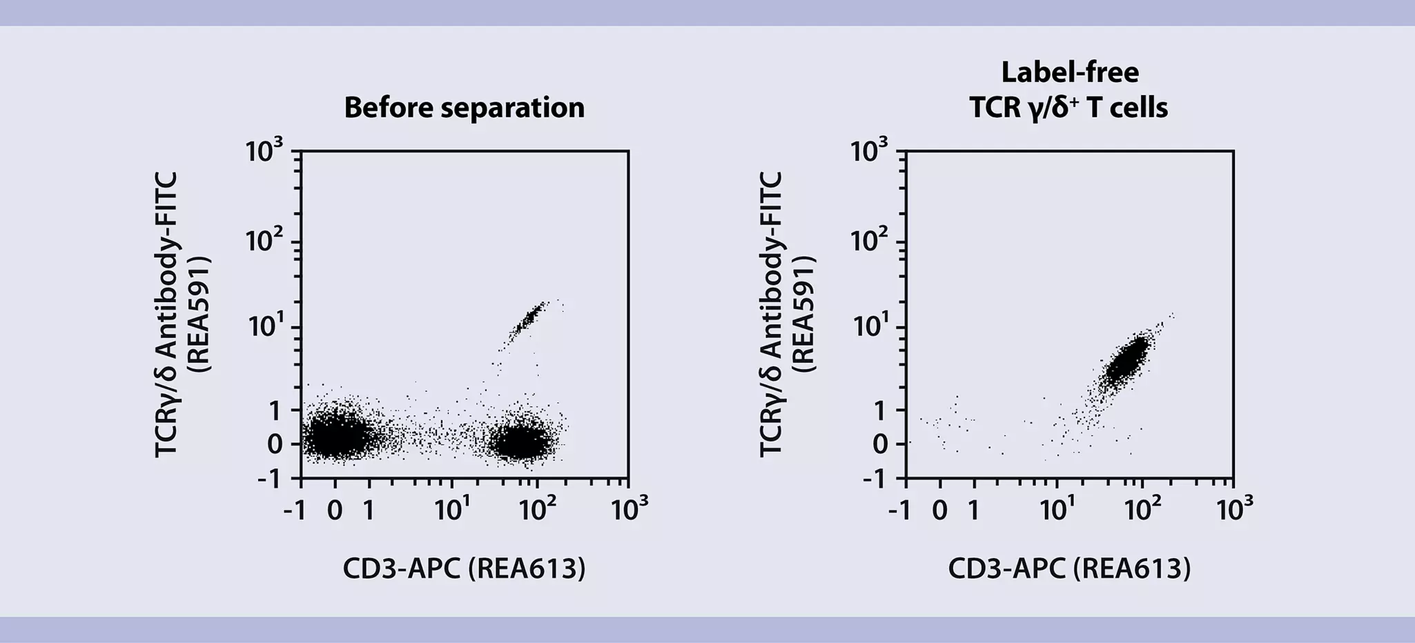
The illustration shows that a very high purity of γδ T cells is achieved starting from PBMCs by using the TCRγ/δ+ T Cell Isolation Kit.

Dot plot illustration showing gamma delta Tcell isolation with REAlease Anti-TCR gd MicroBead Kit, human.
Isolation of γδ T cells with the REAlease® Anti-TCR γ/δ MicroBead Kit, human.

Isolation of γδ T cells with the REAlease Anti-TCR γ/δ MicroBead Kit, human.

The figure indicates that, starting from PBMCs, a very high purity of γδ T cells (95%) is obtained using REAlease Anti-TCR γ/δ MicroBead Kit, human. After the isolation process, the target cells are MicroBead- and label-free.

Fast and easy γδ T cell separation directly from blood products

γδ T cells can be directly isolated from whole blood, buffy coat, and Leukopak® using our StraightFrom® REAlease® Technology.

The StraightFrom REAlease Kits offer the possibility to positively select γδ T cells directly in the starting material, offering an alternative solution without density gradient centrifugation or red blood cell lysis. After the isolation, γδ T cells are MicroBead- and label-free, suitable for TCRγ/δ investigation.

The magnetic isolation process can be achieved manually using the appropriate MACS® Columns or in a semi-automated process by utilizing the MultiMACS™ Cell24 Separator Plus. For a fully automated process use the autoMACS® NEO Separator.

For γδ T cell sorting, the MACSQuant® Tyto® Cell Sorter offers new possibilities with a microchip-based technology.

Dot plot illustration showing the isolation of gamma delta T cells with the StraightFrom® Buffy Coat REAlease®  Anti-TCR γ/δ MicroBead Kit, human.
Isolation of γδ T cells with the StraightFrom® Buffy Coat REAlease® Anti-TCR γ/δ MicroBead Kit, human.

Isolation of γδ T cells with the StraightFrom Buffy Coat REAlease Anti-TCR γ/δ MicroBead Kit, human.

The image depicts that a very high purity of γδ T cells (94%) is achieved using StraightFrom Buffy Coat REAlease Anti-TCRγ/δ MicroBead Kit, human. After the isolation process, the target cells are MicroBead- and label-free.

Overview of all available solutions to isolate γδ T cells from PBMCs or directly from blood products.

Product Name and Material numberIsolationStarting materialTarget cellsCell capacity
REAlease® Anti-TCR γ/δ MicroBead Kit, humanPositive –
Bead- and label-free cells
PBMCs(pan γδ) Vδ1 and Vδ2 T cells1×109
StraightFrom® Whole Blood REAlease® Anti-TCRγ/δ MicroBead Kit, humanPositive –
Bead- and label-free cells
Whole blood(pan γδ) Vδ1 and Vδ2 T cells40 mL whole blood
StraightFrom® Buffy Coat REAlease® Anti-TCRγ/δ MicroBead Kit, humanPositive –
Bead- and label-free cells
Buffy coat(pan γδ) Vδ1 and Vδ2 T cells1 buffy coat
StraightFrom® Leukopak® REAlease® Anti-TCRγ/δ MicroBead Kit, humanPositive –
Bead- and label-free cells
Leukopak(pan γδ) Vδ1 and Vδ2 T cells½ Leukopak
TCRγ/δ T Cell Isolation Kit, humanNegativePBMCs(pan γδ) Vδ1 and Vδ2 T cells1×109
Anti-TCRγ/δ MicroBead Kit, humanPositivePBMCs(pan γδ) Vδ1 and Vδ2 T cells1×109
TCR Vδ1 Antibody, anti-human, FITC, REAlease®
TCR Vδ1 Antibody, anti-human, PE, REAlease®
Positive –
Flow sorting
(PE and FITC)
PBMCs
single-cell suspension
Vδ1 T cells100 tests
TCRα/β T cell Depletion Kit, human *DepletionPBMCsRest of PBMCs-
feeder cells
1×10⁹

Table 1: The table describes all available solutions to isolate γδ T cells. γδ T cells can be isolated from PBMCs in positive or untouched manner. Alternatively, γδ T cells can be isolated directly from blood products.

* Also available in GMP grade.

2. Gamma delta (γδ) T cell activation and expansion

Achieve efficient γδ T cell activation and expansion. Cultivate the cells in TexMACS™ Medium supplemented with human AB serum in the presence of MACS® Premium-Grade Cytokines such as Human IL-2 IS, IL-7, and IL-15. Activate and expand γδ T cells with T Cell TransAct™, human, a stimulation reagent that ensures gentle and efficient activation while maintaining high viability.

TexMACS Medium and MACS Cytokines are available in GMP quality to enable a smooth transition from research- to clinical-grade generation of γδ T cells.

Illustration bar chart showing good gamma delta Tcell activation with T Cell TransAct after 1-4 days stimuli.
Gamma delta (γδ) T cells were studied in cell culture for their activation status.

γδ T cells studied in cell culture for their activation status

γδ T cells were isolated with the REAlease Anti-TCR γ/δ MicroBead Kit, human and cultured in TexMACS Medium supplemented with 5% human AB serum in the presence of MACS Premium-Grade Cytokines Human IL-2 IS, and Human IL-15 (100 U/mL each), and T Cell TransAct, human.

After isolation, γδ T cells don't express CD25, CD69, and CD137. Such expression is noted only after stimulation with T Cell TransAct, human after 1 day and 4 days in culture.

Bar chart illustration showing a high gamma delta T cell cytokine secretion with T Cell TransAct.
Gamma delta (γδ) T cells cytokine secretion with T Cell TransAct™.

γδ T cells studied in cell culture for their cytokine secretion

γδ T cells can be assessed for their activation status by measuring cytokine secretion in the culture. Isolated and non-stimulated γδ T cells don't secrete pro-inflammatory cytokines such as TNF-α and IFN-γ after overnight culture, whereas stimulated γδ T cells with T Cell TransAct secrete TNF-α and IFN-γ after 1 and 4 days in culture.

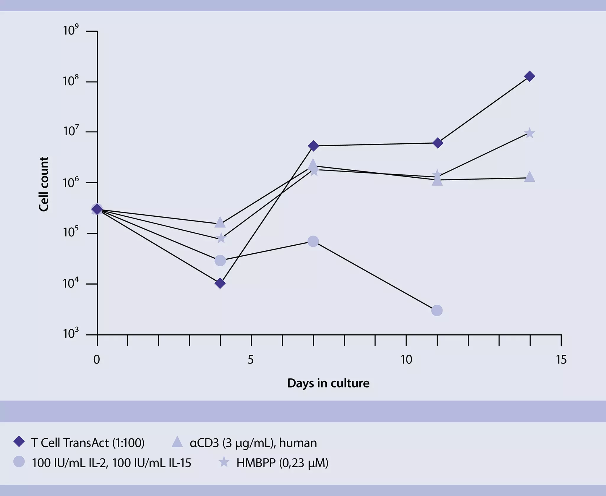
Line chart illustration showing gamma delta Tcell expansion abilities, best achieved using T Cell TransAct.
γδ T cells were tested for their expansion ability when stimulated.

γδ T cells tested for expansion ability

Different methods can be used to expand γδ T cells, depending on the desired phenotype. T Cell TransAct provides primary and costimulatory signals through CD3 and CD28 binding, and can be used to expand γδ T cells. Upon testing different stimulatory reagents which involve only cytokines, CD3, or phospho-antigen HMB-PP ((E)-4-Hydroxy-3-methyl-but-2-enyl pyrophosphate) stimulation, the best expansion rate of 400-fold was achieved with T Cell TransAct.

3. Gene delivery in gamma delta (γδ) T cell transduction

The number of cells expressing a target transgene is influenced by the efficiency of gene transfer. However, viral transduction of γδ T cells is often suboptimal. Using transduction enhancers can help to further improve the efficiency of the gene delivery to γδ T cells. The synthetic, non-toxic, soluble Vectofusin®-1 enhances viral vector transduction efficiency through a straightforward and user-friendly protocol.

4. Gamma delta (γδ) T cell characterization using flow cytometry analysis

Phenotypic characterization of γδ T cells

γδ T cells are heterogeneous cells comprising different cell subpopulations expressing different Vδ and Vγ chains such as Vδ1, Vδ2, Vδ3, and Vγ9 expression.

Our fluorochrome-conjugated REAfinity® Recombinant Antibodies ensure γδ T cell phenotyping with optimized and validated protocols, and gating strategies for fast and easy analysis.

Dot plot illustration on the phenotypic subset characterization of gamma delta T cells with REAlease Anti-TCRγ/δ MicroBead Kit, MS Columns, and a MiniMACS™ Separator.
Identification of Vδ1 and Vδ2 T cells subsets.

Identification of Vδ1 and Vδ2 T cell subsets

γδ cells were isolated from human PBMCs using the REAlease Anti-TCRγ/δ MicroBead Kit and were analyzed by flow cytometry with fluorescently labeled antibodies. The isolation process enriched total γδ cells to 99%, with TCRVδ1+ and TCRVδ2+ subsets successfully enriched to 18.72% and 76.18%, respectively.

Targeted gamma delta (γδ) T cell discovery with spatial imaging

High-plex spatial protein profiling on the MACSima™ System opens the door to a world of possibilities. It allows you to discover target γδ T cells, dive deep into the details of tumor infiltration, and assess the biodistribution of γδ T cells in preclinical settings.

A new publication by Herold et al., 2024 in Journal of Leukocyte Biology, assesses human γδ T cell subsets in their native tissue microenvironments, utilizing the MACSima System and a 54-plex antibody panel.

Product highlight

MACSima System

The MACSima System combines high-plex spatial protein profiling with RNA detection on the same tissue section.

MACSima™ System

Functional profiling of gamma delta (γδ) T cells

Miltenyi Biotec provides flow cytometry solutions for analyzing target cell–killing as well as intracellular and secreted cytokine levels of γδ T cells. Developed for precise and standardized quantification, these solutions prioritize reliability and comparability.

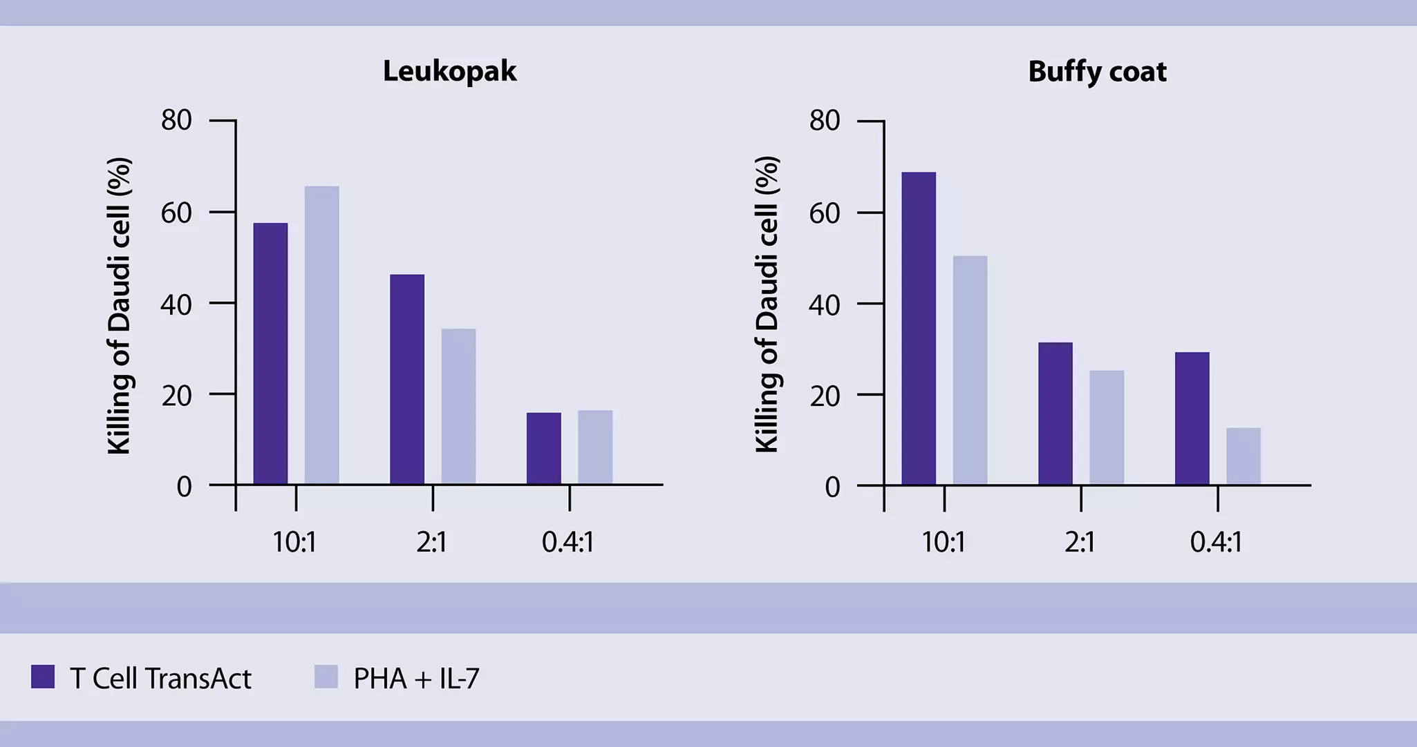
Bar chart illustration showing excellent cytotoxic activity of gamma delta T after isolation with StraightFrom® REAlease® MicroBead Kits and activation with T Cell TransActTM
Measure of cell-killing activity.

Direct measure of the cell-killing activity

The cytotoxic activity of γδ T cells is assessed by coculturing them with a tumor cell line in the presence of appropriate stimuli such as T Cell TransAct, using various effector-to-target cell ratios. After coculture with γδ T cells, the viability of the tumor cells is measured through luciferase assay. In the presence of stimulated γδ T cells, tumor cells are killed effectively.

You have previously chosen International as your location.
What do you want to do?