

Synonyms: S152, S152. LPFS2, T14, TNFRSF7, Tp55
EXTENDED VALIDATION
- Clone:
- M-T271
- Type of antibody:
- Primary antibodies
- Isotype:
- mouse IgG1κ
- Applications:
- FC, MICS, IF, IHC Glossary
- Select a conjugate:
- Select a size:


Other sizes
Related products for CD27 Antibody, anti-human


Flow cytometry applications
APC
APC-Vio 770
Biotin
FITC
PE
PE-Vio 770
PerCP-Vio 700
Vio Bright FITC
VioBlue
VioGreen
Recommended dilution:
1:50
Remark:
Cells should be stained prior to fixation, if formaldehyde is used as a fixative.
Data source:
Miltenyi Biotec
Conjugate:
APC
Used products:
130-114-152, 130-113-626
Description:
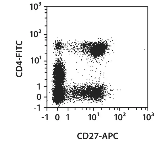
Human peripheral blood mononuclear cells (PBMCs) were stained with CD27 antibodies as well as with CD4 antibodies and analyzed by flow cytometry using the MACSQuant® Analyzer. The Tandem Signal Enhancer has been used to increase binding specificity of tandem-dye–conjugated antibodies. Cell debris and dead cells were excluded from the analysis based on scatter signals and propidium iodide fluorescence or 4',6-diamidino-2-phenylindole (DAPI) fluorescence, as in the case of tandem conjugates.
MICS (MACSima Imaging Cyclic Staining) applications
APC
FITC
PE
Fixation:
frozen – Acetone
Species tested:
human
Recommended dilution:
1:50
Data source:
Miltenyi Biotec
Compatible applications:
IF, IHC
Conjugate:
APC
Used products:
130-113-626, 130-114-152
Testing grade:
Extended validation for CD27 Antibody, anti-human
Specificity
In order to compare the epitope specificity of an antibody, the clone being used is compared with other known clones recognizing the same antigen in a competition assay.
Other clones
Overlap in epitope recognition with M-T271
57703
partial
L128
partial
LG.3A10
partial
LG.7F9 (h/m/r)
partial
REA499
partial
O323
high
Cells were incubated with an excess of purified unconjugated CD27 (M-T271) antibody followed by staining with fluorochrome-conjugated antibodies from other known clones targeting the same antigen. Fluorescence intensity was then used to assess epitope overlap: a minimal or absent signal indicated high epitope overlap, an intermediate signal suggested partial overlap, and a strong signal reflected low overlap.
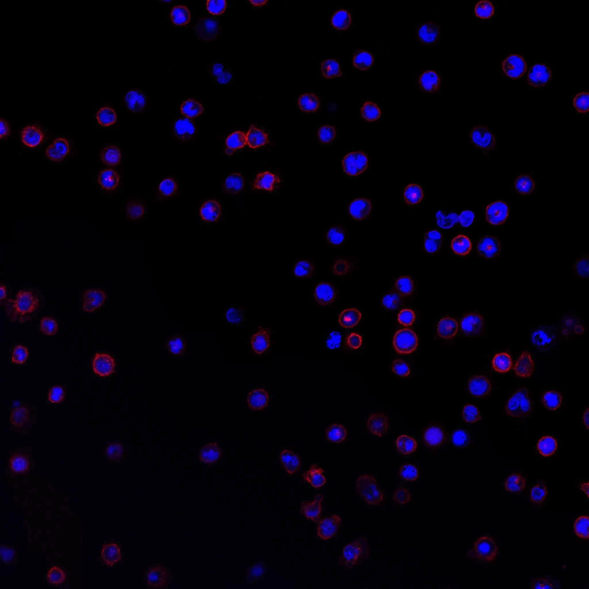
To ensure antibody specificity, the target gene is knocked out in a suitable cell line using the CRISPR/Cas9 system and the knockout is confirmed by sequencing of the target locus. The antibody is considered to bind specifically to the intended epitope if no antibody binding to the knockout cells can be detected. The antibody staining is controlled by fluorescence microscopy and/or flow cytometry.
WT
KO

Fluorescence microscopy image of CD27 knockout cells. Wild type (WT, left) and knockout cells (KO, right) were stained with CD27-PE (M-T271, red) and counterstained with DRAQ5 (blue) as DNA stain.

Overlay histogram showing flow cytometric analysis of CD27 knockout cells. Wild type (red) and knockout cells (blue) were stained with CD27-PE, clone (M-T271). Flow cytometry was performed with the MACSQuant® Analyzer. Cell debris, dead cells and cell doublets were excluded from the analysis based on scatter signals and propidium iodide fluorescence.
Sensitivity
Selected fluorochrome conjugated antibodies from Miltenyi Biotec were compared to commercially available hybridoma clones in flow cytometry analysis.
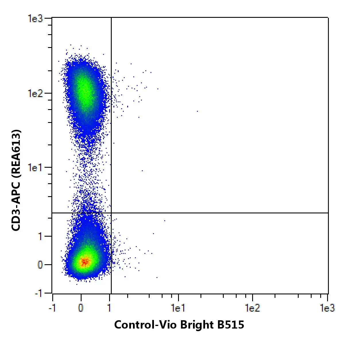

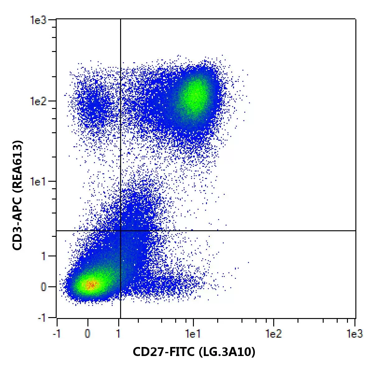

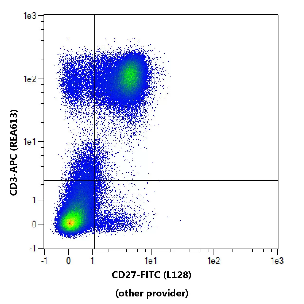
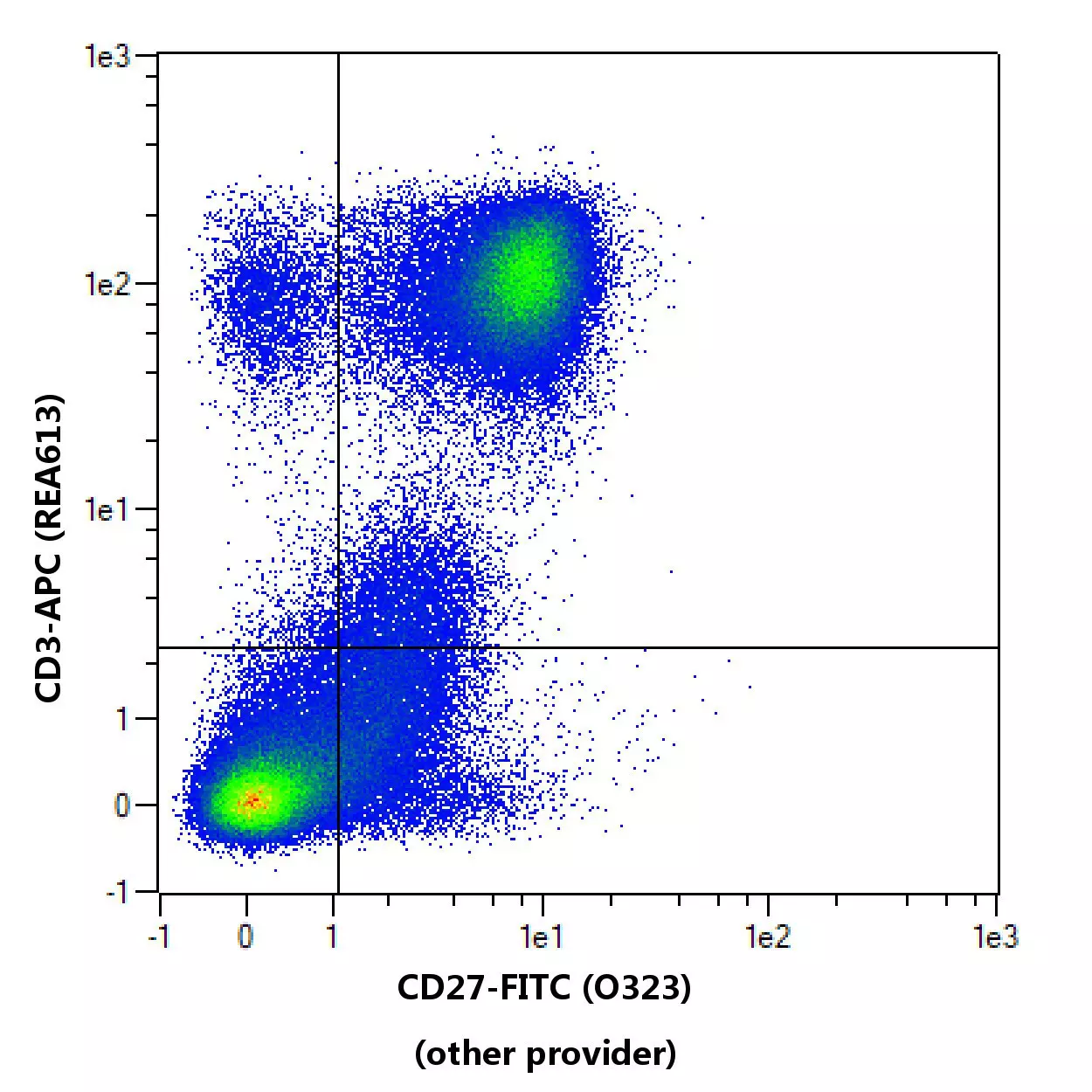

Flow cytometric comparison of different clones for CD27. Human peripheral blood mononuclear cells (PBMCs) were stained with CD27antibodies and with a suitable counterstaining. As a control, CD27 antibody staining was omitted and cells were measured in the same channels. Flow cytometry was performed with the MACSQuant® Analyzer. Cell debris, dead cells, and cell doublets were excluded from the analysis based on scatter signals and 4',6-diamidino-2-phenylindole (DAPI) fluorescence. No FcR Blocking Reagent was used. The recommended titers of respective antibodies from different suppliers were used.
To provide an indication on how an antibody performs after fixation of cells, in-house data on staining results before and after fixation with 3.7% formaldehyde using Miltenyi Biotec antibodies are provided. Different experimental settings may lead to different results.
No fixation
Post fixation

Comparison of staining pattern on non-fixed and fixed cells using CD27 (M-T271). The performance of the antibody after fixation was tested by comparing the staining pattern on fresh (no fixation) versus fixed (post fixation) cells.
Specifications for CD27 Antibody, anti-human
Overview
Clone M-T271 recognizes CD27, a member of the tumor necrosis factor receptor family. CD27 is expressed at differing levels on memory B cells, a fraction of plasma cells, and on naive and memory T cells, but is not expressed on naive B cells or effector T cells. It is also expressed on NK cells. CD27 therefore represents a useful marker to distinguish certain T and B cell subsets from each other.
S152, S152. LPFS2, T14, TNFRSF7, Tp55
Detailed product information
Technical specifications
Clone
M-T271
Clonality
monoclonal
Isotype
mouse IgG1κ
Isotype control
Isotype Control Antibody, mouse IgG1
Host
mouse
Type of antibody
Primary antibodies
Species reactivity
human, non-human primate
Cross-reactivity
rhesus monkey (Macaca mulatta), cynomolgus monkey (Macaca fascicularis)
Antigen
CD27
Molecular mass of antigen [kDa]
27
Distribution of antigen
B cells, NK cells, red blood cells, T cells, plasma cells, thymocytes
Entrez Gene ID
RRID
AB_2751156, AB_2751226, AB_2751155, AB_2751221, AB_2784097, AB_2751157, AB_2751160, AB_2751222, AB_2751220, AB_2784096, AB_2751161, AB_2751223, AB_2733628, AB_2733629, AB_2751224, AB_2751158, AB_2751225, AB_2751159, AB_2751154
Resources for CD27 Antibody, anti-human
Certificates
To access a Certificate of Analysis (CoA) follow the link below and enter the specific lot of your product.
Should you wish to request additional documentation, such as a Safety Data Sheet (SDS) in other languages than English or Swedish, an SDS-replacement document, or a Certificate of Origin (CoO), please direct your inquiry to your local technical support team.
 Powered by Bioz
You have previously chosen International as your location.
What do you want to do?