
EXTENDED VALIDATION
 97/100 Bioz rating
- Clone:
- Bio3-18E7
- Type of antibody:
- Secondary antibodies
- Isotype:
- mouse IgG1
- Applications:
- FC, IHC, ICC Glossary
- Select a conjugate:
- Select a size:


Other sizes
Related products for Biotin Antibody


Flow cytometry applications
APC
APC-Vio 770
FITC
PE
PE-Vio 615
PE-Vio 770
PerCP
PerCP-Vio 700
Vio B515
Vio Bright B515
Vio Bright FITC
VioBlue
VioGreen
Recommended dilution:
1:50
Remark:
The antibody is suited for staining of formaldehyde-fixed cells.
Data source:
Miltenyi Biotec
Conjugate:
APC
Used products:
130-113-288, 130-113-850
Description:
Human peripheral blood mononuclear cells (PBMCs) were labeled with CD303 (BDCA-2)-Biotin and fluorescently stained with Anti-Biotin antibodies as well as with CD304 (BDCA-4/Neuropilin-1) antibodies. The Tandem Signal Enhancer has been used to increase binding specificity of tandem-dye–conjugated antibodies. For all other conjugates the FcR Blocking Reagent has been used to avoid Fc receptor–mediated antibody labeling. Cells were analyzed by flow cytometry using the MACSQuant® Analyzer. Cell debris and dead cells were excluded from the analysis based on scatter signals and propidium iodide fluorescence or 4',6-diamidino-2-phenylindole (DAPI) fluorescence, as in the case of tandem conjugates.

Immunohistochemistry applications
Alkaline Phosphatase
Data source:
Miltenyi Biotec
Conjugate:
Alkaline Phosphatase
Used products:
130-092-612
Immunocytochemistry applications
Alkaline Phosphatase
Data source:
Miltenyi Biotec
Conjugate:
Alkaline Phosphatase
Used products:
130-092-612
Extended validation for Biotin Antibody
Sensitivity
Selected fluorochrome conjugated antibodies from Miltenyi Biotec were compared to commercially available hybridoma clones in flow cytometry analysis.
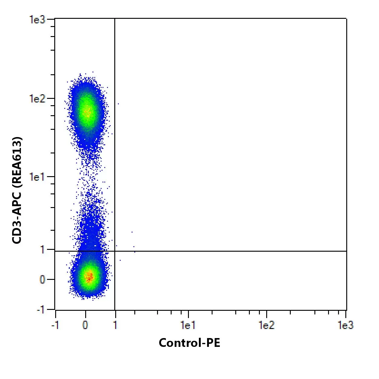

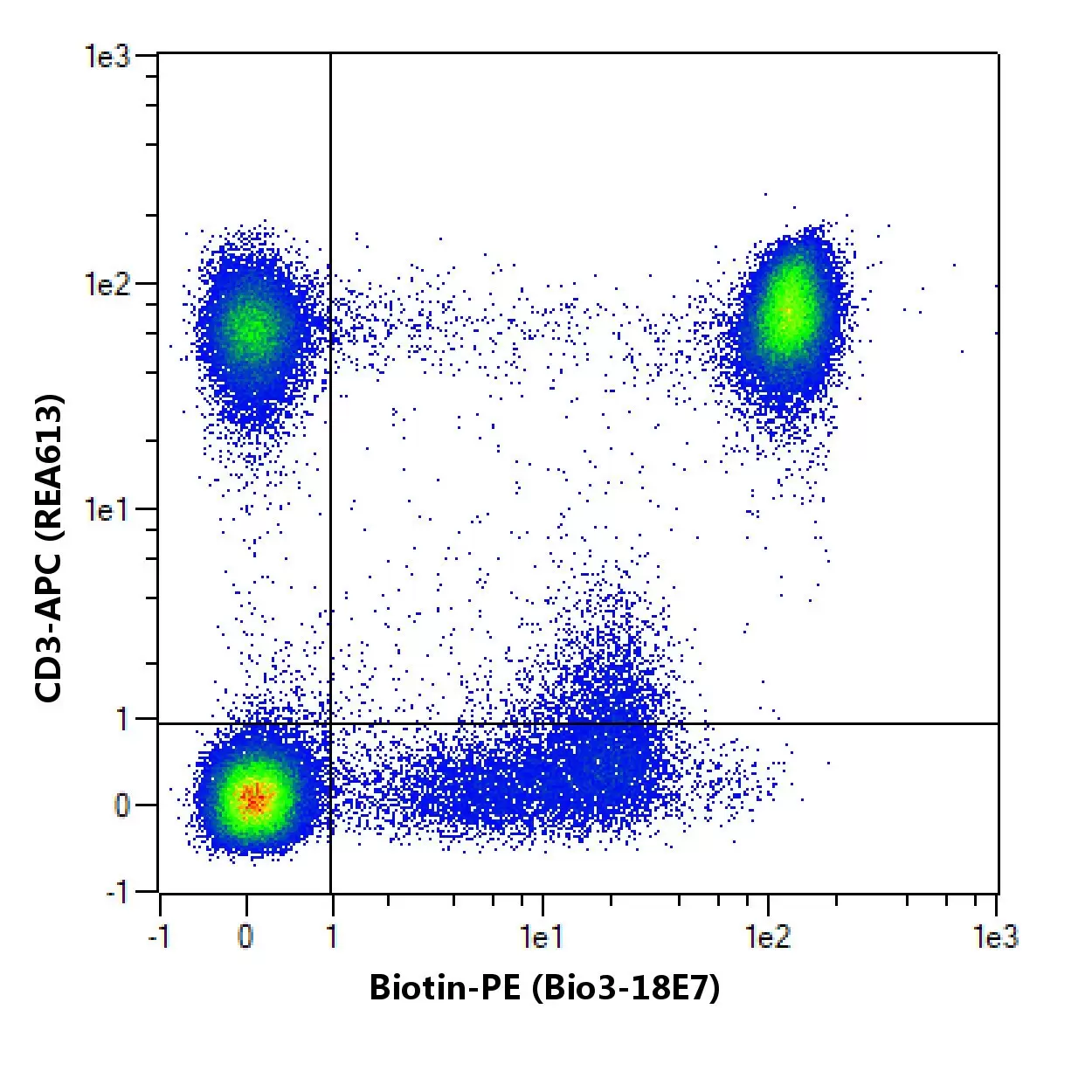
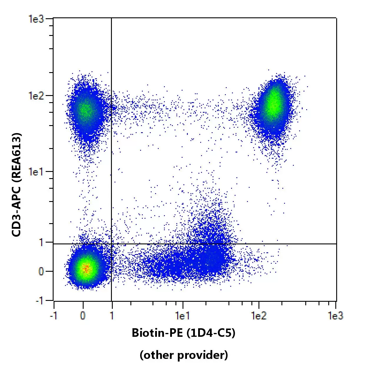
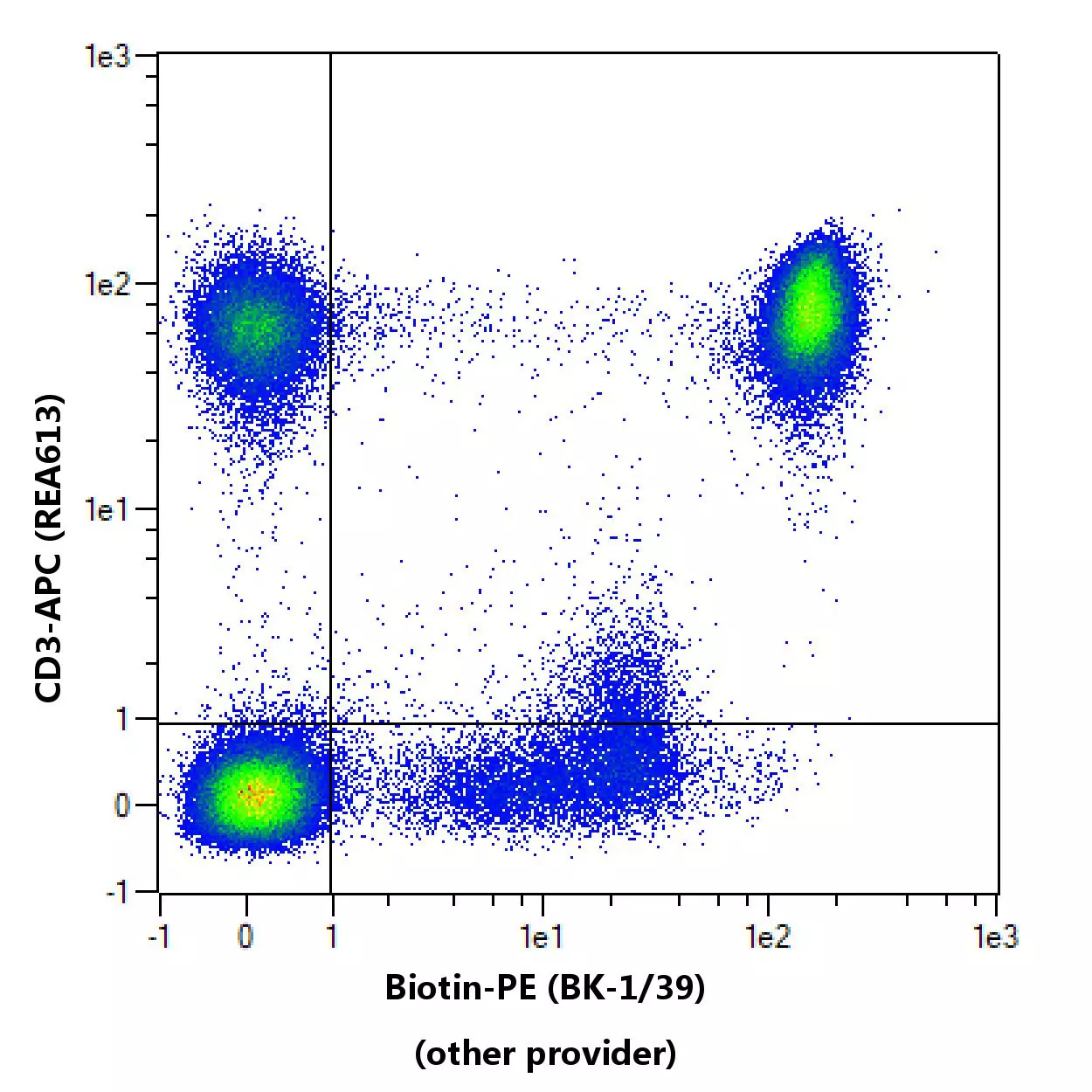
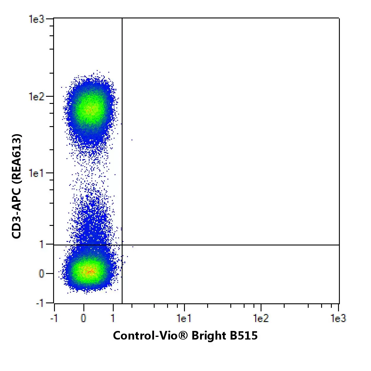
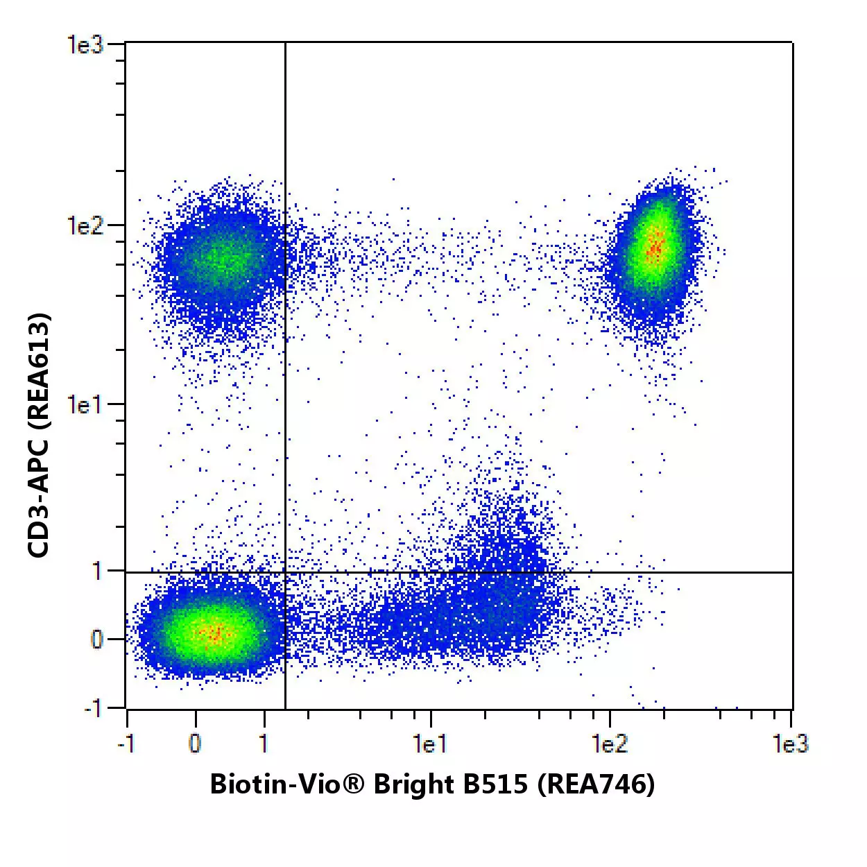
Flow cytometric comparison of different clones for Biotin. Human peripheral blood mononuclear cells (PBMCs) loaded with CD4-Biotin were stained with Biotin antibodies and with a suitable counterstaining. As a control, unloaded human peripheral blood mononuclear cells (PBMCs) were stained with the antibody Biotin and an appropriate counterstain. Flow cytometry was performed with the MACSQuant® Analyzer. Cell debris, dead cells, and cell doublets were excluded from the analysis based on scatter signals and 4',6-diamidino-2-phenylindole (DAPI) fluorescence. No FcR Blocking Reagent was used. The recommended titers of respective antibodies from different suppliers were used.
To provide an indication on how an antibody performs after fixation of cells, in-house data on staining results before and after fixation with 3.7% formaldehyde using Miltenyi Biotec antibodies are provided. Different experimental settings may lead to different results.
No fixation
Post fixation

Comparison of staining pattern on non-fixed and fixed cells using Biotin (Bio3-18E7). The performance of the antibody after fixation was tested by comparing the staining pattern on fresh (no fixation) versus fixed (post fixation) cells.
Specifications for Biotin Antibody
Overview
Anti-Biotin fluorochromes are ideal for fluorescent staining of cells separated with Anti-Biotin MicroBeads, Streptavidin MicroBeads, or with the Anti-Biotin MultiSort Kit. They can also be used to stain cells labeled with cocktails of biotinylated antibodies, for example for quality control when using MACS® Cell Isolation Kits for the isolation of untouched cells. The Anti-Biotin antibody does not bind to free biotin which is often present in cell culture media.
Detailed product information
Technical specifications
Clone
Bio3-18E7
Clonality
monoclonal
Isotype
mouse IgG1
Isotype control
Isotype Control Antibody, mouse IgG1
Host
mouse
Type of antibody
Secondary antibodies
Species reactivity
any
Antigen
Biotin
RRID
AB_2733913, AB_2726353, AB_2726778, AB_2726349, AB_2726072, AB_2733929, AB_871546, AB_2733998, AB_2732953, AB_2726350, AB_2726348, AB_2726076, AB_2726694, AB_2726075, AB_2726352, AB_2726073, AB_2733611, AB_2733612, AB_2733999, AB_2726351, AB_2726071, AB_2726074, AB_2733928, AB_2726077, AB_2733912, AB_2732952, AB_2726354
Resources for Biotin Antibody
Certificates
To access a Certificate of Analysis (CoA) follow the link below and enter the specific lot of your product.
Should you wish to request additional documentation, such as a Safety Data Sheet (SDS) in other languages than English or Swedish, an SDS-replacement document, or a Certificate of Origin (CoO), please direct your inquiry to your local technical support team.
 Powered by Bioz
You have previously chosen International as your location.
What do you want to do?