MACSima® Imaging Cyclic Staining (MICS): a multiomics approach to highly multiplexed spatial biology imaging
The principle of cyclic staining

Miltenyi Biotec’s MICS (MACSima™ Imaging Cyclic Staining) technology enables the simultaneous analysis of hundreds of markers on a single sample. Based on fluorescence microscopy, it uses the principle of cyclic staining with different fluorochrome-conjugated antibodies (protein) or RNAsky™ Detection Probes (RNA) to acquire microscopy data for a multitude of parameters without harming the sample. The process comprises three steps (stain, image, and erase), all performed fully automated by the MACSima System. Both RNA and protein targets are analyzed on the same tissue section, with RNA cycles performed first, followed by protein cycles.
The mechanisms for signal erasure
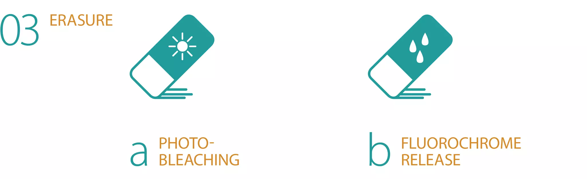
After staining with fluorochrome-conjugated antibodies (protein) or RNAsky Detection Probes (RNA) (01) and image acquisition of the stained sample (02), the fluorescent signal can be erased by either of the two following mechanisms:

(03a) Photobleaching
The fluorescence signal of samples that have been stained with fluorochrome-conjugated antibodies, such as our recombinant REAfinity™ Antibodies, can be erased via photobleaching.
To do this, the MACSima System will move your sample to the bleaching unit, where the area of interest is illuminated for a short time to bleach the attached fluorochromes. After photobleaching, the instrument initiates the next staining cycle in a fully automated fashion.

(03b) Fluorochrome release
Staining of samples with REAdye_lease™ (and REAlease®) fluorochrome-conjugated antibody complexes or RNAsky Detection Probes allows for signal erasure via a controlled release of fluorochromes.
For controlled fluorochrome release, the MACSima System simply adds the release reagent to your sample after the imaging cycle. This erasure mechanism is gentle and particularly efficient as the signal is erased from the entire sample at once.
After fluorochrome release, the instrument initiates the next staining cycle in a fully automated fashion. For maximum flexibility, both erasure mechanisms can be freely combined during one experiment.

Want to learn more about spatial biology and MICS technology?
Go through our MACS Handbook chapters dedicated to spatial biology
You have previously chosen International as your location.
What do you want to do?