Human CD4+CD25+ regulatory T cell isolation, in vitro suppression assay, and analysis
In vitro human regulatory T cell suppression assay
This application protocol describes every step from human Treg and Tresp cell isolation to their co-cultivation in an in vitro suppression assay using the Treg Suppression Inspector and their flow cytometric analysis.
Protocol
Human CD4+CD25+ regulatory T cell isolation, in vitro suppression assay, and analysis
3 h
Isolation of CD4+CD25+ Treg cells
2 h
Staining of Treg and Tresp cells and flow cytometric analysis
30 min
Preparation of in vitro suppression assay
5 d
Cell harvest from in vitro suppression assay
2 h
Flow cytometric analysis of in vitro suppression assay
Protocol

This protocol includes detailed instructions for preparing necessary reagents and materials and then carrying out each step.
Separation buffer
1× PBS (pH 7.2), 0.5% BSA, 2 mM EDTA
or
6×1.5 L autoMACS® Running Buffer – MACS® Separation Buffer
Preparation of in vitro suppression assay
Suppression medium
The suppression medium can be stored for up to 7 days under sterile conditions at 4 °C.
500 mL TexMACS™ Medium
+ 5% human AB serum
+ 1% PenStrep
Isolation of PBMCs
- When working with anticoagulated peripheral blood or buffy coat, PBMCs should be isolated by density gradient centrifugation, for example, using Ficoll-Paque™ according to the manufacturer’s instructions.
- Collect a sample of PBMCs for subsequent flow cytometric analysis. See "Flow cytometric analysis".
Magnetic labeling of non-CD4+ cells
- All steps described below for the magnetic labeling and depletion of non-CD4+ cells, as well as the labeling and selection of CD25+ cells, are performed using the CD4+CD25+ Regulatory T Cell Isolation Kit, human.
- Treg and Tresp cells are simultaneously isolated with the CD4+ CD25+ Regulatory T Cell Isolation Kit, human from one blood sample. Treg cells (CD4+ CD25+) are the final positive fraction and Tresp cells (CD4+ CD25–) are the final negative fraction.
- Volumes for magnetic labeling given below are for up to 1×10⁷ total cells. When working with fewer than 1×10⁷ cells, use the same volumes as indicated. When working with higher cell numbers, scale up all reagent volumes and total volumes accordingly (e.g. for 2×10⁷ total cells, use twice the volume of all indicated reagent volumes and total volumes).
- Determine cell number.
- Centrifuge cell suspension at 300×g for 10 minutes. Discard supernatant.
- Resuspend cell pellet in 90 μL of separation buffer per 1×10⁷ total cells.
- Add 10 μL of CD4+ T Cell Biotin-Antibody Cocktail per 1×10⁷ total cells.
- Mix well and incubate for 5 minutes in the refrigerator (2–8 °C).
- Add 20 μL of Anti-Biotin MicroBeads per 1×10⁷ total cells.
- Mix well and incubate for 10 minutes in the refrigerator (2–8 °C).
- Adjust the volume to 500 μL with separation buffer.
▲ Note: Resuspend up to 1×10⁸ cells in 500 μL of separation buffer. For higher cell numbers, scale up buffer volume accordingly.
Magnetic depletion of non-CD4+ cells
- The first step of Treg cell isolation is a depletion of non-CD4+ cells. Here, an LD Column is used, which has a capacity of 1×10⁸ labeled cells and 5×10⁸ total cells.
- When using the CD4+CD25+ Regulatory T cell Isolation Kit, human it is not recommended to process more than 1.3×10⁸ total cells on an LD Column. When exceeding this cell number, it is strongly recommended to split the sample and use additional LD Columns.
- Place LD Column(s) in the magnetic field of a MidiMACS™ Separator.
- Prepare column by rinsing with 2 mL of separation buffer. Always wait until the column reservoir is empty before proceeding to the next step.
- Apply cell suspension onto the column.
- Collect unlabeled cells that pass through and wash column twice with 1 mL of separation buffer each. Collect total effluent; this is the unlabeled pre-enriched CD4+ cell fraction which is needed for further Treg and Tresp cell isolation.
- Determine cell number.
Magnetic labeling of CD25+ cells
- Volumes for magnetic labeling given below are for an initial starting cell number of up to 1×10⁷ total cells. For higher initial cell numbers, scale up all volumes accordingly.
- Centrifuge cell suspension at 300×g for 10 minutes. Discard supernatant.
- Resuspend cell pellet in 90 μL of separation buffer per 1×10⁷ total cells.
- Add 10 μL of CD25 MicroBeads II per 1×10⁷ total cells.
- Mix well and incubate for 15 minutes in the refrigerator (2–8 °C).
- Wash cells by adding 1–2 mL of separation buffer and centrifuge at 300×g for 10 minutes. Discard supernatant.
- Resuspend up to 1×10⁸ cells in 500 μL of separation buffer.
▲ Note: For higher cell numbers, scale up buffer volume accordingly.
Magnetic separation of CD25+ cells
- The second step during Treg and Tresp cell isolation is the positive selection of CD25+ cells. Here, two consecutive MS Columns are used, with a capacity of 1×10⁷ labeled cells. To not exceed the capacity, it is recommended to determine the frequency of CD25+ cells in the cell suspension by flow cytometry beforehand.
- To assess the purity of the isolated Treg and Tresp cells, a flow cytometric analysis must be performed. Refer to "Surface immunofluorescent staining of Treg and Tresp cells and flow cytometric analysis" for a detailed protocol.
- Place an MS Column in the magnetic field of a MiniMACS™ Separator.
- Prepare column by rinsing with 500 μL of separation buffer.
- Apply cell suspension onto the column. Collect flow-through containing unlabeled Tresp cells (CD4+CD25–).
- Wash column three times with 500 μL of separation buffer each. Collect unlabeled cells that pass through and combine with the effluent from step 3.
- Remove column from the separator and place it on a suitable collection tube.
- Pipette 1 mL of separation buffer onto the column. Immediately flush out magnetically labeled cells by firmly pushing the plunger into the column.
- To increase purity of CD4+CD25+ cells, the eluted fraction can be enriched over a second MS Column (recommended). Repeat the magnetic separation procedure described in steps 1 to 6 using a new MS Column. The isolated Treg and Tresp cells are now ready-to-use for in vitro suppression assay.
General
- To determine the purity of the isolated Treg and Tresp cells, perform the flow cytometric analysis for the final positive (Treg cells) and negative fraction (Tresp cells). It is also recommended to analyze the starting fraction (collect a sample after "Isolation of PBMCs").
- Volumes given below are for up to 1×10⁷ cells. When working with fewer cells, use the same volumes as indicated. When working with higher cell numbers, scale up all reagent volumes and total volumes, accordingly (e.g. for 2×10⁷ nucleated cells, use twice the volume of all indicated reagent volumes and total volumes).
Immunofluorescent staining
- Determine cell number.
- Centrifuge cell suspension at 300×g for 10 minutes. Discard supernatant.
- Resuspend up to 1×10⁷ nucleated cells per 70 μL of separation buffer.
- Add 10 μL of CD4 Antibody, anti-human, VioBlue® and 20 μL of CD25 Antibody, anti-human, PE.
- Mix well and incubate for 10 minutes in the dark in the refrigerator (2–8 °C).
- Wash cells by adding 1–2 mL of separation buffer per 1×10⁷ cells and centrifuge at 300×g for 10 minutes at 4 °C. Discard supernatant.
- Resuspend cell pellet in a suitable amount of buffer for analysis by flow cytometry.
Purity analysis of Treg and Tresp cells by flow cytometry
- To assess the purity of the isolated Treg and Tresp cells, cells are analyzed by flow cytometry. The analysis should be performed with the cell sample taken in "Isolation of PBMCs" and the cell samples taken in "Magnetic separation of CD25+ cells".
- Identify lymphocytes according to forward scatter (FSC) and sideward scatter (SSC, data not shown).
- Exclude dead cells from the analysis by using a live/dead cell exclusion marker (e.g. propidium iodide) (data not shown).
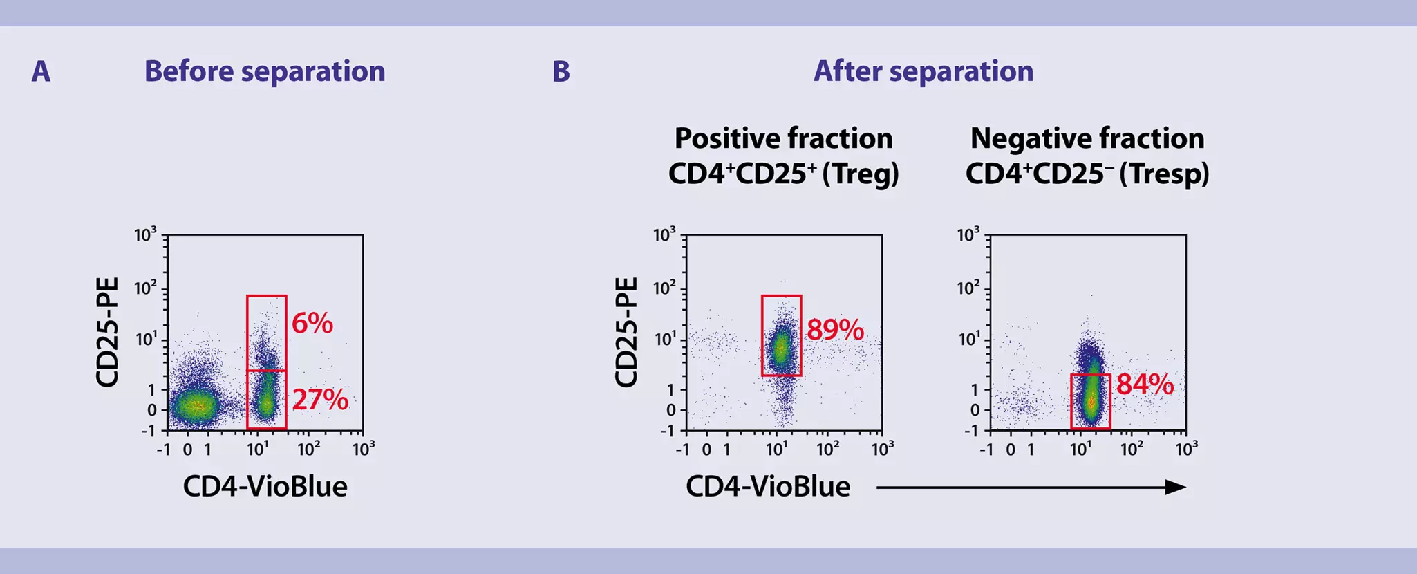
- Analyze the lymphocytes further for their expression of CD4 (x-axis) and CD25 (y-axis) to assess the frequency of CD4+CD25+ Treg cells and of CD4+CD25- Tresp cells in the starting fraction (figure 1A), as well as in the isolated fractions (figure 1B).
 View details
General information
- In this protocol one MACSiBead™ Particle per cell (bead-to-cell ratio 1:1) is used for stimulation.
- For in vitro suppression, Treg cells, Tresp cells, and the Treg Suppression Inspector (amount of MACSiBead Particles) are co-cultured in different ratios in a 96-well flat-bottom plate (table 1). To improve the final flow cytometric resolution, cell numbers can be scaled up accordingly (i.e. 2×10⁵ Tresp cells per well, upscaling of Treg cells and MACSiBead Particles accordingly).
Table 1: Number of Treg cells, Tresp cells, and Treg Suppression Inspector (MACSiBead Particles) per well of a 96-well flat-bottom plate.
| Ratio Tresp:Treg | Tresp | Treg | Treg Suppression Inspector |
| 1:0 | 5×10⁴ | – | 5×10⁴ |
| 1:1 | 5×10⁴ | 5×10⁴ | 1×10⁵ |
| 2:1 | 5×10⁴ | 2.5×10⁴ | 7.5×10⁴ |
| 4:1 | 5×10⁴ | 1.3×10⁴ | 6.3×10⁴ |
| 8:1 | 5×10⁴ | 6×10³ | 5.6×10⁴ |
| 0:1 | – | 5×10⁴ | 5×10⁴ |
| 1:0 | 5×10⁴ | – | – |
| 0:1 | – | 5×10⁴ | – |
| Total | 3×10⁵ | 2×10⁵ | 4×10⁵ |
| Total triplicates | 9×10⁵ | 6×10⁵ | 1.2×10⁶ |
Fluorescent labeling of Tresp cells
- To monitor the proliferation of Tresp cells during the in vitro suppression assay, the cells have to be stained with a fluorescent dye which allows tracking of cell division, e.g., by using the CellTrace™ Violet Cell Proliferation Kit. For more information refer to the manufacturer's instructions. It is also possible to use CSFE or 3H-thymidine incorporation with this in vitro suppression assay protocol. Data shown in this application protocol were acquired with the CellTrace Violet Cell Proliferation Kit.
Cell preparation
- Determine the total number of Treg cells and Tresp cells. For one assay, 9×10⁵ Tresp cells and 6×10⁵ Tregs are needed (table 1). If suppression assay is performed with higher cell numbers, scale up the number of cells accordingly.
- Transfer required volume of cell suspension to suitable tubes.
- Add 5–10 volumes of suppression medium to the cells and centrifuge at 300×g for 10 minutes. Aspirate supernatant completely.
- Resuspend Tresp cells (9×10⁵) in 1800 μL and Treg cells (6×10⁵) in 1200 μL of suppression medium.
▲ Note: The concentration in both cell suspensions is now 5×10⁵ cells/mL. - Pipette the appropriate volumes of Treg and Tresp cell suspension in a 96-well flat-bottom plate. Refer to table 2 in "Preparation of Treg Suppression Inspector, human" for the respective volumes.
Preparation of Treg Suppression Inspector, human
- MACSiBead Particles are bigger in size than MicroBeads and sediment rapidly. It is therefore mandatory to bring the MACSiBead Particles in suspension by vortexing prior to use.
- Resuspend Treg Suppression Inspector thoroughly by vortexing.
- Transfer 60 μL to a suitable tube.
▲ Note: Concentration of Treg Suppression Inspector is 2×10⁷ MACSiBead Particles per mL. - Add 300–600 µL of suppression medium and centrifuge at 300×g for 5 minutes. Aspirate supernatant completely.
- Resuspend Treg Suppression Inspector in 120 μL of suppression medium. The reagent is now ready to use.
▲ Note: Concentration of prepared Treg Suppression Inspector is now 1×10⁷ MACSiBead Particles per mL. - Pipette the appropriate volumes of Treg Suppression Inspector (amount of MACSiBead Particles) to the Treg/Tresp co-cultured cells into the 96-well flat-bottom plate. Refer to table 2 for the respective volumes.
- Fill up wells to a total volume of 210 µL with suppression medium (table 2).
Table 2: Pipetting scheme for one assay with a total volume of 210 µL of cell suspension per well and a concentration of 5×10⁵ cells/mL.
| Ratio Tresp:Treg | Tresp (5×10⁵ cells/mL) | Treg (5×10⁵ cells/mL) | Treg Suppression Inspector (1×10⁷ MACSiBead Particles/mL) | Culture medium |
| 1:0 | 100 µL | – | 5 µL | 105 µL |
| 1:1 | 100 µL | 100 µL | 10 µL | – |
| 2:1 | 100 µL | 50 µL | 7.5 µL | 53 µL |
| 4:1 | 100 µL | 25 µL | 6.5 µL | 79 µL |
| 8:1 | 100 µL | 12.5 µL | 6 µL | 92 µL |
| 0:1 | – | 100 µL | 5 µL | 105 µL |
| 1:0 | 100 µL | – | – | 110 µL |
| 0:1 | – | 100 µL | – | 110 µL |
| Total volume | 600 µL | 387.5 µL | 40 µL | 654 µL |
| Total volume triplicates | 1800 µL | 1200 µL | 120 µL | ~ 2 mL |
Incubation
Incubate the suppression assay at 37 °C and 5–7% CO2 for 5 days.
▲ Note: If a proliferation dye (e.g. CellTrace or CFSE) was used, refer to "Flow cytometric analysis" for a detailed description of final flow cytometric analysis.
▲ Note: If 3H-thymidine was used: After 4 days add the appropriate volume of 3H-thymidine to each well and incubate at 37 °C and 5–7% CO2 for 16 hours. Measure 3H-thymidine incorporation, e.g., by using a liquid scintillation counter.
Immunofluorescent staining
- Harvest the cells by transferring the cell suspension of each well into a separate 5 mL tube.
- Wash cells by adding 1–2 mL of separation buffer for up to 1×10⁷ cells and centrifuge at 300×g for 10 minutes at 4 °C. Discard supernatant.
- Resuspend cells in 90 μL of separation buffer.
- Add 10 μL of CD4-VioBlue® and 20 μL of CD25-PE.
- Mix well and incubate for 10 minutes in the dark in the refrigerator (2–8 °C).
- Wash cells by adding 1–2 mL of separation buffer per 1×10⁷ cells and centrifuge at 300×g for 10 minutes at 4 °C. Discard supernatant.
- Resuspend cell pellet in a suitable amount of separation buffer for analysis by flow cytometry.
Flow cytometric analysis
- Identify lymphocytes according to FSC and SSC.
- Exclude dead cells from the analysis by using a live/dead cell exclusion marker, e.g., propidium iodide (PI).
- Gate CD4+ cells according to CD4 expression (y-axis) and FSC (x-axis).
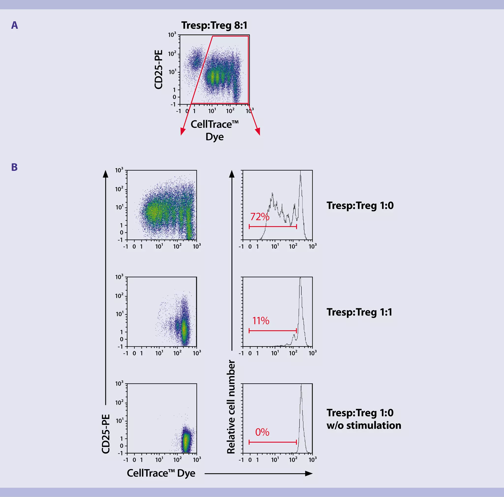
- CD4+CD25+ Treg cells can be distinguished from CD4+CD25– Tresp cells according to CD25 expression (y-axis) and CellTrace™ (x-axis) (figure 2A). Gate CellTrace-positive cells for further cell proliferation analysis (figure 2A, red gate).
- Apply the gating strategy to all samples. Analyze the frequency of proliferating cells using a histogram plot with the fluorescence intensity of CellTrace on the x-axis (figure 2B) and by determining CellTrace dilution (figure 2B, red bar).
Final flow cytometric evaluation
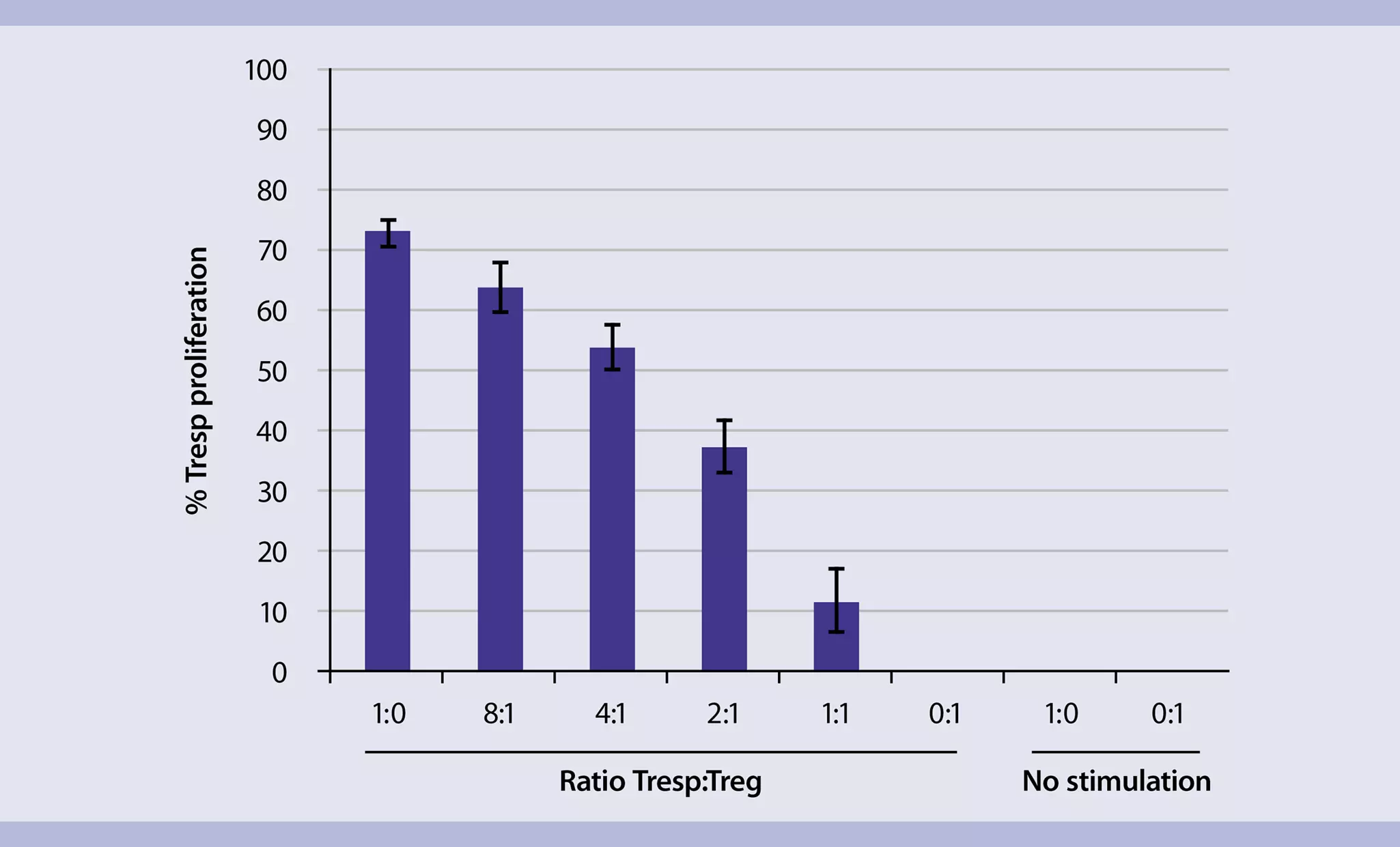
The data obtained by flow cytometry can be summarized in a graphic as depicted in figure 3.
You have previously chosen International as your location.
What do you want to do?