- Use a MHC MACSimer FleX Kit for peptide loading with the peptide of interest or use pre-loaded MACSimers.
- Depending on sample composition (e.g. whole blood, isolated PBMC, enriched T cells) and the individual application field, the optimum amount of peptide-loaded MHC MACSimers for T cell staining can greatly vary. It is recommended to perform an application-specific titration assay prior to final experiment setup.
- Volumes given below are for up to 106 nucleated cells. When working with fewer than 106 cells, use the same volumes as indicated. When working with higher cell numbers, scale up all reagent volumes accordingly (e.g. for 2×10⁶ nucleated cells, use twice the volumes of all indicated reagents).
- Prepare an MHC MACSimer master mix using both MACSimers in a suitable titer with a total volume of 50 µL per staining of 106 nucleated cells.
- Prepare an antibody staining cocktail containing all antibodies with a total volume of 100 µL per sample (see table 1 in tab Materials).
- Pipette 100 µL cells into each reaction tube.
- Stain cells with 1 µL Viobility™ 405/520 Fixable Dye per reaction tube and mix well.
- Incubate cells for 15 minutes in the dark at room temperature (+20 to +25 °C).
- Wash cells with 1 mL staining buffer per reaction tube and centrifuge at 300×g for 10 minutes. Aspirate supernatant completely.
- Resuspend cells in 50 µL MHC MACSimer master mix and incubate for 10 minutes in the dark at room temperature.
- Wash cells with 1 mL staining buffer per reaction tube and centrifuge at 300×g for 10 minutes. Aspirate supernatant completely.
- Resuspend cells in 100 µL antibody staining cocktail per reaction tube. Incubate for 10 minutes in the dark at room temperature.
- Wash cells with 1 mL staining buffer per reaction tube and centrifuge at 300×g for 10 minutes. Aspirate supernatant completely.
- Resuspend cells in 200 µL staining buffer. Acquire 180 µL of cells using a MACSQuant® Analyzer 16.
▲ Note: Use a suitable compensation for the measurement.
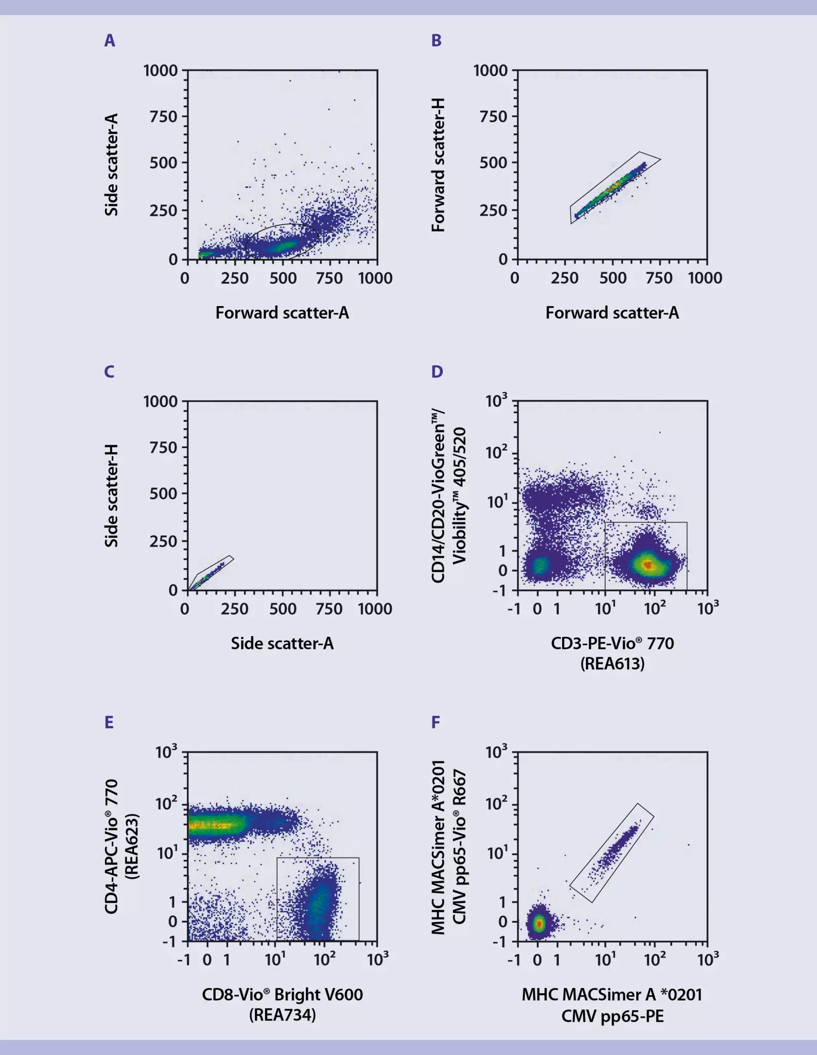
 View details