Recombinant SARS-CoV-2 Spike-Prot (HEK)-Biotin, Streptavidin, PE, Streptavidin, PE-Vio® 770, and fluorochrome-conjugated antibodies as well as 7-AAD Staining Solution are available as single products or all-in-one in the SARS-CoV-2 Spike B Cell Analysis Kit, anti-human (# 130-128-022).
Analysis of SARS-CoV-2–specific B cells using recombinant SARS-CoV-2 proteins
This application protocol describes the preparation of tetramer solutions, staining, and flow cytometric analysis of SARS-CoV-2–specific B cells using recombinant SARS-CoV-2 spike proteins.
Protocol
Preparation of PEB buffer
Prepare a solution containing phosphate-buffered saline (PBS), pH 7.2, 0.5% bovine serum albumin (BSA), and 2 mM EDTA by diluting MACS® BSA Stock Solution (# 130-091-376) 1:20 with autoMACS® Rinsing Solution (# 130-091-222). Keep buffer cold (2−8 °C). Degas buffer before use, as air bubbles could block the column.
Preparation of spike-tetramer solutions
The following protocol is an example when using Streptavidin, PE and Streptavidin, PE-Vio 770. Other streptavidin conjugates can be used as well. Always check the concentration of the stock beforehand and adapt the protocol accordingly.
For the preparation of spike-tetramer solutions, Recombinant SARS-CoV-2 Spike-Prot (HEK)-Biotin was conjugated to Streptavidin, PE and Strepdavidin, PE-Vio 770 with a ratio of 4 mol to 1 mol, respectively (refer to table 1).
▲ Note: Always prepare spike-tetramer solutions fresh before the use. They can be stored for up to 24 hours at 2–8 °C protected from light.
- Resuspend Recombinant SARS-CoV-2 Spike-Prot (HEK)-Biotin in deionized, sterile-filtered water to a final concentration of 0.1 mg/mL (e.g. use 250 µL water for 25 µg protein).
▲ Note: To avoid repeated freeze-thaw cycles prepare working aliquots from the stock solution of Recombinant SARS-CoV-2 Spike-Prot (HEK)-Biotin. Store the working aliquots at –70 °C. Once thawed, the protein can be stored for up to three days at 2–8 °C. - Dilute Streptavidin, PE as well as Streptavidin, PE-Vio 770 to a concentration of 50 µg/mL in PEB buffer.
▲ Note: Check the concentration of the streptavidin conjugates and dilute to 50 µg/mL, if needed. - Mix Recombinant SARS-CoV-2 Spike-Prot (HEK)-Biotin, Streptavidin, PE or Streptavidin, PE-Vio 770, and PEB buffer carefully as indicated in table 1 and incubate for 15 minutes at room temperature.
Each test indicated in table 1 corresponds to 5×10⁶ B cells (or 107 PBMCs). When working with less than 5×10⁶ B cells (or 107 PBMCs), use the same volumes as indicated for one test. When working with more than 5×10⁶ B cells (or 107 PBMCs), scale up all reagent volumes accordingly.
Table 1: Volumes of spike-tetramer solutions using Streptavidin, PE
| Spike-tetramer-PE | |||
| No. of tests | Recombinant SARS-CoV-2 Spike-Prot (HEK)-Biotin (0.1 mg/mL) | Streptavidin, PE (50 µg/mL) | PEB buffer |
| 1 | 3 µL | 0.6 µL | 1.4 µL |
| 5 | 15 µL | 3 µL | 7 µL |
| 10 | 30 µL | 6 µL | 14 µL |
| 20 | 60 µL | 12 µL | 28 µL |
Table 2: Volumes of spike-tetramer solutions using Streptavidin, PE-Vio 770
| Spike-tetramer-PE-Vio 770 | |||
| No. of tests | Recombinant SARS-CoV-2 Spike-Prot (HEK)-Biotin (0.1 mg/mL) | Streptavidin, PE-Vio 770 (50 µg/mL) | PEB buffer |
| 1 | 6 µL | 1.2 µL | 2.8 µL |
| 5 | 30 µL | 6 µL | 14 µL |
| 10 | 60 µL | 12 µL | 28 µL |
| 20 | 120 µL | 24 µL | 56 µL |
▲ Note: If using an IgG Antibody, the other antibodies should not be IgG as isotype. Therefore, the use of REAfinity™ Antibodies for this panel design is not recommended, as REAfinity Antibodies are all IgG isotype.
Preparation of the antibody staining cocktail
- For each test (5×10⁶ B cells or 107 PBMCs), mix 2 μL of each fluorochrome-conjugated antibody, 5 μL of 7-AAD Staining Solution, 5 μL of spike‑tetramer-PE, and 10 μL of spike-tetramer-PE-Vio 770.
- Fill up to 100 μL with PEB buffer.
▲Note: Try different volumes of tetramers conjugates in order to achieve the best double staining results (refer to section Flow cytometric analysis).
Table 3: Suggested antibody panel
| Channel | Fluorochrome | Clone |
| V1 | IgG Antibody, anti-human, VioBlue® | IS11-3B2.2.3 |
| V2 | IgA Antibody, anti-human, VioGreen™ | IS11-8E10 |
| B1 | CD27 Antibody, anti-human, Vio Bright FITC | M-T271 |
| B2 | Spike-tetramer-PE | |
| B3 | 7-AAD Staining Solution | 7-AAD |
| R1 | IgM Antibody, anti-human, APC | PJ2-22H3 |
| R2 | CD19 Antibody, anti-human, APC-Vio 770 | LT19 |
| B4 | Spike-tetramer-PE-Vio 770 |
(Optional) Enrichment of CD19+ cells
B cells can be enriched from single-cell suspension of peripheral blood mononuclear cells (PBMCs) using the REAlease® CD19 MicroBead Kit (# 130-117-034) accordingly to the data sheet including removal of magnetic labeling.
Labeling of B cells
- Resuspend up to 5×10⁶ B cells (up to 107 PBMCs) in 100 μL of antibody staining cocktail. Incubate for 30 minutes at 2–8 °C.
- Wash cells by adding 1–2 mL of PEB buffer and centrifuge at 300×g for 10 minutes. Aspirate supernatant completely.
- Resuspend cell pellet in a suitable amount of PEB buffer and proceed to flow cytometric analysis.
Flow cytometric analysis
- Prior to acquisition of samples in a flow cytometer, e.g., the MACSQuant® Analyzer 10, make sure the lasers are properly calibrated (by using, e.g., MACSQuant Calibration Beads) and compensation of the spillover of fluorochrome emissions has been established in advance. Prior to acquisition adjust also forward scatter (FSC)/side scatter (SSC) parameters to properly locate all leukocyte populations in the plots.
Data analysis
Antigen-specific B cells are relatively rare cells. The detection of such cells requires the acquisition of a large number of events. Pre-enrichment of B cells using, e.g., the REAlease CD19 MicroBead Kit (# 130-117-034), prior to flow cytometry will decrease the total number of cells which need to be acquired in order to detect rare events.
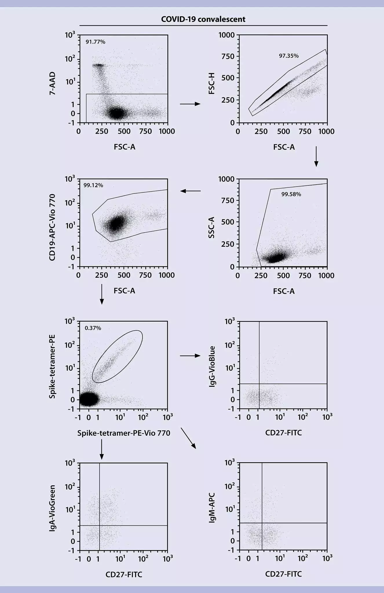
- Exclude dead cells from the analysis using 7-AAD Staining Solution/FSC to gate viable cells.
- Gate on viable cells and use FSC-H/FSC-A parameters to exclude doublets.
- Gate on singlets and use SSC/FSC parameters to exclude erythrocytes and debris.
- Gate on nucleated cells and use CD19 to identify pan B cells. Alternatively, gate on memory B cells by selecting CD19+ CD27+ cells.
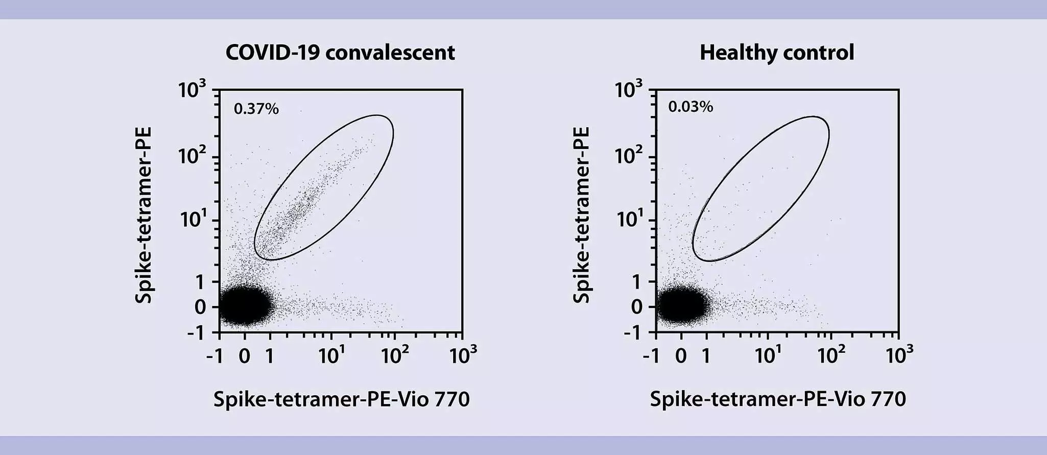
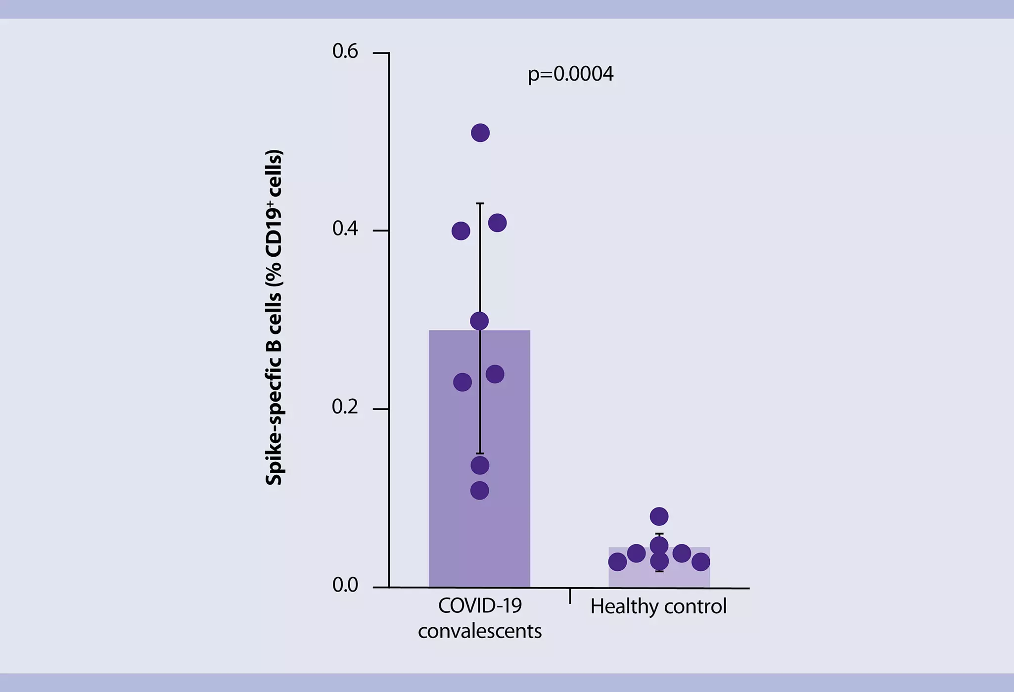
- Gate on pan B cells and use double staining with two spike-tetramer solutions to gate on spike-specific B cells on the diagonal of the dot plot. This gating strategy allows to exclude false-positive cells.
- Gate on antigen-specific B cells and use IgG, IgM, or IgA to detect and quantify each specific isotypes of spike-specific B cells.
You have previously chosen International as your location.
What do you want to do?