Mouse neurons and other neural cell types


1 Introduction 2 Cell types, frequencies, marker expression 3 Miltenyi Biotec applications for neural cells 4 Sample preparation of neural tissue 5 Magnetic separation of neural cells from neural tissue 6 Cultivation of neural cells
1 Introduction
The central nervous system (CNS) comprises two major classes of cells: neurons and glial cells. Neurons receive, process, and transmit information, while glial cells support and protect neurons by maintaining homeostasis, forming myelin, supplying nutrients, destroying pathogens, and removing dead neurons, among other functions. The two processes responsible for the genesis of neurons and glial cells are termed neurogenesis and gliogenesis, respectively. These processes occur at distinct stages of the mammalian development, facilitated by the presence and activity of neural stem cells (NSCs). CNS development begins with the induction of the neuroectoderm to form the neural plate and then the neural tube. The caudal region of the neural tube gives rise to the spinal cord and the rostral region becomes the brain. During these early stages of neural development, cells divide rapidly. Immature neurons migrate from zones of cell proliferation to their final positions and start to extend neurites to form contacts with other cells. After prenatal development, neurogenesis is limited and restricted to the subventricular zone (SVZ) in the lateral wall of the lateral ventricle, and the subgranular zone (SGZ) of the dentate gyrus of the adult mammalian brain. In the peripheral nervous system (PNS), glial cells derive from the neural crest, and comprise Schwann cells, satellite glial cells, enteric glial cells, and olfactory ensheathing cells.
 MACS Handbook:
 Related PDFs:
2 Cell types, frequencies, marker expression
Neural stem cells
Neurogenesis persists in two regions of the mammalian adult brain: the SVZ in the lateral wall of the lateral ventricle and in the SGZ of the dentate gyrus. Adult NSCs are multipotent stem cells that have the capacity to self-renew and differentiate into neurons, astrocytes, and oligodendrocytes. NSCs play a crucial role in the repair and regeneration of the nervous system and express characteristic markers such as, Nestin, Sox2, Pax6, GLAST and glial fibrillary acidic protein (GFAP).
Neurons
The mammalian brain contains between 100 million and 100 billion neurons, depending on the species. Neurons are electrically excitable cells that transmit information through action potentials and neurotransmitters to other neurons or other cell types. Depending on the neurotransmitter and receptors on the receiving cell, the signal may be excitatory or inhibitory. Neurons exhibit extensive morphological and physiological heterogeneity and lack a specific pan cell surface marker, such that pan neuronal cells can only be identified and isolated indirectly by labeling non-neuronal cells.
Astrocytes
Astrocytes are the most abundant cell type of the CNS and exhibit broad morphological and functional heterogeneity. Astrocytes regulate ion and glutamate homeostasis, control the number and function of synapses, contribute to wound healing, form the blood-brain–barrier, and modulate cerebral blood flow. Astroglial cells also act as adult neural stem cells in the subventricular zone of the lateral ventricle’s lateral wall and in the subgranular layer of the dentate gyrus. GLAST is the most abundant glutamate transporter and serves as astrocyte marker in the developing and neonatal mammalian CNS, as well as for radial glia and neural stem cells. The ACSA-2 antigen is specifically expressed on GLAST (ACSA-1)–positive astrocytes and is a second astrocyte-specific surface marker (PMID: 28317180). The antigen targeted by Anti-ACSA-2 is ATP1B2 (PMID: 28373281). Mature astrocytes and radial glia also express GFAP.
Oligodendrocytes
Oligodendrocytes form the myelin sheath around neuronal axons, which facilitates the fast saltatory propagation of action potentials. Several distinct phenotypic developmental stages of oligodendrocytes have been identified, both in vivo and in vitro, based on the expression of specific markers. Morphological criteria alone are often insufficient to characterize each stage. Oligodendrocyte precursor cells (OPCs) are small, round, process-bearing cells with active proliferation and migratory properties. They are identified in vitro with the Anti-A2B5 antibody which recognizes several gangliosides. However, this antibody also labels other cell types in vivo, so more specific markers are used, such as the platelet-derived growth factor receptor alpha (PDGFRα – a transmembrane receptor tyrosine kinase), and the single membrane-spanning chondroitin sulphate proteoglycan NG2 (AN2). These are the most reliable markers for OPCs in vivo and are also used to label adult OPCs (PMID: 18931697). When OPCs begin to differentiate, they express a sulphated surface antigen known as POA (pro-oligodendroblast antigen), which is recognized by the Anti-O4 antibody. During oligodendrocyte development, O4 expression begins on late oligodendrocyte progenitors that are A2B5-positive. While A2B5 expression disappears, O4 expression continues. As the cells differentiate, they synthesize galactocerebroside, myelin basic protein (MBP), proteolipid protein (PLP), myelin oligodendrocyte glycoprotein (MOG), and O1.
Microglia
Microglia (brain macrophages) are the resident immune effector cells of the CNS. Activated microglia also serve as antigen-presenting cells. They are morphologically, immunophenotypically, and functionally related to cells of the monocyte/macrophage lineage and characterized by expression of CD11b in humans and mice (CD11b/c in rat), CD68, F4/80, and MHC class II.
3 Miltenyi Biotec applications for neural cells
Miltenyi Biotec has created dedicated applications to isolate, cultivate, and characterize different neural cell types.
- Isolation and cultivation of neurons from adult mouse brain
- Isolation and cultivation of neurons from neonatal mouse brain
- Isolation and cultivation of CD171 (L1CAM)-positive neurons from neonatal mouse brain
- Isolation and cultivation of astrocytes from adult mouse brain
- Isolation and cultivation of ACSA-2–positive astrocytes from neonatal mouse brain
- Isolation and cultivation of GLAST-positive astrocytes from neonatal mouse brain
- Isolation and cultivation of microglia from adult mouse or rat brain
- Isolation and cultivation of microglia from neonatal mouse or rat brain
- Isolation and cultivation of oligodendrocytes from adult mouse or rat brain
- Isolation and cultivation of O4-positive oligodendrocytes from neonatal mouse or rat brain
- Isolation and cultivation of oligodendrocyte precursor cells (OPCs) from neonatal mouse brain
- Isolation and cultivation of endothelial cells from adult mouse brain
- Isolation of endothelial cells from neonatal mouse brain
A special protocol describing immunofluorescence staining with neural cell antibodies for microscopy, as well as tutorial videos for the isolation of viable astrocytes and microglia can be accessed in the Related resources panel to the right.
4 Sample preparation of neural tissue
Neural cell samples can be obtained from whole brains, a particular brain region, or the spinal cord. Downstream applications to characterize neural cells may require prior sample preparation, such as tissue dissociation, to generate single-cell suspensions. For details, see Chapter mouse brain.
MACS Handbook:
5 Magnetic separation of neural cells from neural tissue
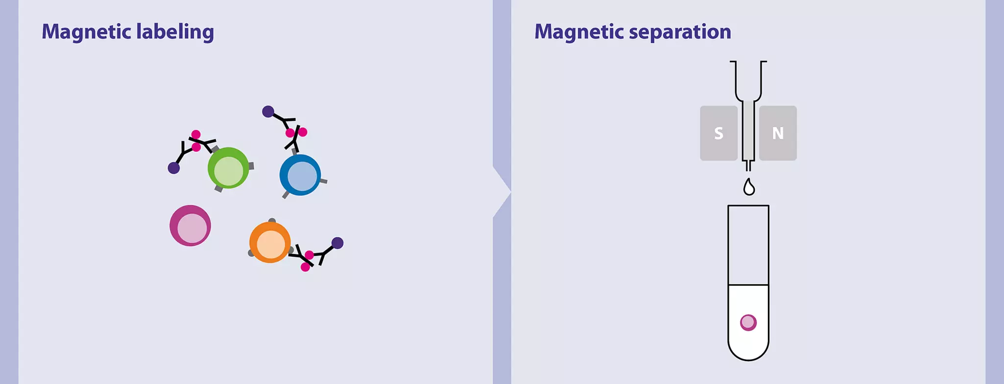
Miltenyi Biotec has developed numerous products for the isolation of distinct neural cell types from single-cell suspensions. Most kits are based on the positive selection of magnetically labeled target cells. Isolation of neurons, however, is done by depleting non-target cells because of the lack of specific neuronal surface markers that would allow positive selection. For details on MACS® Cell Separation Technology, see the MACS Handbook chapter Magnetic cell separation.
MACS Handbook:
5.1 Isolation of neurons and neuronal precursor cells
| Starting material | Isolation strategy | Comments | Automation | Product |
| Neurons | ||||
| Single-cell suspension | Depletion of non-target cells | For neonatal or adult mouse brain dissociated with either the Neural Tissue Dissociation Kit – Postnatal neurons or the Adult Brain Dissociation Kit, mouse, rat | Yes* | Neuron Isolation Kit, mouse |
| Single-cell suspension | Positive selection of target cells | For neonatal mouse brain dissociated with the Neural Tissue Dissociation Kit (T) | Yes* | CD171 (L1CAM) MicroBead Kit, mouse |
| Single-cell suspension | Depletion of non-target cells | For mouse brain tissue older than postnatal day 7 dissociated with the Adult Brain Dissociation Kit, mouse and rat | Yes* | Adult Neuron Isolation Kit, mouse |
| Single-cell suspension | Positive selection of targeted retinal ganglion cells | For rat retina tissue dissociated with the Neural Tissue Dissociation Kit – Postnatal Neurons | Yes | Retinal Ganglion Cell Isolation Kit, rat |
| Neuronal precursor cells | ||||
| Single-cell suspension | Positive selection of target cells | For neonatal mouse or rat brain dissociated with the Neural Tissue Dissociation Kit (T) | Yes* | Anti-PSA-NCAM MicroBeads, human, mouse, rat |
| Single-cell suspension | Positive selection of target cells | For neonatal or postnatal mouse brain tissue dissociated with the Neural Tissue Dissociation Kit (P) | Yes | Anti-Prominin-1 MicroBeads, mouse |
* Automated isolation can be performed on the autoMACS® NEO Separator
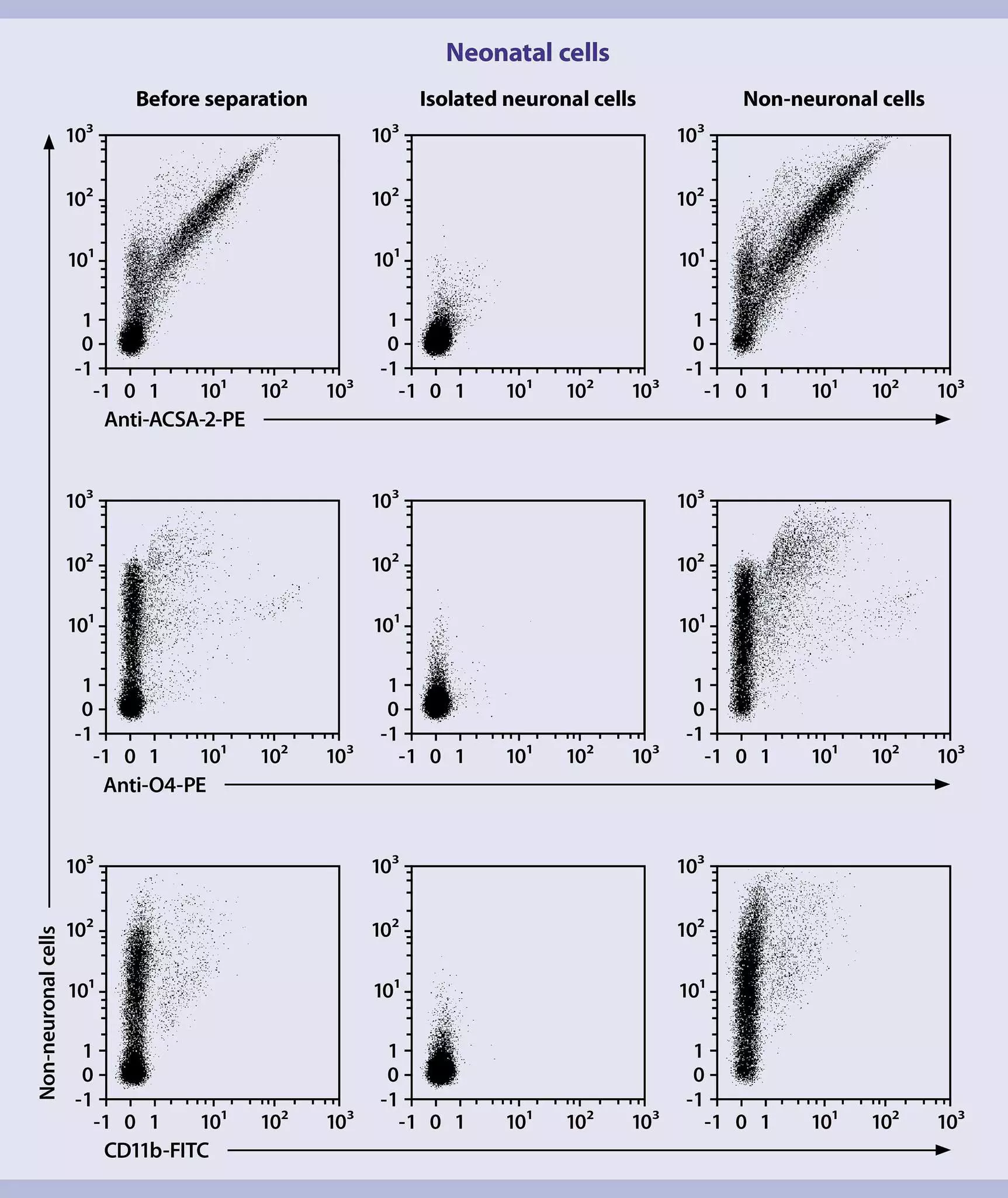
Miltenyi Biotec’s Neuron Isolation Kit, mouse works by depleting non-neuronal cells based on novel non-neuronal cell markers. The kit achieves purities of 97% for animal age ≤P7 and 93% for animal age >P7. The isolated neurons are ready for downstream applications, including cell culture, cellular and molecular analysis, and functional assays.
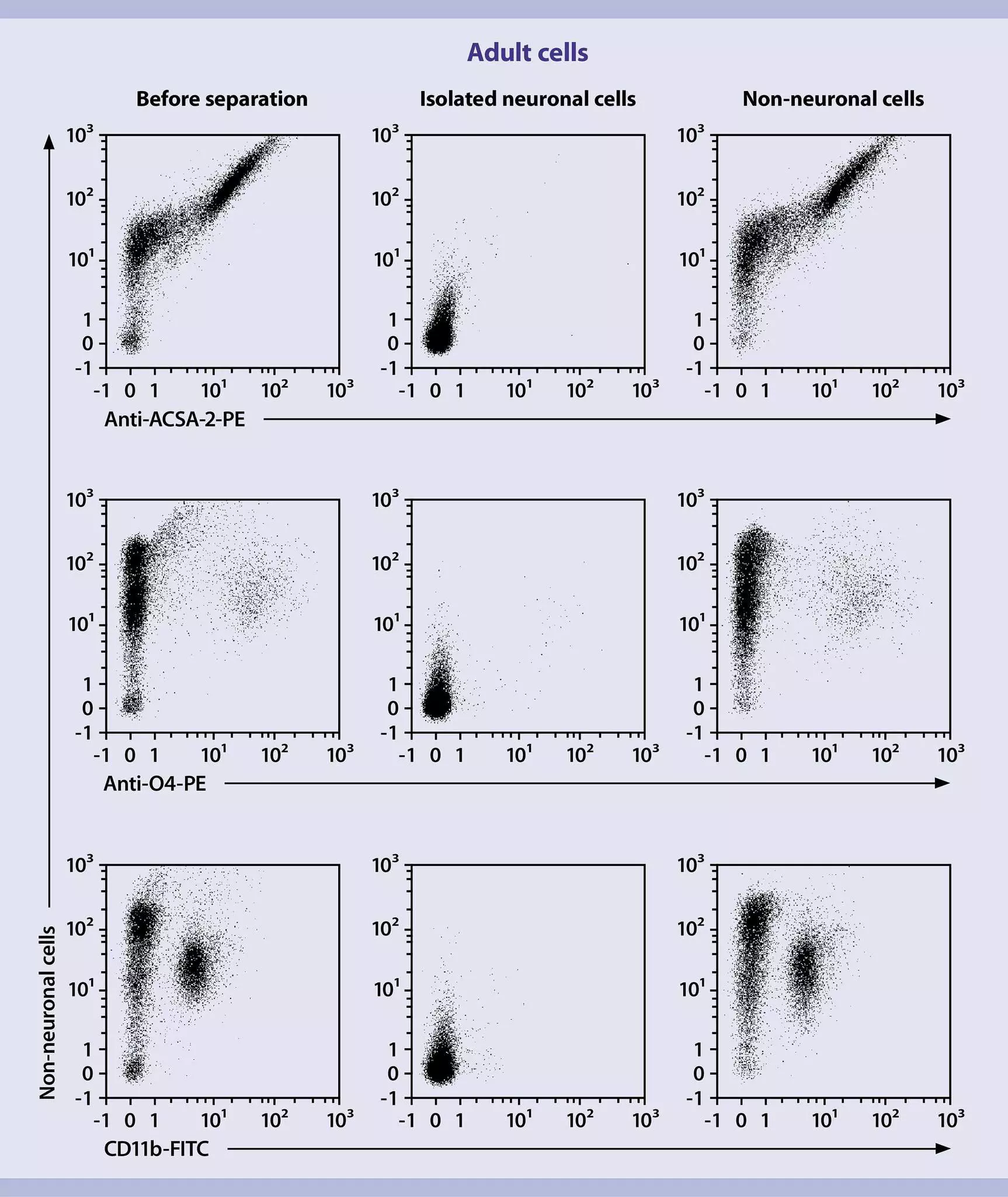
Adult neurons are especially fragile, making the isolation and culture of primary adult neurons challenging. In this regard, isolation and culture are generally limited to embryonic tissues (approx. E16–18, when glial cells are less abundant) or to early postnatal stages before formation of synapses occurs. However, embryonic neurons behave differently from adult neurons in terms of pharmacology, electrophysiology, development, and regenerative and pathological characteristics. Miltenyi Biotec helps to overcome these challenges by offering solutions for adult tissue dissociation and neuronal isolation. The Adult Neuron Isolation Kit, mouse, combined with the Adult Brain Dissociation Kit, mouse and rat and the gentleMACS™ Octo Dissociator with Heaters, allows isolation of viable neurons to high purities from adult mouse brain within half a day.
To assess and further boost purity of isolated neurons, Miltenyi Biotec offers a wide range of antibodies specific to non-neuronal cell surface markers to exclude contamination by glial cells, such as astrocytes, microglia, and oligodendrocytes.
 View details
The CD171 (L1CAM, L1 cell adhesion molecule) antibody and CD171 (L1CAM) MicroBead Kit were developed to detect and isolate L1CAM-positive neurons, such as Purkinje neurons in the cerebellum, from dissociated mouse brain tissue derived from mice younger than eight days.
CD171 (L1CAM) MicroBead Kit, mouse
 View details
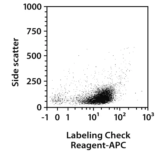
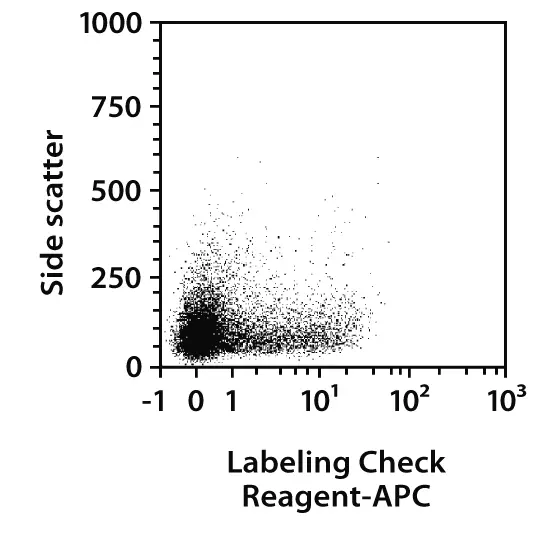
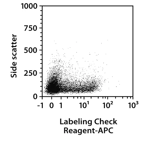
Neuron isolation based on positive selection of CD171. P7 mouse cerebelli were dissociated using the Neural Tissue Dissociation Kit (T). Subsequently, neurons were isolated using the CD171 (L1CAM) MicroBead Kit, mouse. Cells were fluorescently stained with Labeling Check Reagent-APC and analyzed by flow cytometry on the MACSQuant Analyzer. Cell debris and dead cells were excluded from the analysis based on scatter signals and propidium iodide fluorescence.

5.2 Isolation of astrocytes
At a glance: Kits and reagents for the separation of astrocytes
| Starting material | Isolation strategy | Comments | Automation | Product |
| Single-cell suspension | Positive selection of target cells | For dissociated neonatal or adult mouse brain | Yes | Anti-ACSA-2 MicroBead Kit, mouse |
| Single-cell suspension | Positive selection of target cells | For neonatal mouse or rat brain dissociated with the Neural Tissue Dissociation Kit (T) | Yes | Anti-GLAST (ACSA-1) MicroBead Kit, human, mouse, rat |
| PBS-based buffer optimized to remove dead cells during isolation of adult astrocytes | AstroMACS Separation Buffer |
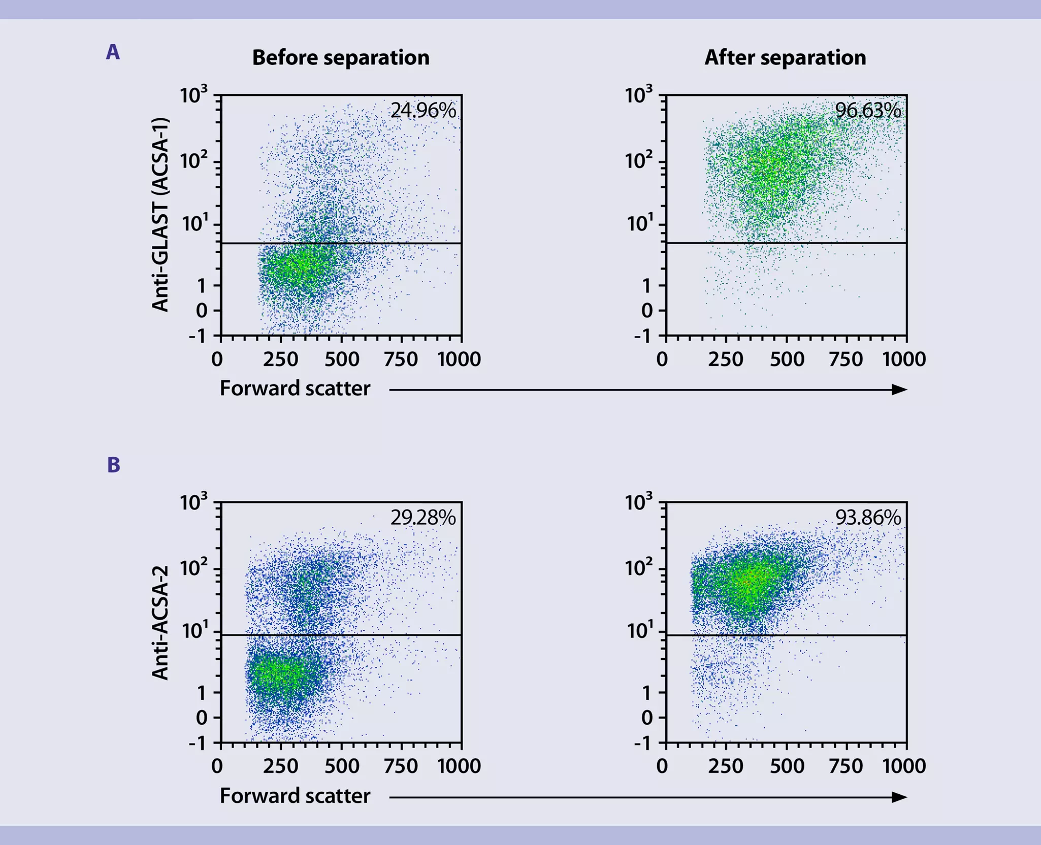
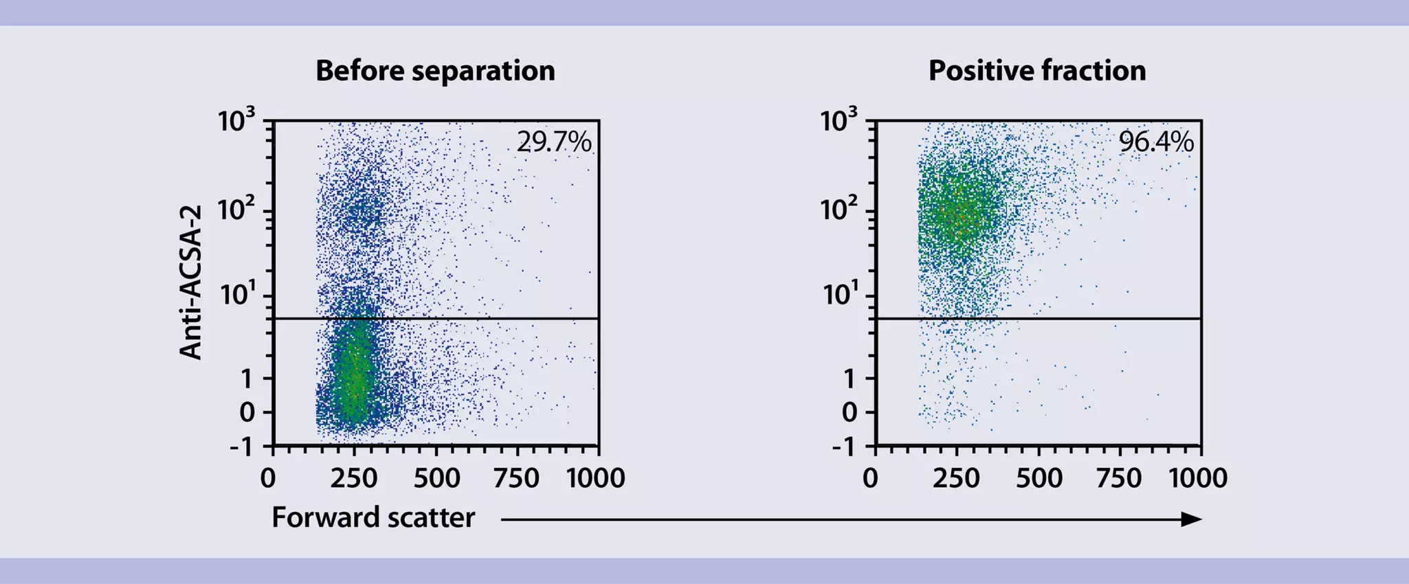
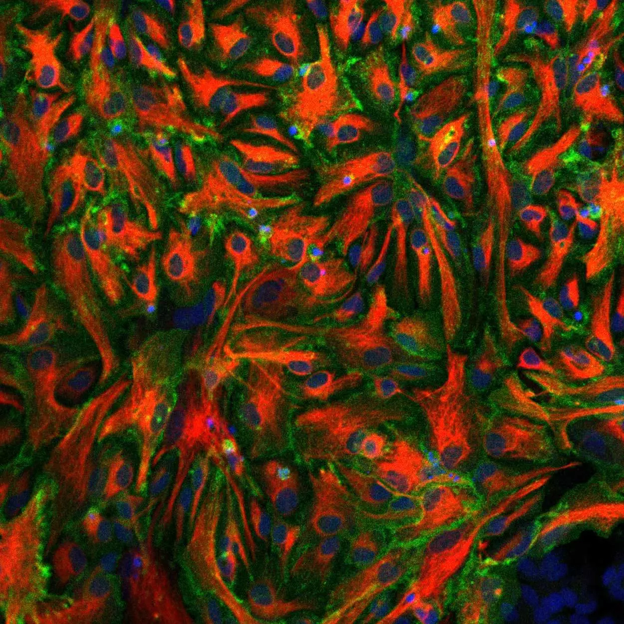
Highly purified viable astrocytes from neonatal and adult mouse brain can be acquired by positive selection using one of the two astrocyte-specific cell surface markers, glutamate-aspartate transporter 1 (GLAST-1, also known as ACSA-1) or astrocyte cell surface antigen-2 (ACSA-2). Both antibodies (Anti-GLAST (ACSA-1) and Anti-ACSA-2) can be used for flow cytometry, immunocytochemistry, and immunohistochemistry analysis.
The Anti-GLAST (ACSA-1) antibody has been developed by Miltenyi Biotec and detects the extracellular epitope of the glutamate transporter GLAST-1 (EAAT1). This antibody was generated by immunization of GLAST-1 knockout mice with GLAST-expressing 1881 cells. The Anti-GLAST antibody labels all GFAP, GS, BLBP-positive astrocytes, reactive astrocytes, radial glia, Müller glia, and Bergmann glia, and shows superior performance in astrocyte detection and separation (PMID: 22374709).
The Anti-ACSA-2 antibody is a novel monoclonal antibody generated by immunizing rats with astrocytes isolated from GFAP-EGFP transgenic mice. Immunohistochemical and flow cytometry analyses demonstrate the high specificity of Anti-ACSA-2 for astrocyte lineage cells (PMID: 28317180). The ACSA-2 epitope was identified recently as ATP1B2 and the authors recommend the Anti-ACSA-2 antibody as “a first-choice method for astrocyte isolation and characterization.” (PMID: 28373281). Unlike GLAST, the ACSA-2 epitope is papain-resistant. Thus, it can be used for astrocyte separation and analysis after papain-based tissue dissociation, as well as for astrocyte isolation from adult mouse brain.
In addition, Miltenyi Biotec offers the Astrocyte Isolation Starter Kit that comprises the reagents necessary to dissociate and isolate GLAST+ astrocytes from rats or mice younger than postnatal day 7.
 Related videos:
5.3 Isolation of oligodendrocyte lineage cells
At a glance: Kits and reagents for the separation of oligodendrocyte lineage cells
| Starting material | Isolation strategy | Comments | Automation | Product |
| Oligodendrocyte precursor cells (OPCs) | ||||
| Single-cell suspension | Positive selection of target cells | Use neonatal mouse brain | Yes | CD140a (PDGFRα) MicroBead Kit, mouse |
| Single-cell suspension | Positive selection of target cells | Use neonatal mouse brain; re-expression of antigen necessary | Yes | Anti-AN2 (NG2) MicroBeads, human, mouse |
| Single-cell suspension | Positive selection of target cells | Use neonatal mouse brain | Yes | Anti-A2B5 MicroBeads, human, mouse, rat |
| Premature oligodendrocytes | ||||
| Single-cell suspension | Positive selection of target cells | Use neonatal or adult mouse or rat brain | Yes | Anti-O4 MicroBeads, human, mouse, rat |
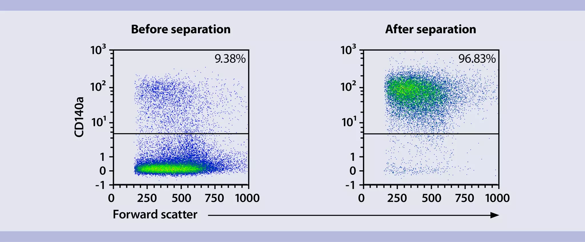
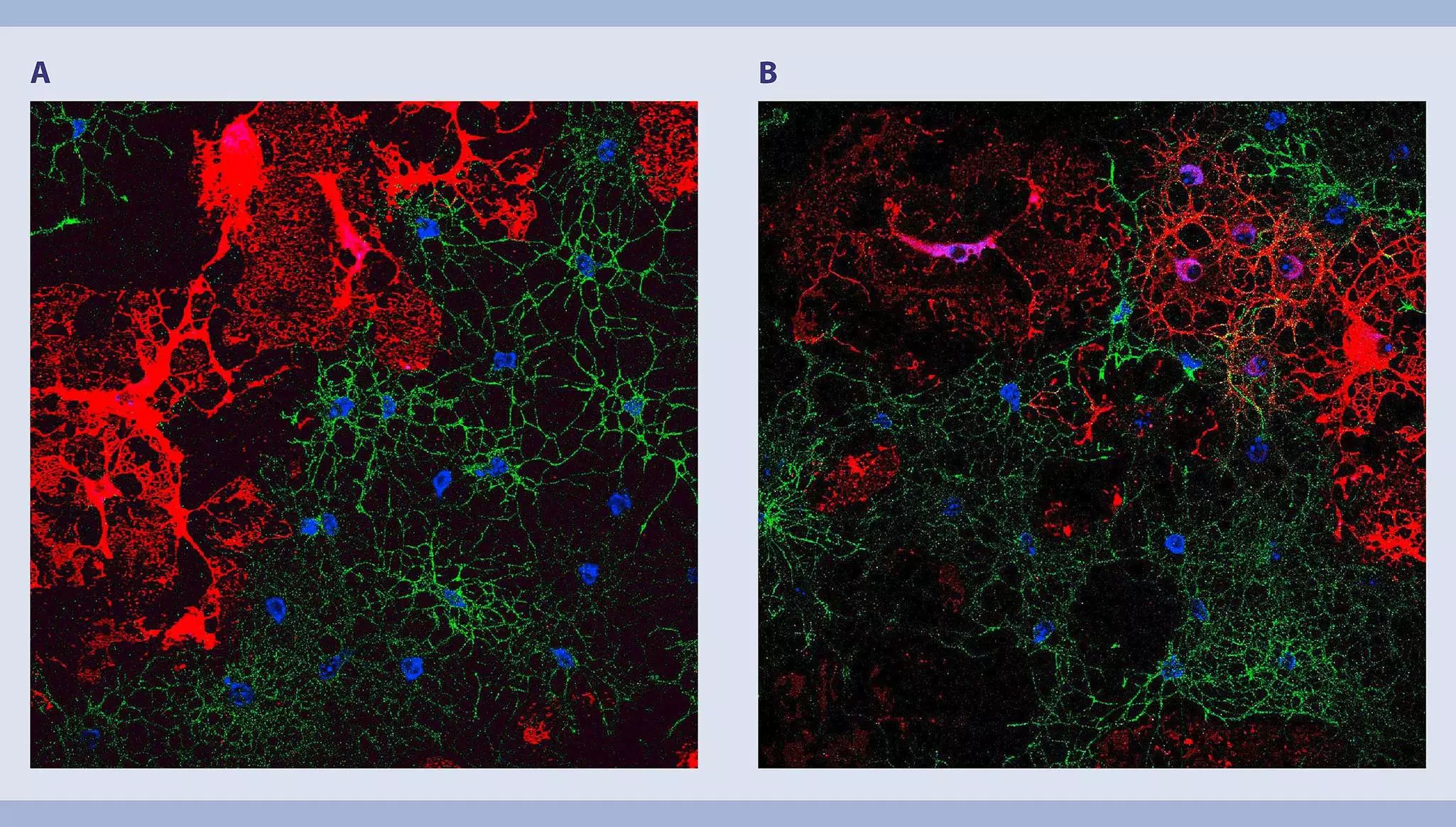
Miltenyi Biotec offers a range of MicroBeads and kits for the isolation of oligodendrocyte lineage cells. Anti-A2B5 MicroBeads, human, mouse, rat enable selection of glial progenitor cells, whereas the CD140a (PDGFRα) MicroBead Kit and Anti-AN2 (NG2) MicroBeads serve to isolate oligodendrocyte precursors.
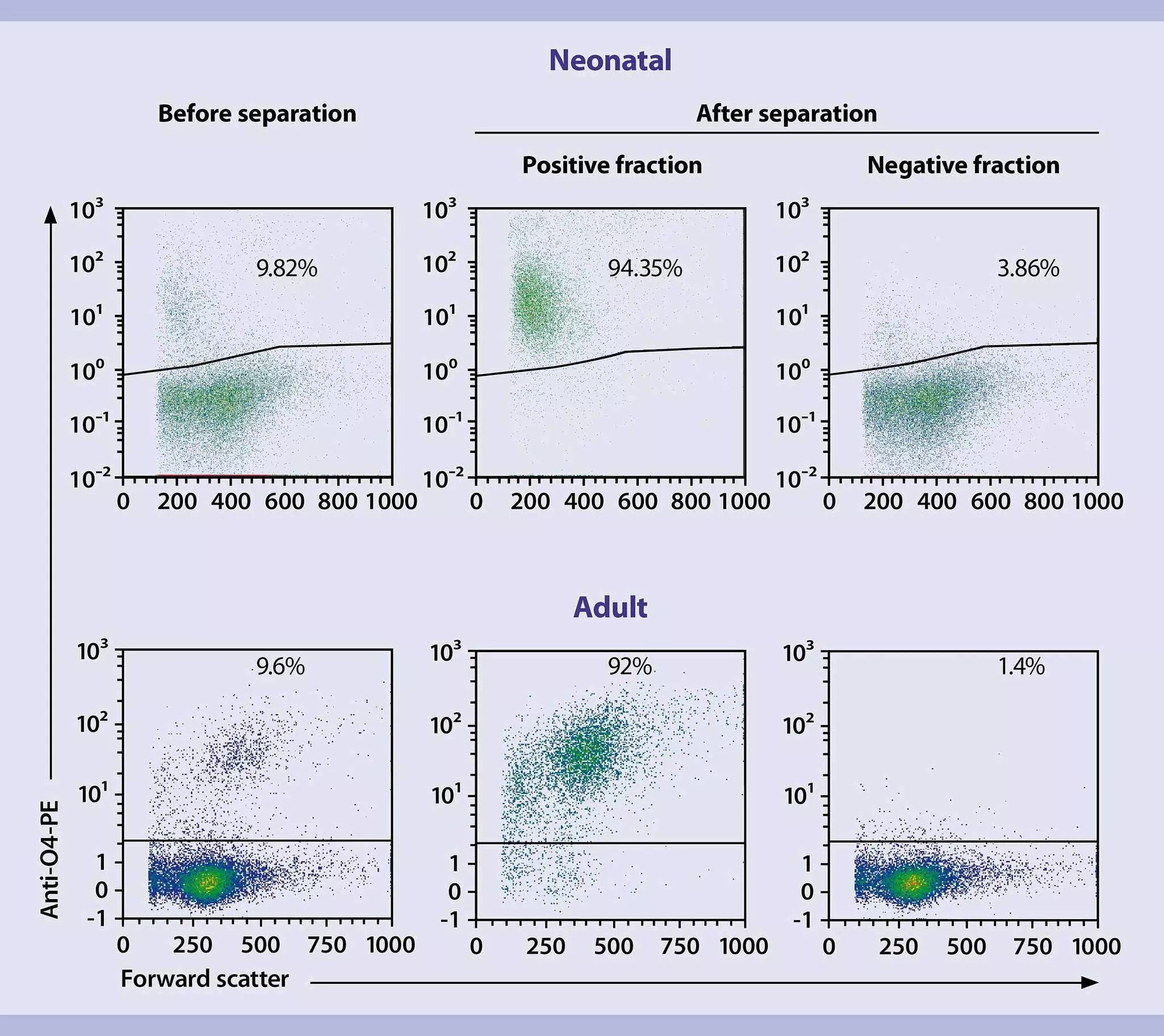
Premature oligodendrocytes can be isolated from neonatal and adult mouse brain tissue with the Anti-O4 MicroBeads, human, mouse, rat.
5.4 Isolation of microglia
| Starting material | Isolation strategy | Comments | Automation | Product |
| Single-cell suspension | Positive selection of target cells | Use neonatal or adult mouse brain | Yes | CD11b (Microglia) MicroBeads, human and mouse |
| Single-cell suspension | Positive selection of target cells | Use neonatal or adult rat brain | Yes | CD11b/c (Microglia) MicroBeads, rat |
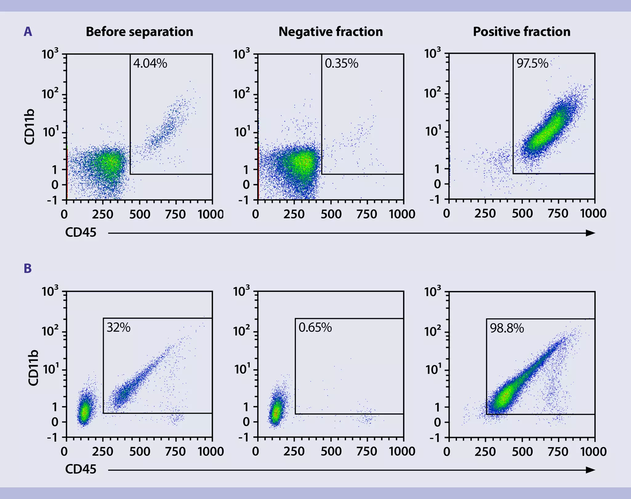
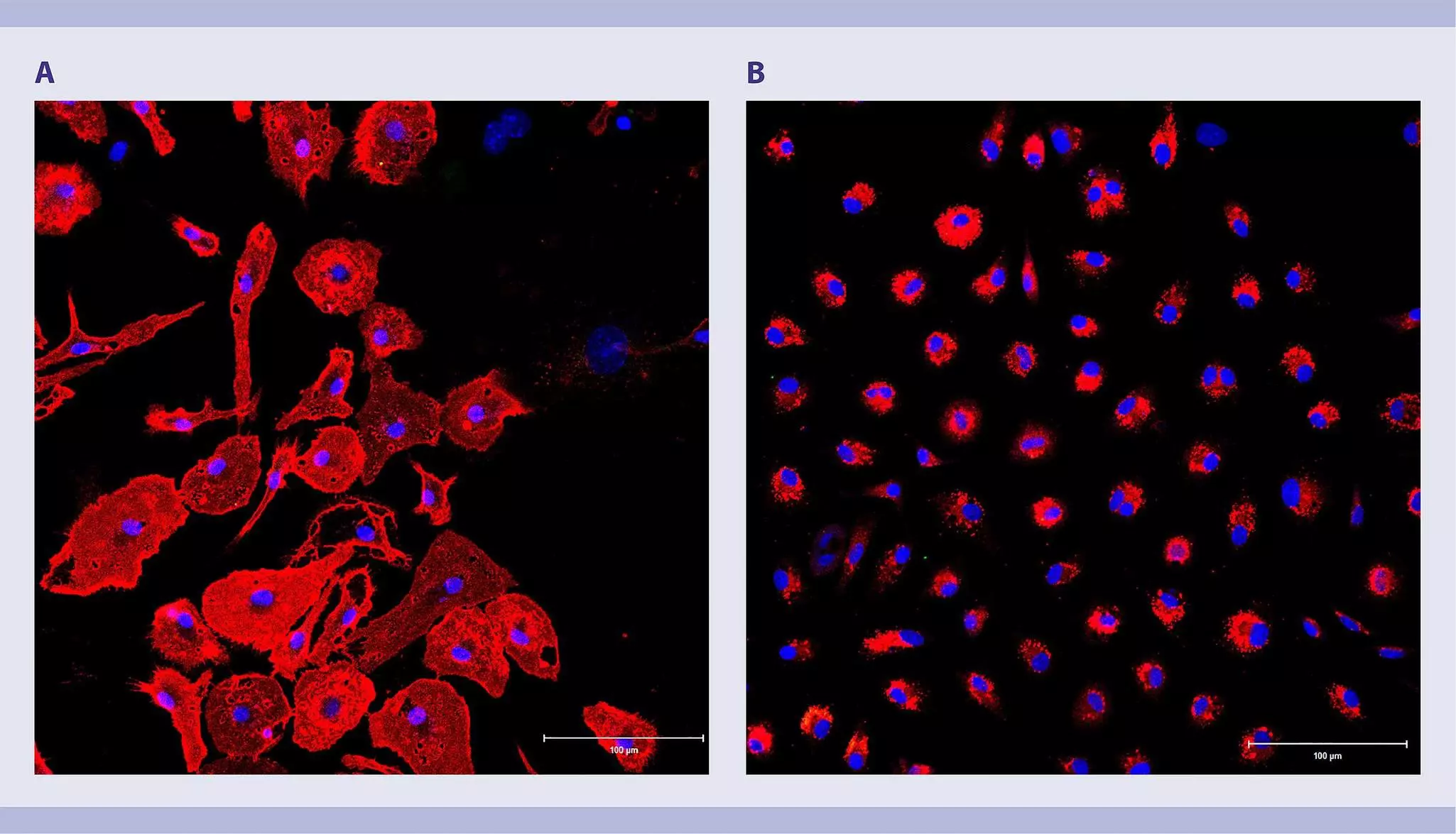
Highly purified (99%) and viable microglial cells can be separated from both neonatal and adult mouse or rat brain tissue with CD11b (Microglia) MicroBeads, human and mouse or CD11b/c (Microglia) MicroBeads, rat. In combination with the MultiMACS™ Cell24 Separator Plus or autoMACS NEO Separator, these kits enable fast microglial cell isolation from up to 24 samples in parallel.
Related videos:
6 Cultivation of neural cells
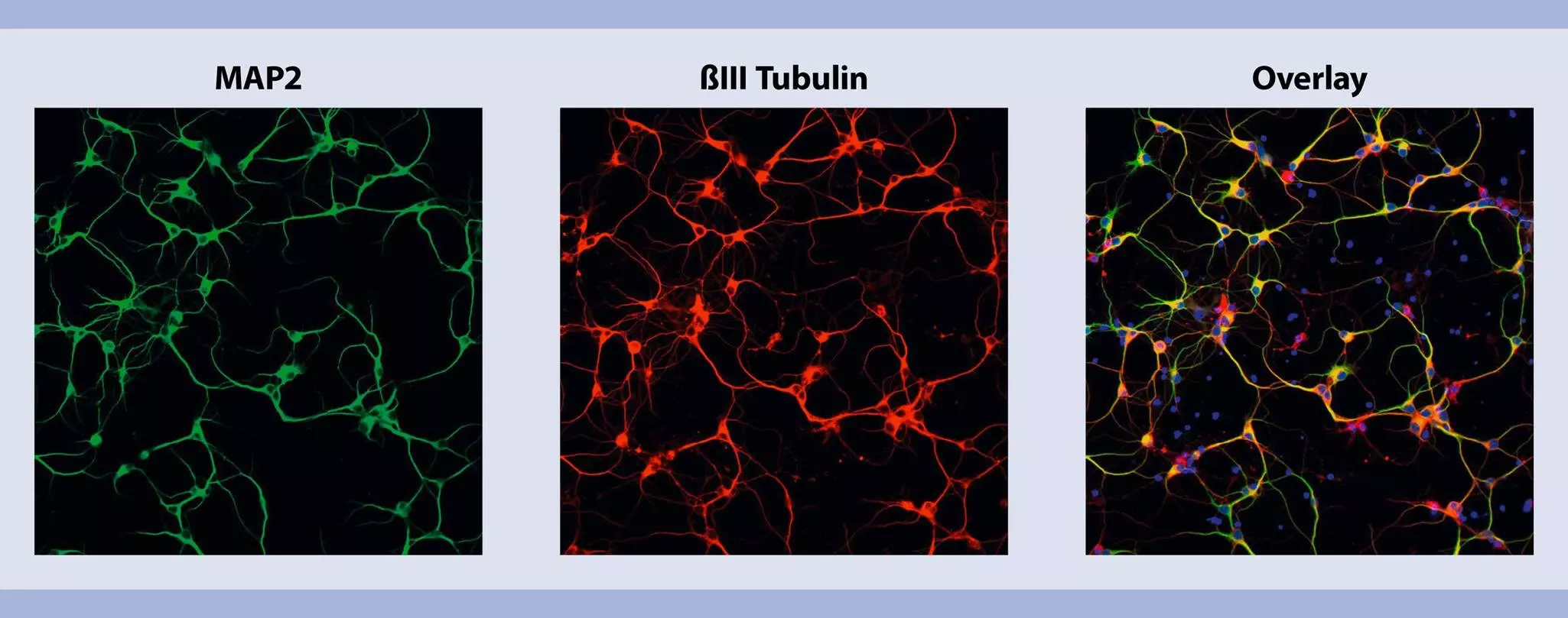
The fragile nature of neural cells makes the cultivation of primary cells a challenging task. To overcome this hurdle, Miltenyi Biotec has developed serum-free MACS Neuro Medium and MACS NeuroBrew-21 supplement, designed for the optimal growth and long-term viability of neural cells from the CNS and PNS after dissociation and isolation.
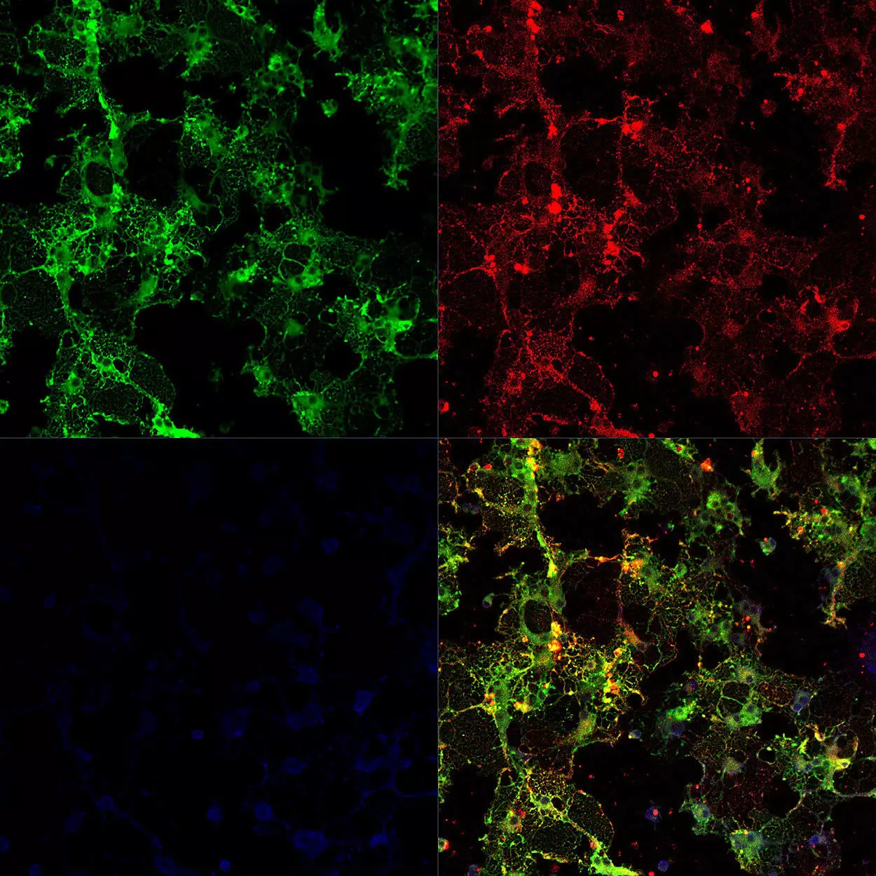
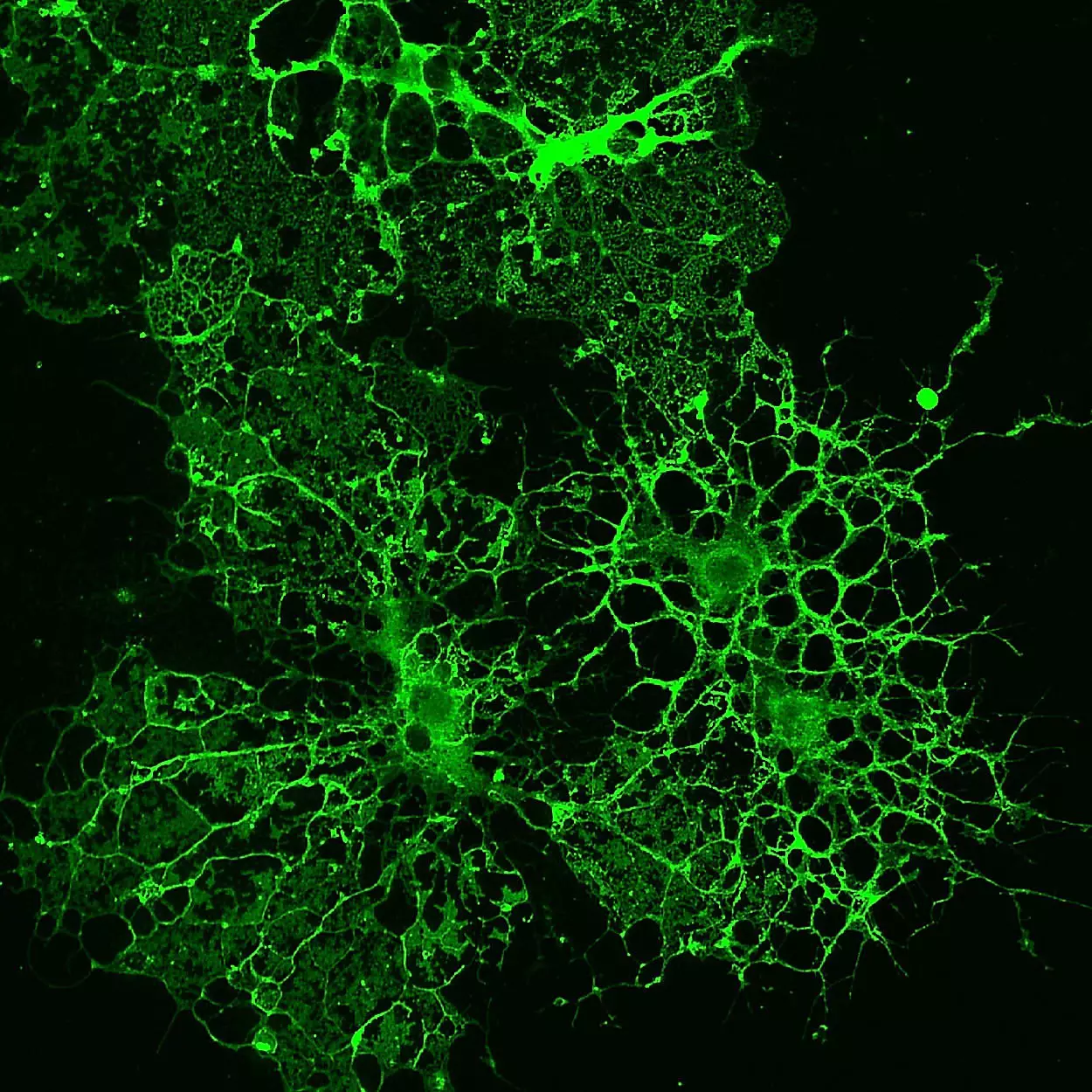
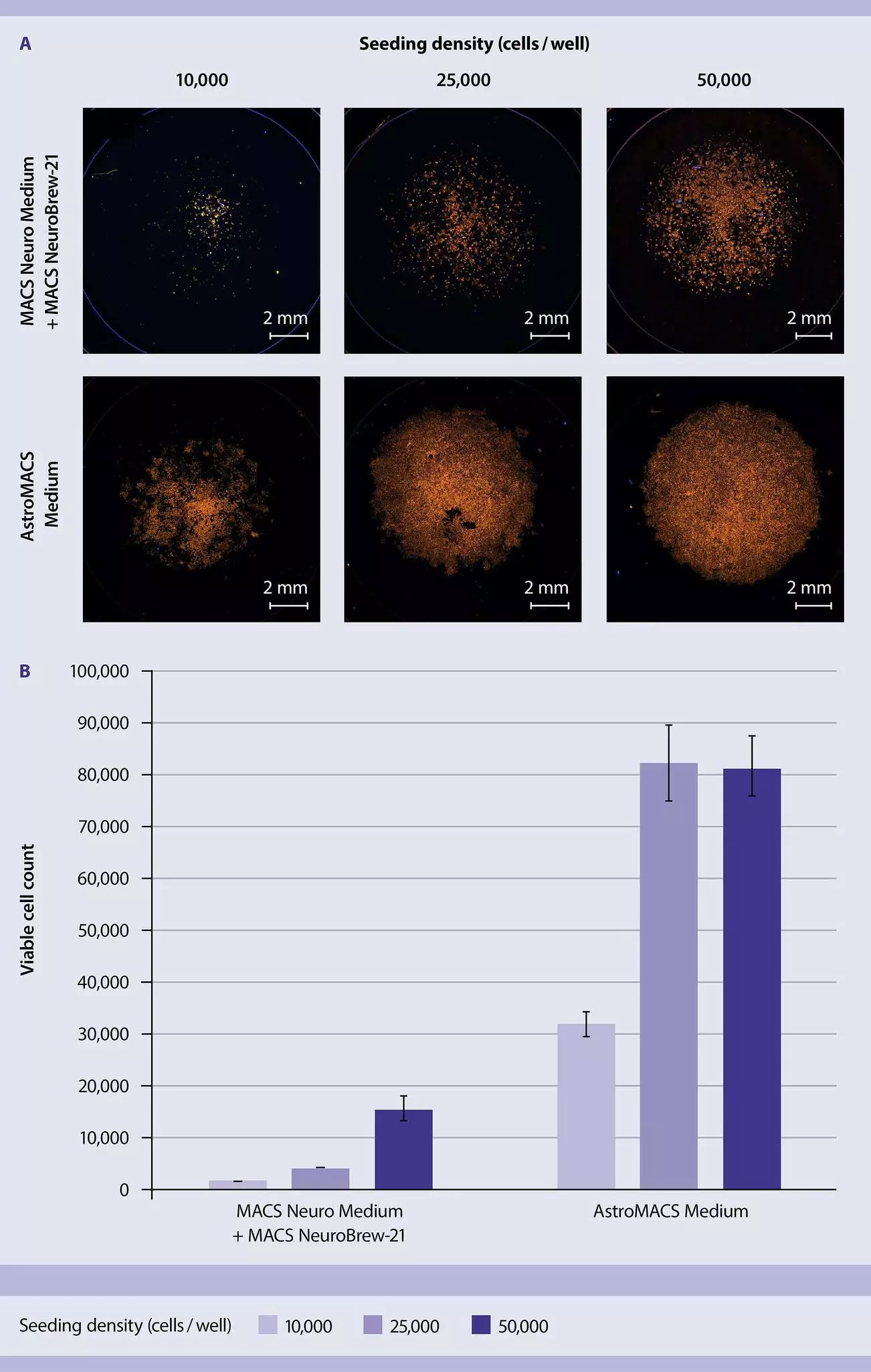
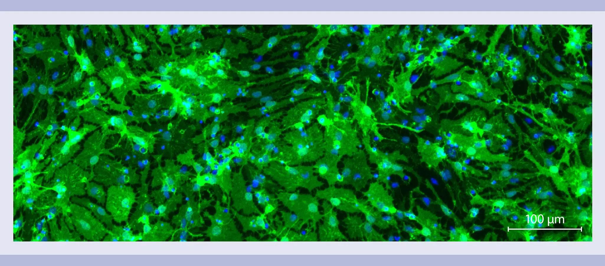
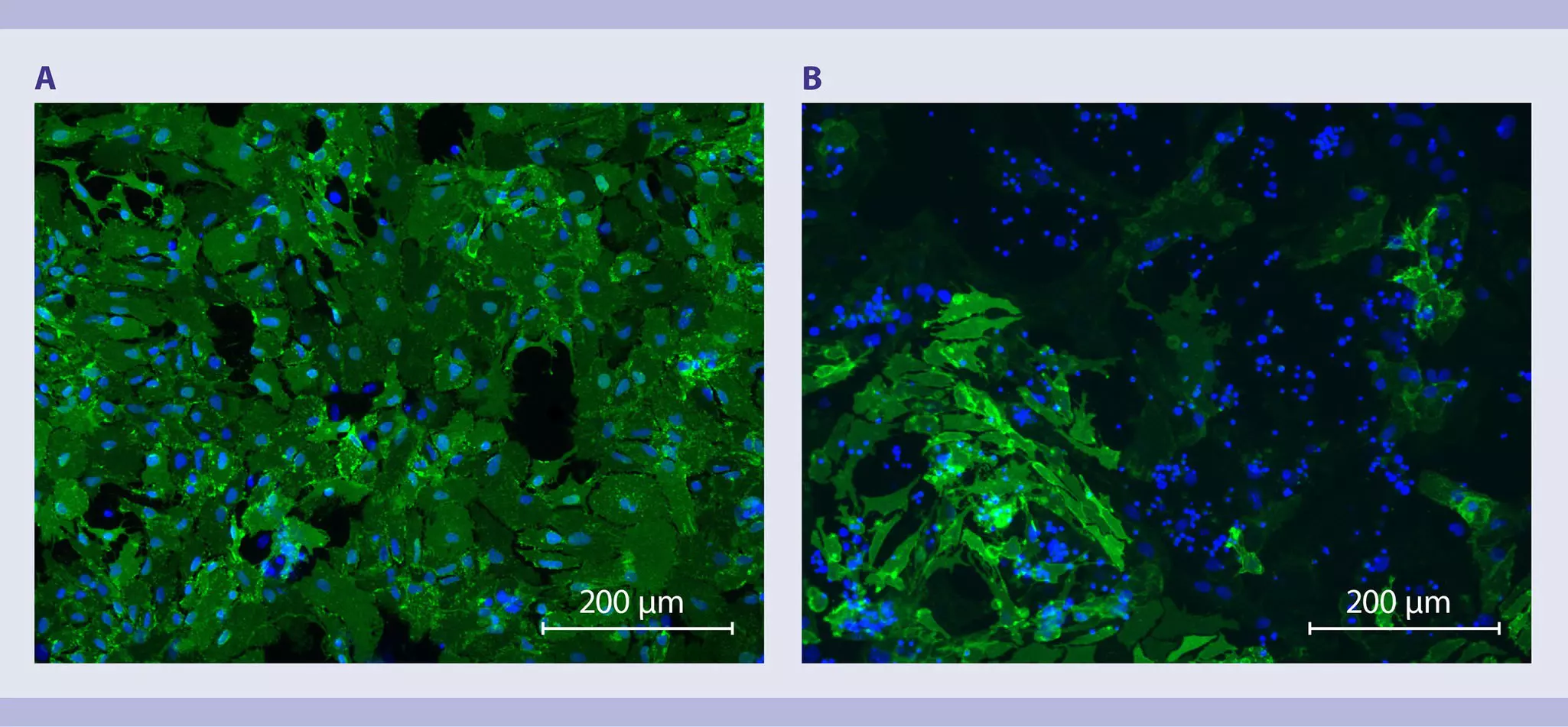
In addition, Miltenyi Biotec has developed a serum-free culture media for astrocytes, the AstroMACS Medium, which supports neonatal astrocyte growth even at very low seeding densities. AstroMACS Medium also enhances cultivation of adult astrocytes, especially when AstroMACS Separation Buffer is used before cell separation to remove dead cells and provide a healthy culture environment.
For more information about Miltenyi Biotec media optimized for neural cells, see chapter Cell culture.
Miltenyi Biotec offers an extensive portfolio of cytokines for neural cell differentiation and maintenance, including human BDNF, CNTF, EGF, FGF-2, and GDNF, which can be purchased in premium, research or MACS GMP Grade.
MACS Handbook:
You have previously chosen International as your location.
What do you want to do?