Immunophenotyping of virus-specific T cells from human blood using flow cytometry
MBTP 28 = Miltenyi Biotec-tested panel 28
This application protocol describes the flow cytometric analysis of virus-specific T cells after enrichment of IFN-γ+ cells from human blood. IFN-γ+ cells were enriched using the CliniMACS® Cytokine Capture System (IFN-gamma) to obtain viable single cell suspensions ready for downstream applications, such as flow cytometric analysis.
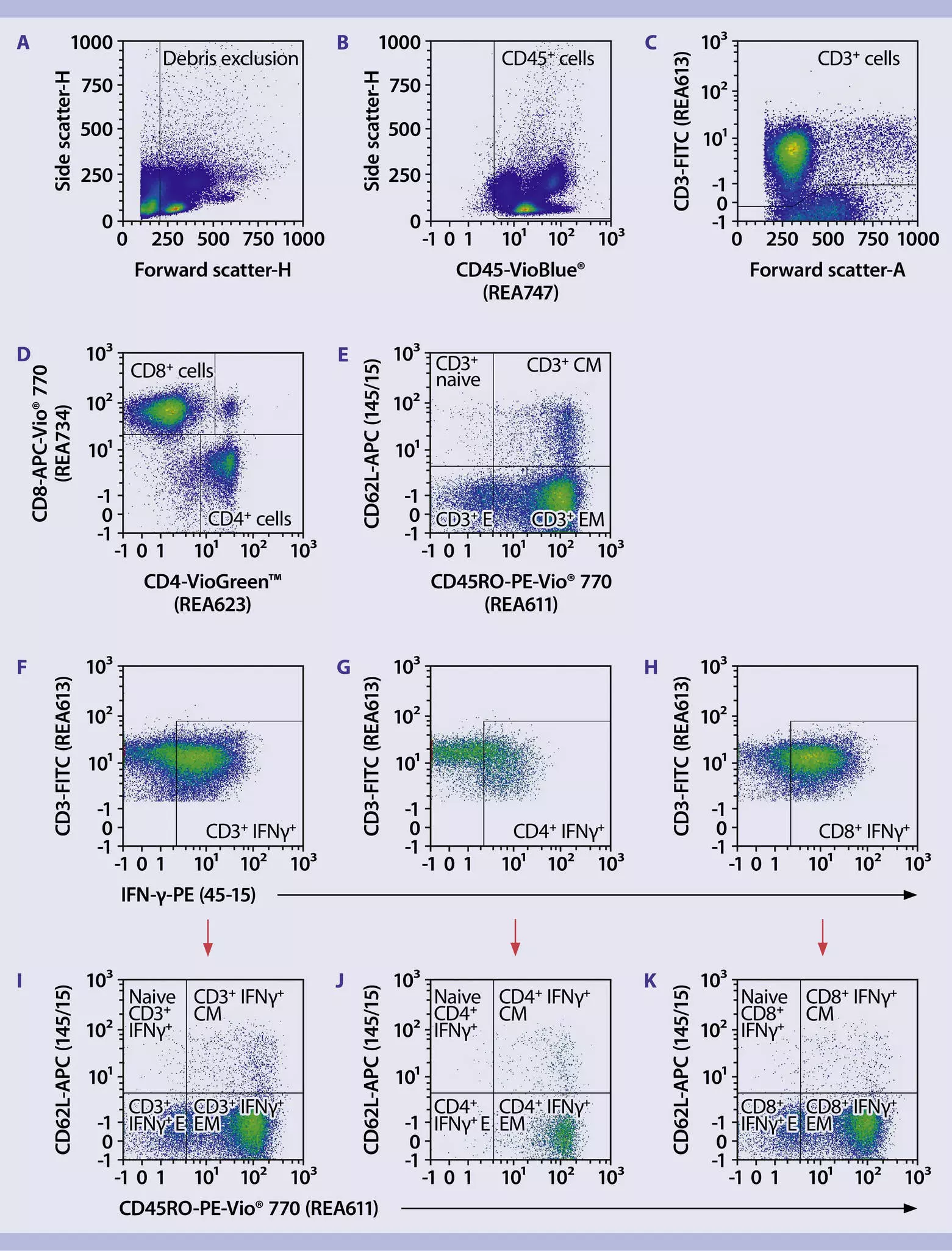
- Volumes given below were for up to 106 nucleated cells. When working with fewer than 106 cells, use the same volumes as indicated. When working with higher cell numbers, scale up all reagent volumes and total volumes accordingly (e.g. for 2 × 10⁶ nucleated cells, use twice the volume of all indicated reagent volumes and total volumes).
- Cell number was determined.
- Cells were centrifuged at 300 × g for 10 minutes. Supernatant was aspirated completely.
- A master mix with all fluorochrome-conjugated antibodies except IFN-γ Antibody, anti-human, PE was prepared according to the dilution listed in the respective antibody data sheets.
▲Note: IFN-γ is an intracellular marker and will be detected after fixation and permeabilization of cells. - 100 µL antibody master mix was added to the cells.
- Cells were resuspended and incubated for 10 minutes in the dark at +4 °C.
- Cells were washed by addition of 1 mL of PEB buffer and centrifuged at 300 × g for 5 minutes at +4 °C. Supernatant was aspirated completely.
- Intracellular staining was performed.
Intracellular staining
- Reagents were prepared freshly. Failure to do so may lead to sub-optimal results.
- The required total buffer volumes were calculated beforehand; volumes will depend on the number of cells to be analyzed as well as the number of tests to be performed.
- Cells previously stained for surface markers were resuspended in 250 µL of Inside Fix from the Inside Stain Kit.
- Cells were mixed well and incubated for 20 minutes at room temperature.
- Cells were centrifuged for 5 minutes at 300 × g.
- Supernatant was aspirated completely and the pellet was resuspended in 1 mL of PEB buffer.
- Cells were centrifuged for 5 minutes at 300×g and supernatant was aspirated completely.
- IFN-γ Antibody, anti-human, PE was diluted in Inside Perm according to the dilution mentioned in the respective data sheet to a final volume of 100 µL and added to the cells.
- Cells were incubated for 10 minutes at room temperature.
- 1 mL of Inside Perm buffer was added and cells were centrifuged for 5 minutes at 300 × g. Supernatant was aspirated completely.
- Cells were resuspended in 0.5–1 mL of PEB buffer and flow cytometric analysis was performed using the MACSQuant® Analyzer.
8 products found
|
For research use only
FC, MICS, IF, IHC
100 tests in 200 µL
(excl. tax)
For research use only
FC
100 tests in 200 µL
(excl. tax)
For research use only
FC, MICS, IF, IHC
100 tests in 200 µL
(excl. tax)
For research use only
ICFC
100 tests in 200 µL
(excl. tax)
For research use only
FC
100 tests in 200 µL
(excl. tax)
For research use only
1 mL
(excl. tax)
For research use only
FC
100 tests in 200 µL
(excl. tax)
For research use only
FC
100 tests in 200 µL
(excl. tax)
You have previously chosen International as your location.
What do you want to do?