NK cells


1 Introduction 2 NK cells from spleen and lymph nodes 3 NK cells from other tissues (lymphoid, non-lymphoid)
1 Introduction
Natural killer (NK) cells are part of the innate immune system. First described for their capacity to directly kill tumor cells without prior sensitization, NK cells are now known to participate in a variety of immune responses, including host protection against viruses, parasites, bacteria, and tumor cells. They also play an important role in stem cell transplantation and pregnancy. NK cells perform their effector functions by either mediating direct cell lysis or by producing a plethora of cytokines. Although typically studied in the mouse spleen, NK cells are also found in other organs, such as the thymus, lung, and liver.
2 NK cells from spleen and lymph nodes
NK cells develop mainly in bone marrow (BM). NK cell development has been progressively dissected, starting from committed precursors (NKP: Lin−/CD122+/NK1.1−/CD49b−) and immature cells (iNK: NK1.1+/CD49b−CD11blow/CD51low) to a mature NK subpopulation (mNK: CD49b+/Ly49s+). NK cells make up 3–4% of total lymphocytes in spleen tissue and less than 1% in lymph nodes.
 MACS Handbook:
2.1 Cell subsets, frequencies, and marker expression
At a glance: Tissue distribution (PMID: 19234143, 16424180), frequencies (PMID: 23034281, 23649470), and function (PMID: 28261203) of NK cell subsets.
| Cell subset | Frequency | Markers | Distribution/function |
| CD11blowCD27lowNK cells | 1.1–2.75% | CD27, CD11b | • Found predominantly in bone marrow and at very low frequency in all lymphoid and nonlymphoid organs |
| CD11blowCD27highNK cells | 11.5–16.5% | CD27, CD11b | • Found predominantly in bone marrow, lymph nodes, and in a greater proportion in the liver • Exhibit greater effector function and responsiveness to chemokines • Produce larger amounts of cytokines • Show decreased cytotoxicity • Interact with dendritic cells |
| CD11bhighCD27high NK cells | 23.2–49.3% | CD27, CD11b | • Homogeneously distributed • Exhibit greater effector function and responsiveness to chemokines • Express higher levels of killer cell lectin-like receptor subfamily G, member 1 (KLRG1) • Interact with dendritic cells |
| CD11bhighCD27low NK cells | 31.4–58.3% | CD27, CD11b | • Most abundant in blood, spleen, lung, and liver • Increased cytotoxicity against target cells |
A key marker of the NK cell lineage is CD27, a member of the TNF receptor superfamily. Its expression combined with the integrin chain CD11b, defines four sequential developmental stages of NK cells: CD11blowCD27low, CD11blowCD27high, CD11bhighCD27high, and CD11bhighCD27low subsets (PMID: 19234143).
These NK cell subsets differ widely in their tissue distribution (PMID: 16424180). CD11blowCD27high NK cells are predominantly found in bone marrow and lymph node, whereas CD11bhighCD27low NK cells are more abundant in blood, spleen, lung, and liver, and double-positive CD11bhighCD27high NK cells are more homogeneously distributed.
2.2 Miltenyi Biotec application protocols for NK cells from spleen
Miltenyi Biotec has created dedicated application protocols to isolate and analyze NK cells
2.3 Sample preparation of spleen
Main sources for mouse NK cells are single-cell suspensions from spleen and lymph nodes, which are used for many downstream applications, including isolation of cell subpopulations, cell culture, or flow cytometry analysis.
Dissociation can be fully automated using the gentleMACS™ Dissociators and dedicated Tissue Dissociation Kits (e.g. Spleen Dissociation Kit, mouse). A special protocol for the preparation of single-cell suspensions from mouse spleen without enzymatic treatment can be downloaded from the Related resources panel to the right. Alternatively, tissues can be dissociated manually. For details, see related chapters.
MACS Handbook:
2.4 Magnetic cell separation of NK cells from spleen and lymph nodes
Miltenyi Biotec has developed numerous products for the magnetic separation of the various NK cell types and subsets that can be found in mouse lymphoid tissue. For details on MACS® Cell Separation Technology, see the MACS Handbook chapter Magnetic cell separation.
MACS Handbook:
2.4.1 Isolation of NK and NKT cells from spleen and lymph nodes
At a glance: Products for the magnetic isolation of NK cells from mouse spleen and lymph nodes.
| Starting material | Isolation strategy | Comments | Automation | Product |
| Single-cell suspensions from spleen and lymph nodes | Depletion of non-target cells | Isolation of all NK cells via depletion of all non-NK cells | Yes* | NK Cell Isolation Kit, mouse |
| Positive selection of target cells | CD49b is expressed on NK cells and a small population of T cells (CD4+CD3+ TCRα/β+). CD49b is less mouse strain–specific than other NK cell markers (e.g. NK1.1) and is expressed by the most common inbred mouse strains | Yes* | CD49b (DX5) MicroBeads, mouse | |
| Positive selection of target cells | NKp46 seems to be exclusively expressed on NK cells. Expression has been shown on BALB/c, SJL, CBA/CA, C57BL/6, NOD, DBA/2, and B6.129 mice | Yes* | Anti-NKp46 MicroBead Kit, mouse | |
| Sequential separation | Isolation of NK1.1+ iNKT cells via depletion of all NK1.1+iNKT cells followed by enrichment via anti-NK1.1-APC and Anti-APC MicroBeads | Yes* | NK1.1+ iNKT Cell Isolation Kit, mouse |
*Automation options range from fully automated benchtop solutions such as the autoMACS® NEO Separator to high-throughput platforms such as the MultiMACS™ Cell24 Separator Plus or MultiMACS X.
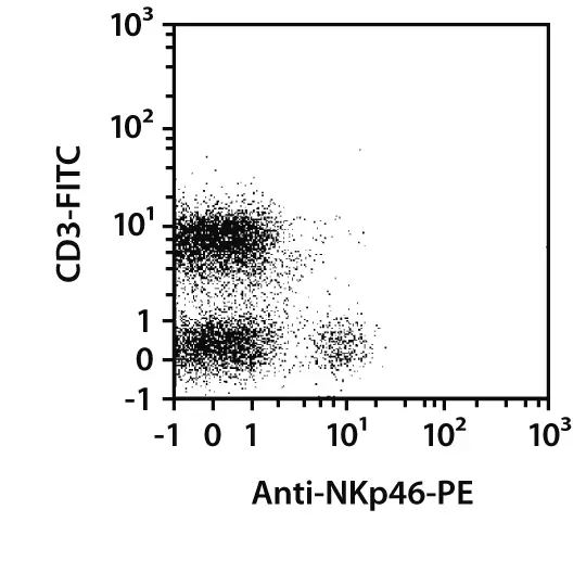
The NK Cell Isolation Kit, mouse enables isolation of untouched NK cells from single-cell suspensions of murine spleen. Non-NK cells (i.e. T cells, dendritic cells, B cells, granulocytes, macrophages, and erythroid cells) are magnetically labeled with a cocktail of biotin-conjugated antibodies and Anti-Biotin MicroBeads and then depleted to generate highly pure unlabeled NK cells. The kit is optimized to provide outstanding purities of NK cells isolated from C57BL/6J and BALB/c mice.
NK cell isolation from single-cell suspensions of spleen
 View details
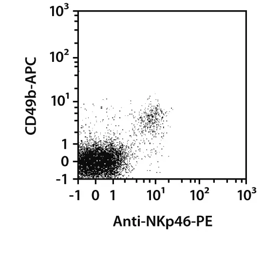
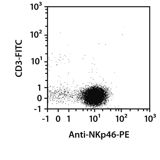
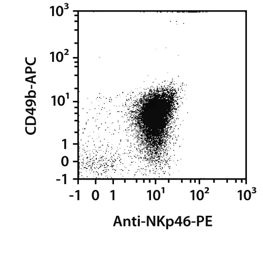
NK cell isolation from single-cell suspensions of spleen. NK cells were isolated from BALB/c mice using the NK Cell Isolation Kit, mouse, an LS Column, and a MidiMACS™ Separator. The cells were fluorescently stained with CD3-FITC, CD49b-APC, and Anti-NKp46-PE. Cells were analyzed by flow cytometry using the MACSQuant® Analyzer 10. Cell debris and dead cells were excluded from the analysis based on scatter signals, CD45-VioGreen™, and DAPI fluorescence.
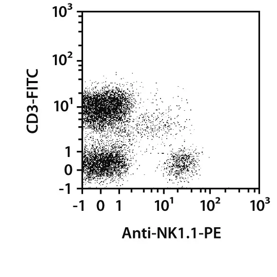
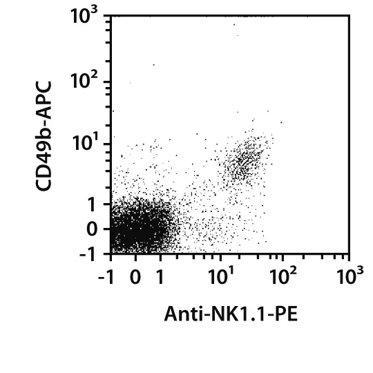
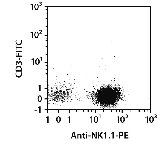
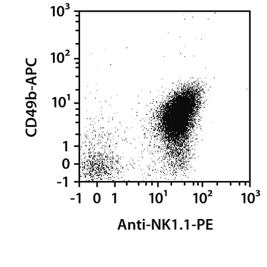
NK cell isolation from single-cell suspensions of spleen. NK cells were isolated from C57BL/6 mice using the NK Cell Isolation Kit, mouse, an LS Column, and a MidiMACS Separator. The cells were fluorescently stained with CD3-FITC, CD49b-APC, and Anti-NK1.1-PE . Cells were analyzed by flow cytometry using the MACSQuant Analyzer 10. Cell debris and dead cells were excluded from the analysis based on scatter signals, CD45-VioGreen, and DAPI fluorescence.
The Anti-NKp46 MicroBead Kit, mouse and CD49b (DX5) MicroBeads, mouse are used for positive selection or depletion of mouse NK cells from single-cell suspensions of lymphoid and non-lymphoid tissues or from peripheral blood.
Positive selection of NK cells from mouse spleen
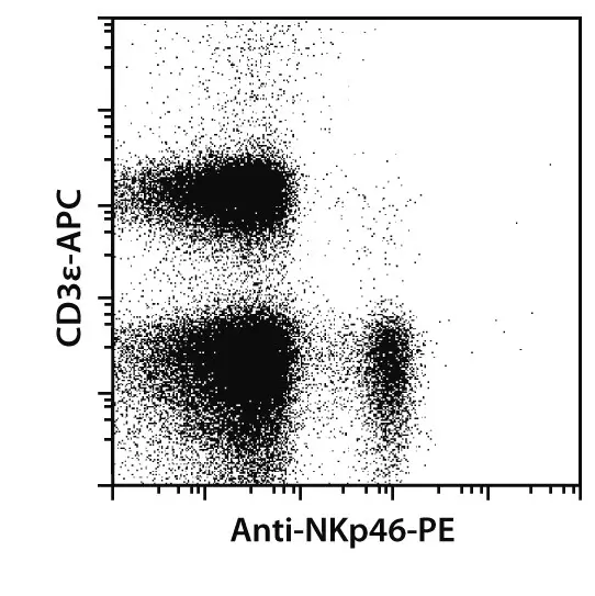
Isolation of NK cells by positive selection. NKp46+ NK cells were isolated from dissociated spleen from BALB/c mice using the Anti-NKp46 MicroBead Kit, mouse, two MS Columns, and a MiniMACS™ Separator. Cells were fluorescently stained with CD3ε-APC and Anti-NKp46-PE and analyzed by flow cytometry using the MACSQuant Analyzer.
Specific separation of CD49b (DX5)+ cells
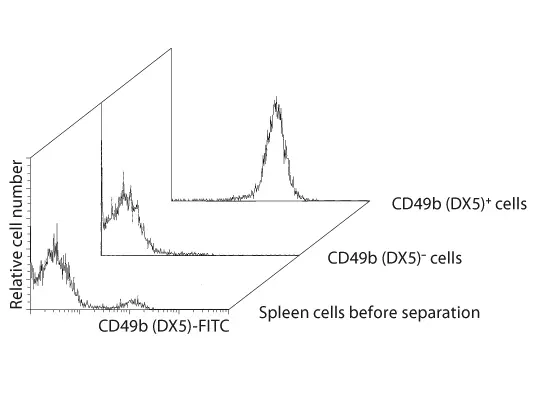
Specific separation of CD49b (DX5)+ cells. Mouse NK cells were isolated from a single-cell suspension of mouse spleen using CD49b (DX5) MicroBeads, a MiniMACS Separator, and an MS Column.
 Related PDFs:
 Related videos:
2.5 Characterization of NK cells from spleen and lymph nodes by flow cytometry
NK cell subsets and their differentiation status can be determined by flow cytometry based on their expression of cell surface markers and transcription factors and their secretion of cytokines. Miltenyi Biotec offers a vast portfolio of conventional monoclonal antibodies and REAfinity™ Recombinant Antibodies for comprehensive analysis.
 Related products:
2.5.1 Flow cytometry panels
At a glance: Antigens for the flow cytometric analysis of mouse NK cells.
| Basic NK lineage | Activating receptors, adhesion and effector functions | Inhibitory receptors | Transcription factors | Ly-49 lectins | Secreted factors | Other relevant markers |
| NK1.1 | β-2 Integrins (CD11a-CD18, CD11b-CD18, CD11c-CD18) | LAIR-1 | CD278 (ICOS) | Ly-49A | IFN-γ | CD25 |
| CD127 | CD335 (NKp46) | CD94/NKG2A | EOMES | Ly49A/D | TNF-α | CD27 |
| CD49b | DNAM-1 | SIGLEC-A | GATA3 | Ly-49C/F/I/H | IL-12 (p40/p70) | TIM-3 |
| CD117 | 2B4/CD244 | KLRG1 | RORγ (t) | Ly-49C/I | GM-CSF | TIGIT |
| CD11b | CD314 (NKG2D) | NKRP1-B, -D | T-bet | Ly-49E/F | CD279 (PD1) | |
| CD122 | NKG2C | 2B4 (CD244) | TOX | Ly-49F | PDL2 | |
| NKG2E | Helios | Ly-49G2 | PLZF | |||
| CD25 | Icaros | Ly-49H | FcεR1γ | |||
| TRAIL (CD253) | Ly-49I | FcεRIα | ||||
| Granzyme B | Ly-49D | |||||
| CD69 | Ly-49G | |||||
| CD62L | ||||||
| CD178 (FasL) | ||||||
| CD107a (LAMP-1) | ||||||
| Perforin | ||||||
| LAG-3 |
Miltenyi Biotec has developed numerous products for the analysis of NK cells and cell subsets found in mouse spleen and lymph nodes. REAfinity Recombinant Antibodies combined with a wide range of Vio® Dyes, which are characterized by high fluorescence intensities and low spillover, enable successful NK cell analysis. The following panel was tested on a single-cell suspension of splenocytes from C57BL/6 mice.
At a glance: Antibody panel for the analysis of NK cell subsets from C57BL/6 mouse spleen.
| Marker | Clone |
| Viobility™ 405/520 Fixable Dye | - |
| Anti-NK1.1-Pe-Vio770 | PK136 (soon REA632) |
| CD11b-VioBlue | REA592 |
| CD335 (NKp46)-FITC | REA815 |
| CD27-APC | REA499 |
| CD45-PE-Vio 615 | REA737 |
| Anti-KLRG1-PE | REA1016 |
| CD3-APC-Vio 770 | REA641 |
| CD19-APC-Vio 770 | REA749 |

 View details
Related PDFs:
3 NK cells from other tissues (lymphoid, non-lymphoid)
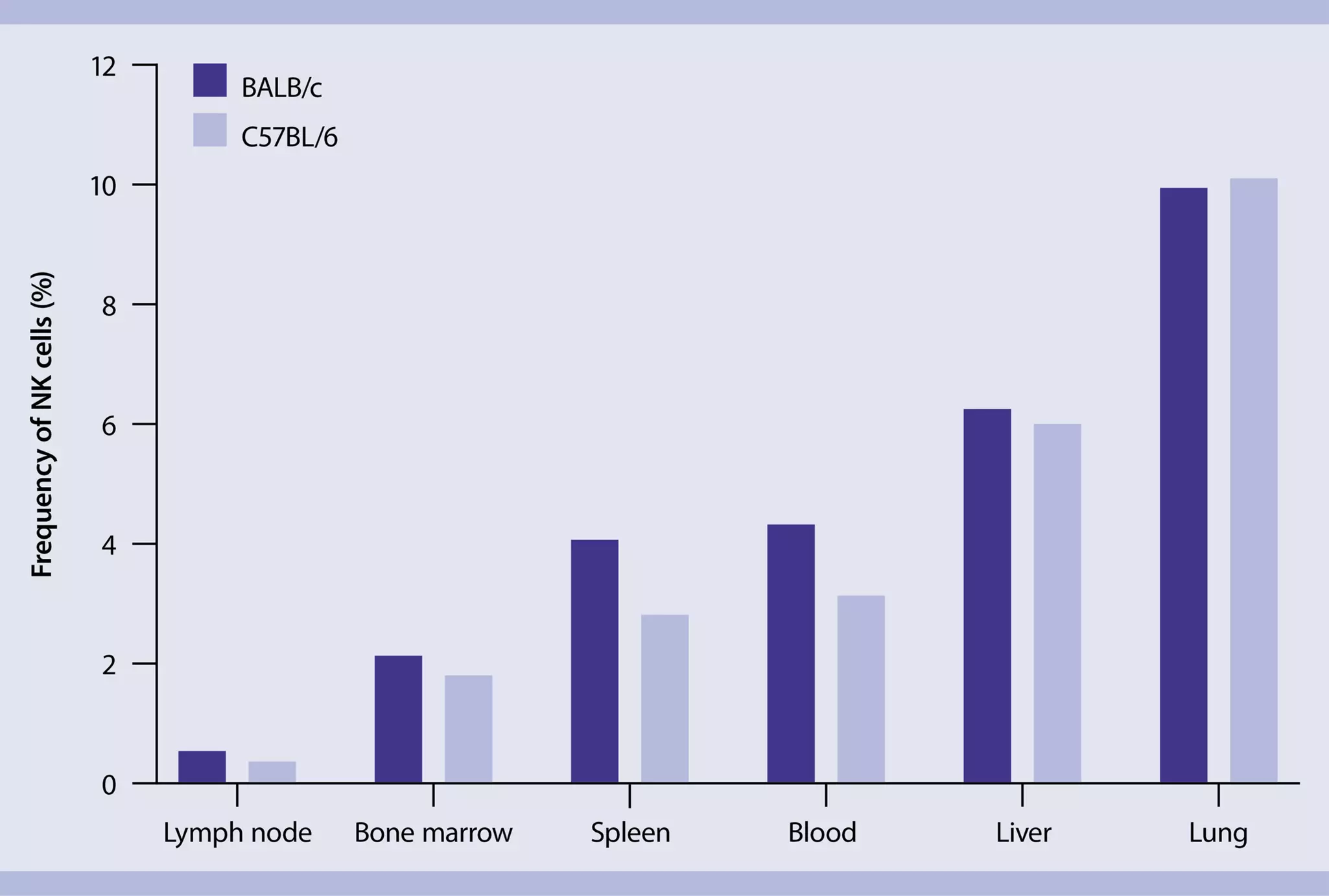
NK cells are widespread throughout lymphoid and non-lymphoid tissues. In most of these tissues, NK cells represent only a minor fraction of total lymphocytes, from 2% in mouse spleen to 10% in mouse lung (PMID: 18425107). Moreover, there are variations among different mouse strains.
3.1 Tissue-specific marker expression
Presence of NK cells in peripheral organs is presumably the result of chemokine-mediated migration from peripheral blood into these organs (PMID: 17979846). NK cells express several chemokine receptors, including CCR2, CCR5, CXCR3, and CX3CR1, which facilitate the targeting of specific NK cells into the relevant chemokine-producing organ.
At a glance: Expression patterns of NK cell markers in different mouse tissues.
| Spleen | Liver | Lung | Bone marrow | Uterus | |
| CD3 | - | - | - | - | - |
| CD49b (DX5) | + | - | + | + | -/+ |
| CD49a | - | + | + | ||
| CD27 | - | low | -/+ | - | |
| NKp46 | + | + | + | + | + |
| CD11b | + | -/low | high | -/+ | + |
| TRAIL | + | ||||
| Ly49 | + | -/low | high | + | high |
| CD94/NKG2A | + | + | high | + | |
| KLRG1 | low | + | + |
3.2 Sample preparation of different tissues
NK cell phenotyping requires complex analysis based on multiple antigens. To enable accurate and reliable analysis of NK cells from tissues, all epitopes needed to identify NK cell subsets must be preserved during tissue dissociation. To that end, Miltenyi Biotec offers kits for the dissociation of various tissues – including lamina propria, liver, lung, and skin. These kits were optimized to preserve the epitopes during the preparation of single-cell suspensions. For details on sample preparation, see MACS Handbook chapters on mouse cell sources and the specific Tissue Dissociation Kits.
MACS Handbook:
3.3 Magnetic cell separation of NK cells from other tissues (lymphoid, non-lymphoid)
Miltenyi Biotec has developed numerous products for the magnetic separation of cell types and cell subsets found in various mouse tissues.
For details on MACS® Cell Separation Technology, see the MACS Handbook chapter Magnetic cell separation.
MACS Handbook:
3.3.1 Isolation of NK cells from other tissues (lymphoid, non-lymphoid)
Depending on the mouse strain and NK cell phenotype, NK cells can be magnetically enriched after tissue dissociation, by positive selection with CD49b (DX5) MicroBeads, mouse or the Anti-NKp46 MicroBead Kit, mouse.

Related PDFs:
You have previously chosen International as your location.
What do you want to do?