Human embryonic stem cells and human iPS cells


1 Introduction 2 Pluripotent stem cell subtypes 3 Somatic cell reprogramming 4 Maintenance of pluripotent stem cell lines 5 PSCs in diverse applications 6 PSC differentiation 6.1 Formation and dissociation of embryoid bodies 6.2 PSC differentiation with ready-to-use kits 6.3 PSC differentiation using cytokines, and small molecules 6.4 PSC Differentiation using organoid models 6.5 PSC differentiation using transcription factors 6.6 Isolation of PSC-derived cells during and after differentiation 6.7 Characterization of the PSC-derived cells
1 Introduction
Pluripotent stem cells (PSCs) are self-renewing stem cells with the remarkable ability to differentiate into any cell type of the embryo. They have the ability to differentiate into all cell types derived from the ectoderm (PMID: 32451506), mesoderm (PMID: 36254806), and endoderm (PMID: 32341565) germ layers, a characteristic known as pluripotency.
The extensive developmental potential of PSCs positions them as prime candidates for applications in regenerative medicine. Here, lineage-specific progeny cells derived from PSCs can potentially be used to replace degenerated or diseased cells and tissues (PMID: 24434846). Moreover, PSCs serve as a powerful tool for studying human diseases and development. In vitro differentiation of PSCs can partially recapitulate human development in vivo. By comparing PSCs from healthy and diseased donors, valuable insights into the pathology of human diseases can be gathered. Coupled with modern genome editing techniques, this approach opens the possibility of modeling disease-specific genotypes/mutations. Finally, PSCs have opened new avenues for drug and toxicity screening (PMID: 23746976, PMID: 33091434).
 Related PDFs:
2 Pluripotent stem cell subtypes
PSCs can be classified based on their origin as human embryonic stem cells (ES cells) or induced pluripotent stem (iPS) cells.

Both cell types grow as cell lines that form characteristic adherent colonies. In terms of morphology, marker expression, developmental potential, and experimental handling, ES and iPS cells are essentially indistinguishable.
3 Somatic cell reprogramming
Reprogramming involves the conversion of differentiated somatic cells into a pluripotent state. This breakthrough process was first achieved in 2006 using mouse somatic cells (PMID: 16904174). In 2007, the same group extended their success to generate iPS cells from adult human dermal fibroblasts, employing four defined transcription factors: Oct3/4, Sox2, Klf4, and c-Myc. The generated iPS cells exhibited similarities to human ES cells in various aspects, including morphology, proliferation, gene expression, and differentiation potential (PMID: 18035408).
Miltenyi Biotec offers reprogramming tools for fibroblast reprogramming into iPS cells based on non-integrative mRNA transfection technology. Among current technologies, mRNA reprogramming has the highest efficiency and is considered the safest and fastest method for de novo generation of iPS cell lines (PMID: 25437882).
 Related videos:
3.1 Establishment of a fibroblast culture for reprogramming
Establishing high-quality cultures of proliferating fibroblasts is key to the success of reprogramming experiments. The Whole Skin Dissociation Kit, human in combination with the gentleMACS® Octo Dissociator with Heaters enables gentle and efficient dissociation of human skin biopsies into single-cell suspensions and the subsequent generation of fibroblast cultures from patient samples. Unlike traditional outgrowth cultures, starting with a single-cell suspension ensures consistent monolayer cultures. This approach yields enough fibroblasts for reprogramming within 5–8 days after plating, depending on biopsy size and cell number required for the planned reprogramming method.
At a glance: Kits and reagents for the generation of a fibroblast culture
| Use | Product |
| Tissue dissociation | Whole Skin Dissociation Kit, human |
| Tissue dissociator | gentleMACS Tissue Dissociator Family |
| Removing cell aggregates | MACS® SmartStrainers and Pre-Separation Filters |
3.2 mRNA reprogramming of fibroblasts
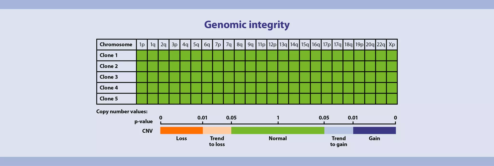
mRNA reprogramming technology excels in both conversion efficiency and kinetics, presenting a noteworthy advancement for swift, safe, and effective reprogramming of mature human cells. Unlike DNA- and virus-based methods, it eliminates the risk of genomic integration and associated mutagenesis (PMID: 20888316, 25437882). StemMACS™ iPSC mRNA Reprogramming Kit, human (includes the xeno-free medium, StemMACS Repro-Brew XF, human) enables quick and efficient iPS cell generation from human fibroblasts in just 14 days. With this comprehensive kit, all essential products for your reprogramming workflow are included in one convenient package.
A chemically defined, xeno-free culture matrix, such as MACSmatrix Laminin 511, plays a pivotal role in achieving reproducibility and consistency in stem cell workflows. This recombinant laminin is fully compatible with the StemMACS iPSC mRNA Reprogramming Kit, human, and has been rigorously tested to ensure reliable results. As an E8 fragment, MACSmatrix Laminin 511 offers excellent coating efficiency, delivering broader coverage while helping reduce costs. Supplied as a ready-to-use liquid, it can be conveniently stored at 2–8°C, enhancing usability and workflow efficiency.
We offer an ideal solution for optimal cell maintenance after reprogramming, StemMACS PSC-Brew XF, human. This xeno-free medium is specifically designed for the culture of PSCs, promoting robust proliferation while ensuring the stability and preservation of the pluripotent phenotype.
At a glance: Kits and reagents for fibroblast reprogramming into iPS cells
| Use | Product |
| Human fibroblast reprogramming into iPS cells | StemMACS iPSC mRNA Reprogramming Kit, human |
| Coating reagets | MACSmatrix Laminin 511 |
| Supporting the reprogramming of the fibroblasts into iPS cells | StemMACS Repro-Brew XF, human (available as a part of the StemMACS iPSC mRNA Reprogramming Kit, human, and as stand-alone product) |
| Culturing the generated iPS cells | StemMACS PSC-Brew XF, human |

 View details
3.3 iPS isolation
Post-reprogramming, isolating iPS cells is vital to guarantee a uniform and fibroblast-free cell population. This process not only ensures homogeneity but also facilitates the selection of top-tier clones for subsequent applications. Various methods, such as manual picking, magnetic separation, or cell sorting, can be employed to effectively isolate iPSC colonies.
3.3.1 Isolation via MACS® Technology
Magnetic isolation offers a streamlined method for obtaining pure iPSC populations, avoiding labor-intensive manual iPSC colony picking and maintaining cell viability and functionality (PMID: 30204478). Additionally, this approach allows for automation and scalability, making it a robust tool for high-throughput iPSC research and clinical applications.
With magnetic cell separation, reprogrammed cells can be isolated in bulk (PMID: 23734247). Multiple cell lines can be processed simultaneously, generating large amounts of cells in a short time, and the process can be automated. Furthermore, bulk cells can be subjected to subcloning, if clonal cultures are required. For details on MACS Cell Separation Technology, see the MACS Handbook chapter Magnetic cell separation.
At a glance: Kits and reagents for the magnetic separation of reprogrammed iPS cells
| Use | Product |
| Positive selection of target cells | Anti-TRA-1-60 MicroBeads, human |
| Automation of cell isolation | Automated cell separation systems |
| Improves hPSC performances under challenging culture conditions | StemMACS PSC-Support XF, human |
 MACS Handbook:
Related PDFs:
3.3.2 Isolation using flow cytometry cell sorting
Utilizing a cell sorting method post-reprogramming enables the precise and sensitive isolation of homogeneous iPS cells populations, ideal for subsequent applications.
At a glance: Kits and reagents for sorting the reprogrammed iPS cells
| Use | Product |
| Antibody for labeling the target cells | TRA-1-60 Antibody, anti-human, REAfinity® |
| Instrument for sorting of the target cells | MACSQuant® Tyto® Cell Sorter |
| Supplement to improve hPSC performances under challenging cultural conditions | StemMACS PSC-Support XF, human |
MACS Handbook:
4 Maintenance of pluripotent stem cell lines
Propagation of PSCs demands special attention and qualified reagents to preserve their pristine features. Different cultivation principles have evolved over time, each with unique advantages and disadvantages.
4.1 Feeder-free culture
At a glance: Media, supplements and matrix for feeder-free culture of PSCs
| Protduct category | Comments | Product |
| Media | Research grade | StemMACS PSC-Brew XF, human |
| Supplement | Improves hPSC performances under challenging cultural conditions | StemMACS PSC-Support XF, human |
| Media | Research grade | StemMACS iPS-Brew XF, human |
| Media | GMP grade | iPS-Brew GMP Basal medium |
| Supplement | For use with iPS-Brew GMP Basal Medium | iPS-Brew GMP Supplement R |
| Recombinant protein | Matrix for robust expansion and maintenance of human PSCs | MACSmatrix Laminin 511 |
Miltenyi Biotec offers StemMACS iPS-Brew XF, human, a research-grade medium designed for robustness and reproducibility, providing a reliable foundation for iPSC culture. For seamless translation into clinical applications, the iPS-Brew GMP Medium is available in both 500 mL bottle and 2-liter bag formats, ensuring scalability and compliance with GMP standards. The 2-liter bag format is specifically designed to streamline workflows and facilitate efficient transition to cell therapy manufacturing.
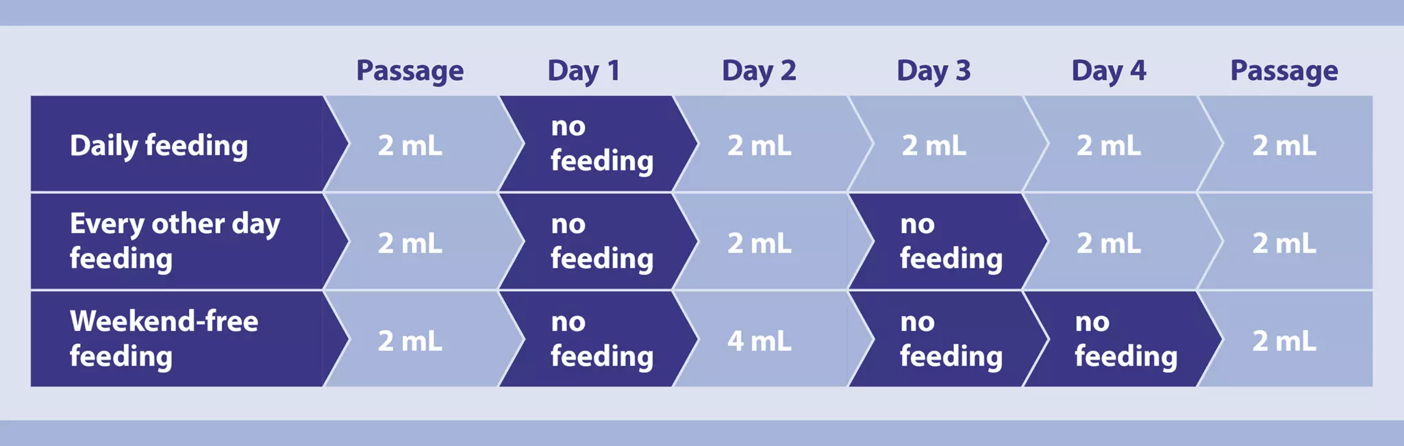
Traditionally, culture protocols called for daily media replacement. This was especially the case with the first generation of feeder-free PSC culture media. Any deviation from the standard protocol could lead to increased PSC spontaneous cell differentiation, slower growth rates, or down-regulation of pluripotency markers. Initial passages often appear fine, but changes can occur during long-term culture with reduced feeding. Updated media formulations enable greater flexibility in feeding schedules. This feature is attributed to the substitution of native FGF-2 with thermostabilized versions of the growth factor.
StemMACS PSC-Brew XF, human, is a new generation of cell culture medium for highly efficient maintenance and expansion of human pluripotent stem cells (hPSC). It has a xeno-free and serum-free formulation that ensures reduced variability and standardized performances during culture. The carefully balanced composition and the presence of stable FGF-2 (FGF-2 IS) ensure constant exposure levels and provides flexibility with feeding schedules. FGF-2 IS stands for fibroblast growth factor 2 "Improved Sequence", also termed fibroblast growth factor basic (FGF-b) or basic FGF. Human FGF-2 IS is a variant of human FGF-2 with a proprietary amino acid substitution. Human FGF-2 IS is covered by patent nos. US10,336,799 and EP2930181B1.
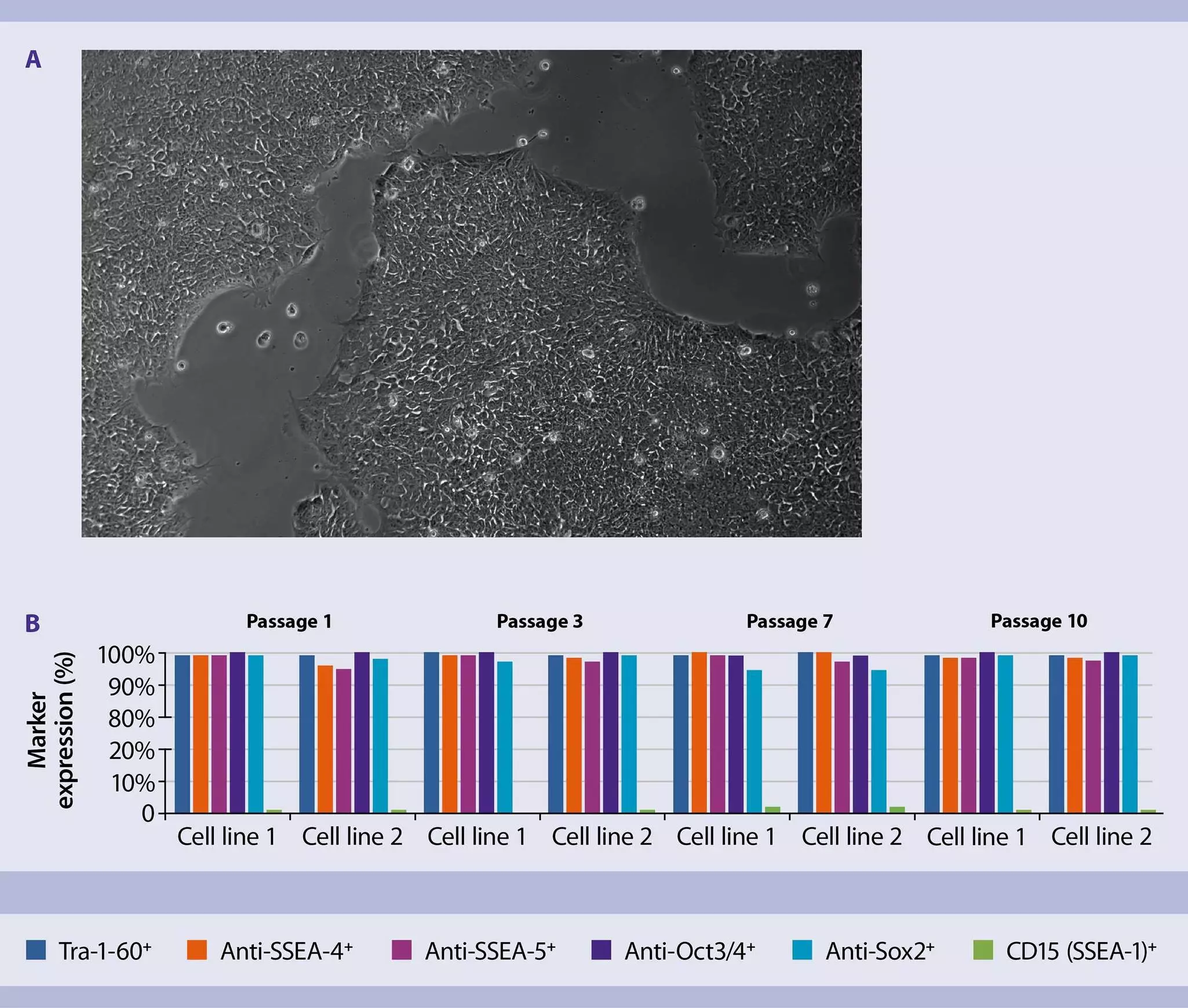
Feeder-free culture systems replace the feeder layer with a coating matrix that facilitates cell attachment. Various extracellular matrices are used, including Matrigel® and its derivatives, which have components of animal origin, and vitronectin or MACSmatrix Laminin 511, which are recombinant, chemically defined matrices (PMID: 24463987). StemMACS PSC-Brew XF, human, and StemMACS iPS-Brew XF, human, are compatible with both defined and undefined cell attachment matrices. However, the use of Laminin-521 is recommended because it maintains the xeno-free culture system and provides superior attachment.
4.2 Splitting pluripotent stem cell cultures
At a glance: Kits and reagents for passaging PSCs
| Use | Comments | Product |
| Routine cluster passaging | Optimized for use with StemMACS PSC Brew XF and StemMACS iPS-Brew XF | StemMACS Passaging Solution XF |
| Cell culture | Eliminates the need for adding any inhibitors | StemMACS PSC-Brew XF, human |
| Increase cell survival at low cellular plating densities | Optimized for use with with StemMACS PSC-Brew XF, human | StemMACS PSC-Support XF, human |
| ROCK inhibition | A selective small molecule | StemMACS Y27632 |
| ROCK inhibition | A selective small molecule | StemMACS Thiazovivin |
Different PSC lines have different growth kinetics. Therefore, the optimal time and ratio for passaging must be determined individually for each cell line. Generally, it is good practice to keep cultures in a subconfluent state, where colonies are separated visibly. Cultures should be split before they reach confluency to avoid fusion of colonies and a rapid drop of growth factor levels.
Detachment of human PSCs must be gentle to maintain maximum viability. StemMACS Passaging Solution XF, human, is designed to minimize cell culture manipulation, eliminating lengthy inactivation, dilution, and centrifugation steps. The ready-to-use formulation facilitates a reproducible and standardized splitting procedure.
Human PSCs show decreased survival and attachment when colonies are dissociated into single cells or small cell clusters. ROCK inhibitors such as StemMACS Y-27632 or StemMACS Thiazovivin counter this effect and increase survival during passaging and thawing after cryopreservation (PMID: 17529971, 20406903).
StemMACS PSC-Brew XF, human, supports cluster passaging seamlessly, eliminating the need for any inhibitors.
4.3 Characterization of pluripotent stem cells
Human PSC lines can vary significantly with respect to their growth and differentiation characteristics. The genetic and epigenetic basis of this heterogeneity is still poorly understood and the subject of ongoing research (PMID: 23673969).
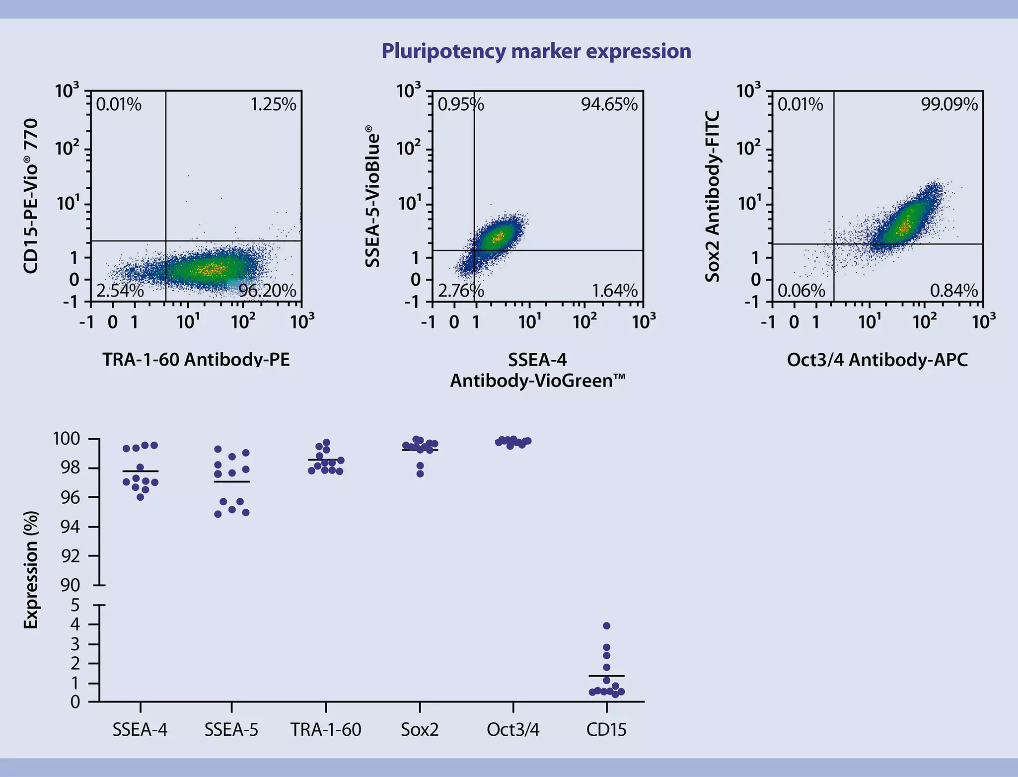
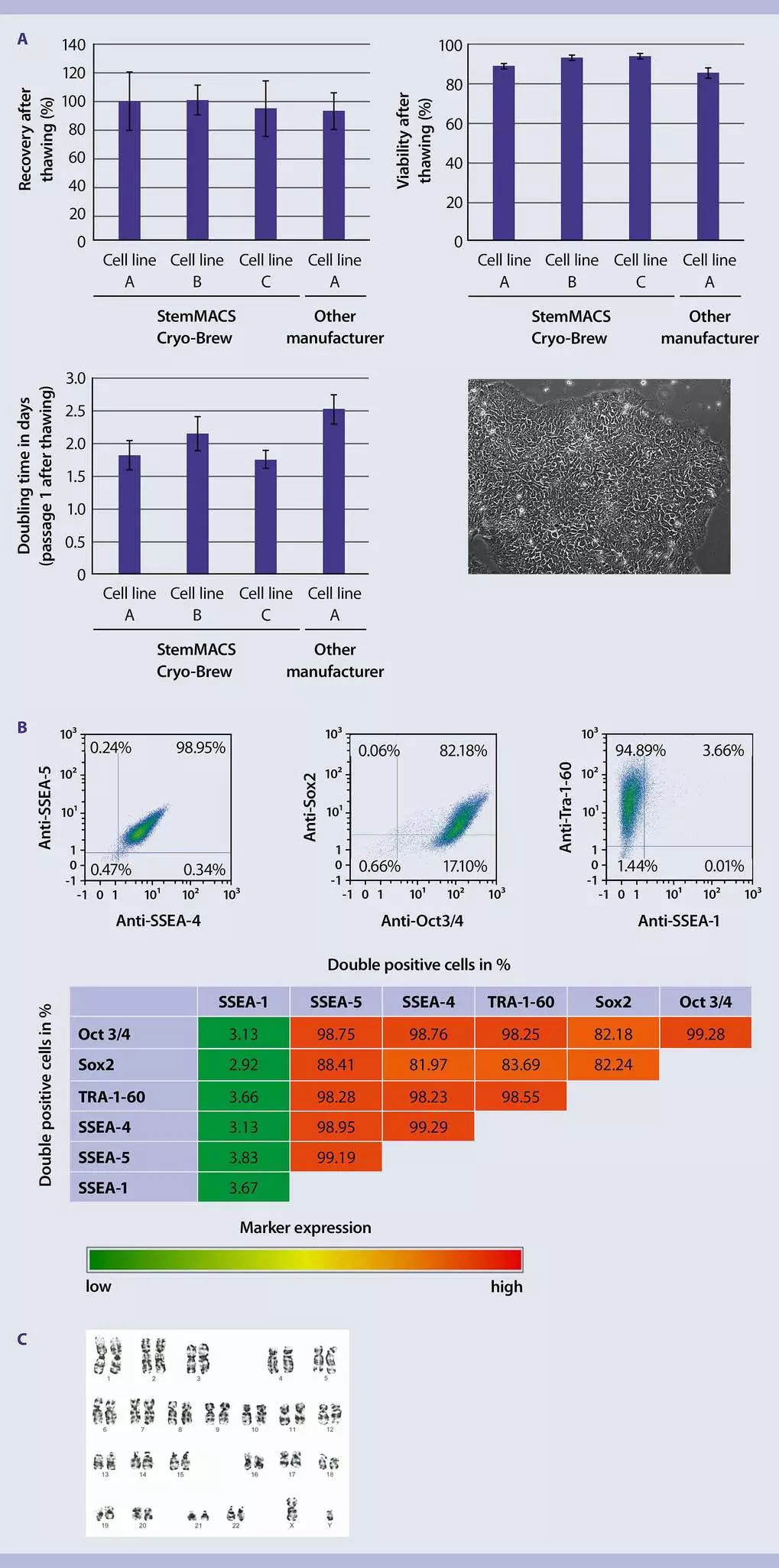
Thorough and comprehensive characterization is therefore mandatory when establishing new ES or iPS cell lines and good practice during propagation of established lines. A typical basic characterization includes assessment of morphology, extra- and intracellular marker expression, pluripotent differentiation potential, and karyotyping.
In addition to antibodies against pluripotency markers, antibodies that detect signs of early differentiation (e.g. Anti-CD15 Antibodies) can complement the analysis. The detection of intracellular antigens has often been hampered by high background signals when conventional mono- or polyclonal antibodies were used for flow cytometric analysis. In contrast, Miltenyi Biotec's REAfinity Recombinant Antibodies ensure highly specific detection with low background signals. For details, see the MACS Handbook chapter Antibodies.
Monitoring the pluripotency and differentiation status of PSC cultures is essential to ensure their quality and reproducibility. While immunofluorescence microscopy provides valuable insights, flow cytometry offers a quantitative approach, enabling precise evaluation of culture composition and facilitating comparisons across cell lines and data points.
The PSC Analysis Cocktail Kit, anti-human, simplifies this process with a robust multicolor panel for flow cytometry, designed in alignment with the International Society for Stem Cell Research (ISSCR) recommendations for marker expression analysis. It enables the simultaneous detection of key intracellular and surface markers, including SSEA-4, TRA-1-60, Oct3/4, and CD15, providing both qualitative and quantitative data for accurate characterization.
The kit’s adaptable design supports the addition of markers like Nanog or SOX2 for more comprehensive analyses. Furthermore, its compatibility with GFP-labeled cell lines broadens its applicability, making it an indispensable tool for advanced PSC research.
MACS Handbook:
4.3.1 Extra- and intracellular marker expression
At a glance: Antibody panel for human PSCs
| Purpose | Specificity | Cell location | Product |
| Characterization | Multiple Antibodies | Intracellular and cell surface | PSC Analysis Cocktail Kit, anti-human |
| Pluripotency | TRA-1-60 | Cell surface | TRA-1-60 Antibody, anti-human, REAfinity |
| Pluripotency | SSEA-4 | Cell surface | SSEA-4 Antibody, anti-human, REAfinity |
| Pluripotency | SSEA-5 | Cell surface | SSEA-5 Antibody, anti-human, REAfinitiy |
| Pluripotency | Oct3/4 Isoform A | Intracellular | Oct3/4 Isoform A Antibody, anti-human/mouse, REAfinity |
| Pluripotency | Sox2 | Intracellular | Sox2 Antibody, anti-human/mouse, REAfinity |
| Differentiation | CD15 | Cell surface | CD15 Antibody, anti-human |
4.3.2 Assessment of pluripotent differentiation potential
At a glance: Kit and antibody panel for the assessment of PSC differentiation potential
| Use | Comments | Product |
| Spontaneous differentiation | Pluripotency assessment by embryoid bodies assay | StemMACS DiffBase XF, human |
| Coating reagents | Coating the cell culture plates with a chemically defined reagent | MACSmatrix Laminin 511 |
| Directed differentiation | Differentiation of PSCs into ectoderm, mesoderm, and endoderm | StemMACS Trilineage Differentiation Kit, human |
| Flow cytometric analysis | Quantitative analysis of differentiated cells with mesoderm identity | CD144 (VE-Cadherin) Antibody, anti-human, REAfinity |
| Flow cytometric analysis | Quantitative analysis of differentiated cells with mesoderm identity | CD140b Antibody, anti-human, REAfinity |
| Flow cytometric analysis | Quantitative analysis of differentiated cells with endoderm identity | CD184 (CXCR4) Antibody, anti-human, REAfinity |
| Flow cytometric analysis | Quantitative analysis of differentiated cells with endoderm identity | Sox17 Antibody, anti-human, REAfinity |
| Flow cytometric analysis | Quantitative analysis of differentiated cells with ectoderm identity | PAX-6 Antibody, anti-human, REAfinity |
| Flow cytometric analysis | Quantitative analysis of differentiated cells with ectoderm identity | Sox2 Antibody, anti-human/mouse, REAfinity |
| Flow cytometric analysis | Quantitative analysis of all viable cells | Propidium Iodide Solution |
Traditional assays to assess the differentiation potential of human PSCs include teratoma formation in mice and embryoid body formation in low-attachment cell culture vessels. In teratoma formation assays, PSCs are injected into mice, where they proliferate and differentiate into the three germ layers for 6 to 30 weeks. Conversely, embryoid body (EB) formation involves cultivating PSC aggregates in suspension, allowing them to undergo spontaneous differentiation for 7 to 21 days. StemMACS DiffBase XF, human, can serve as a convenient medium for assessing hPSC pluripotency through the embryoid body assay.
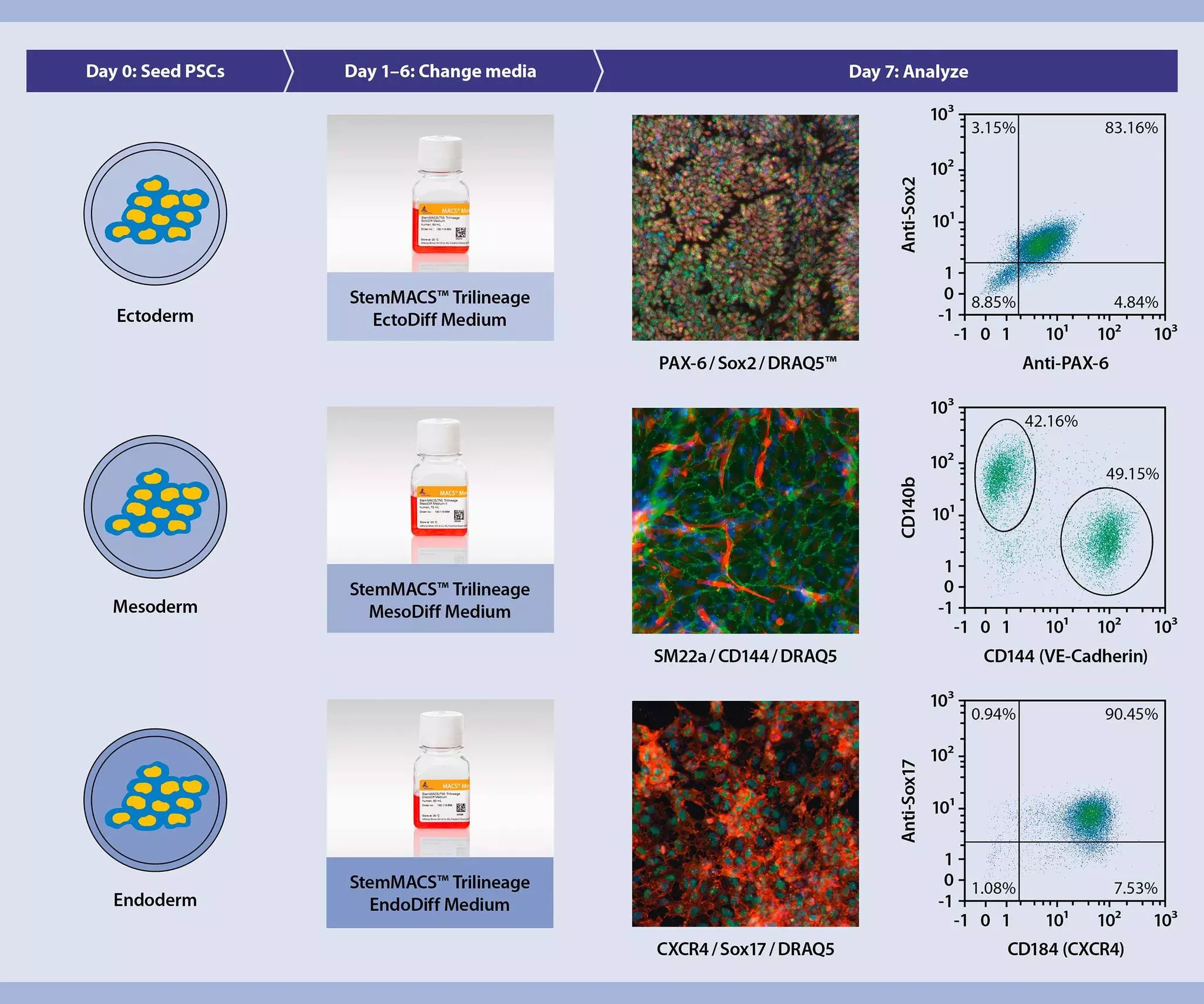
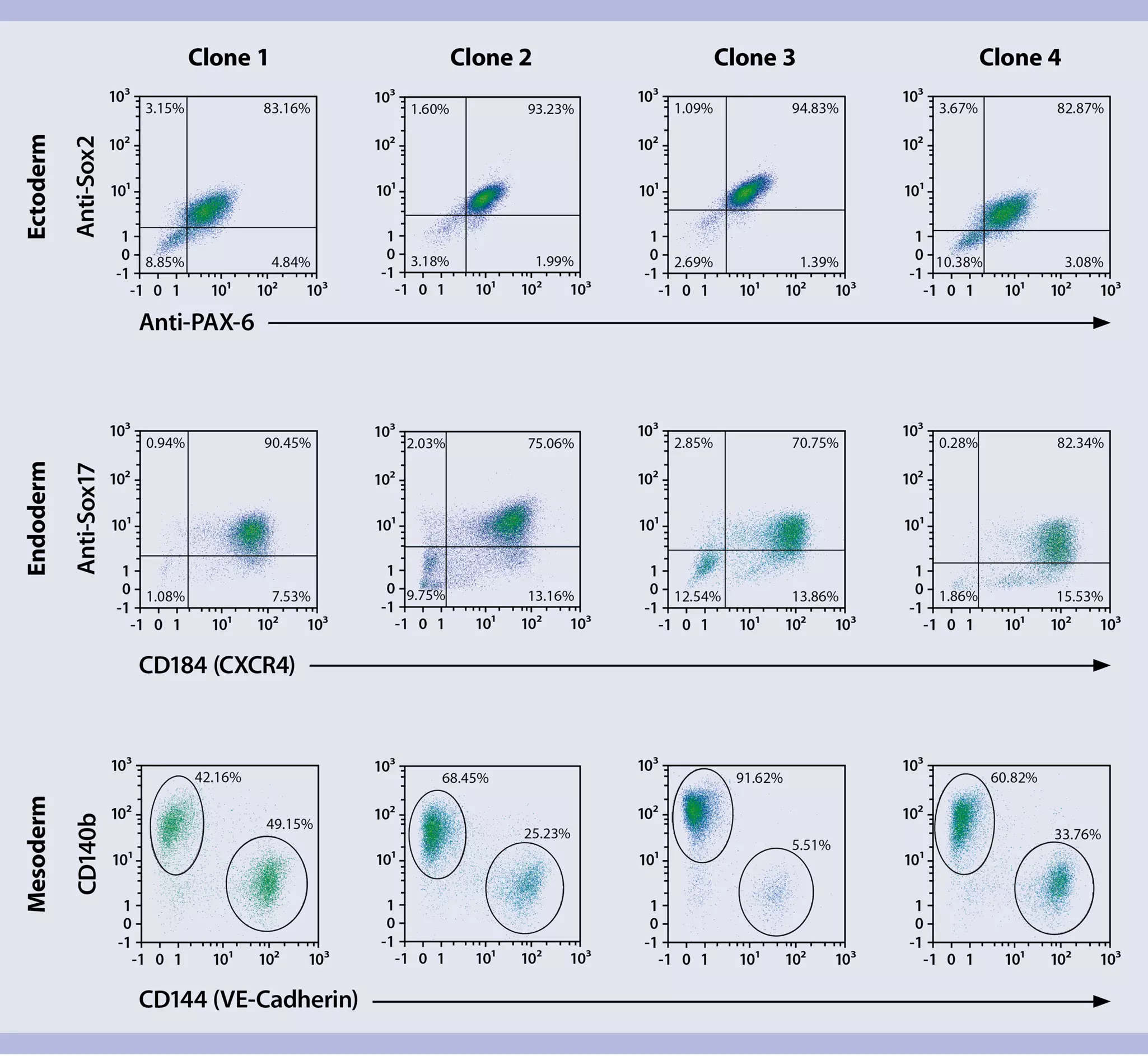
Both traditional assays are time-consuming and difficult to reproduce and quantify. An alternative is directed differentiation into derivatives of the ectodermal, mesodermal, and endodermal lineages. However, to enable the comparison of different cell lines, the same differentiation procedure must be used for all cultures, with highly consistent protocols and reagents. Miltenyi Biotec has developed the StemMACS Trilineage Differentiation Kit, human, which provides standardized, ready-to-use media for differentiation into ectoderm, mesoderm, and endoderm in seven days. Combined with flow cytometry analysis of the resulting differentiated cells, this kit enables a quantitative comparison of the pluripotency of different PSC lines.
Our innovation is further enhanced by expediting analysis through a multicolor flow cytometry panel (Miltenyi Biotec-tested panel 22 (MBTP 22)) featuring REAfinity Recombinant Antibodies. This panel enables swift yet precise qualitative and quantitative assessment of hPSC differentiation potential. It comprises two antibody combinations for each embryonic germ layer (ectoderm, mesoderm, and endoderm), facilitating the detection of both surface and intracellular markers. A scientific poster describing the quantitative assessment of PSC differentiation potential and a protocol for analyzing differentiated samples by flow cytometry can be downloaded from the related resources panel.
4.4 Cryopreservation of pluripotent stem cells
At a glance: Kits and reagents for the cryopreservation of PSCs
| Use | Comments | Products |
| Cell detachment | Optimized for use with StemMACS iPS-Brew XF | StemMACS Passaging Solution XF, human |
| Cryopreservation solution | StemMACS Cryo-Brew |
Optimal storage of PSC lines is critical to maintaining a pool of stable lines, preserved at an early passage number. StemMACS Cryo-Brew is a chemically defined media formulation that preserves PSCs with >90% viability and enables rapid culture initiation after thawing. Adding a ROCK inhibitor to the culture medium after thawing provides optimal attachment and survival because cells recover as single cells or small cell clusters (see 4.2 Splitting pluripotent stem cells cultures). However, adding StemMACS PSC-Brew XF, human, facilitates the swift re-initiation of hPSC cultures post-cryopreservation, without the need to add any ROCK inhibitors.
5 PSCs in diverse applications
At a glance: Media and supplements for supporting PSCs under challenging conditions, e.g. CRISPR-CAS9
| Product category | Comment | Product |
| Media | Research grade | StemMACS PSC-Brew XF, human |
| Supplement | Improves hPSC performances under challenging culture conditions | StemMACS PSC-Support XF, human |
Given their immense potential, PSCs find utility across diverse applications. However, it is crucial to note that PSCs are highly sensitive cells, and the demanding nature of many applications can impose significant stress on them. Therefore, the choice of a high-quality culture medium emerges as a critical determinant for experimental success. A meticulously formulated medium and supplement provide essential nutrients and growth factors, minimizing the risk of cell loss.
The seamless compatibility of StemMACS PSC-Brew XF, human, with StemMACS PSC-Support XF, human, creates an optimal environment tailored to improve performance under challenging conditions, such as gene editing approaches (PMID: 37481731), flow sorting, or after reprogramming.
CRISPR-Cas9 is a valuable tool in PSC research, enabling precise gene editing to modify stem cells for specific experimental or therapeutic purposes. Applied to PSCs, this powerful tool enhances their utility through targeted genetic modifications, driving advancements in personalized medicine and disease modeling.
Miltenyi provides a selection of modified mRNA transfection markers to monitor the success of CRISPR gene editing. These mRNAs are designed for rapid delivery into cells, ensuring a non-toxic, transient, and efficient expression.
At a glance: Modified mRNAs and kits for transfection control
| Use | Product |
| Efficient mRNA delivery | StemMACS mRNA Transfection Kit |
| Positive control for mRNA transfections | StemMACS mCherry mRNA |
| Positive control for mRNA transfections | StemMACS eGFP mRNA |
| Positive control for mRNA transfections | StemMACS™ Nuclear eGFP mRNA |
| Positive control for mRNA transfections | StemMACS™ YPet mRNA |
6 PSC differentiation
Efficient and consistent cell culture outcomes are crucial for the effective differentiation of human PSCs, necessitating dependable and resilient methodologies to sustain cell lines and enhance viability. The implementation of reproducible, feeder-free cultures, up to MACS® GMP-grade, alongside rapid enrichment of lineage-specific cells or multicolor phenotypic flow analysis, not only ensures continuous workflow support for successful cell cultures but also facilitates the flexibility and reproducibility required for subsequent applications.
6.1 Formation and dissociation of embryoid bodies
At a glance: Kits and reagents for the generation and dissociation of embryoid bodies
| Use | Product |
| Generation | StemMACS DiffBase XF, human |
| Dissociation | Embryoid Body Dissociation Kit, human and mouse |
Embryoid bodies (EBs) have been a cornerstone in previous differentiation methods, serving as the standard approach. These three-dimensional multicellular aggregates form when PSCs are cultured in suspension without anti-differentiation factors. In this environment, PSCs naturally undergo spontaneous differentiation, giving rise to a mix of endodermal, ectodermal, and mesodermal cell lineages, ultimately resulting in the formation of EBs (PMID: 17609152).
StemMACS DiffBase XF, human, is a xeno-free medium, optimized and standardized to be used as a base medium tailored for the differentiation of human PSCs. Designed to be applied directly to hPSCs cultured in StemMACS PSC media, StemMACS DiffBase XF, human, can be used to generate EBs without requiring further supplementation with cytokines. Additionally, StemMACS DiffBase XF, human, offers the convenience of assessing hPSC pluripotency via the embryoid body assay. Once generated, EBs can be dissociated into single cells in a reproducible manner using the Embryoid Body Dissociation Kit, human and mouse, combined with the gentleMACS Dissociator. The enzyme cocktail contained in the kit preserves the majority of cell surface epitopes, allowing a reliable analysis or separation of differentiated target cells from the heterogeneous single-cell suspension.
EBs serve as a valuable model for mimicking various aspects of cell differentiation observed during early embryonic development. However, traditional methods of differentiation using EBs often exhibited bias, as exposure to certain growth factors favored the differentiation of specific cell lineages over others (PMID: 17538720). In recent years, there has been a shift towards more precisely defined differentiation protocols. Many of these newer approaches forego the formation of EBs altogether, primarily due to the inherent challenges associated with this method. Instead, researchers now favor adherent culture techniques, where PSCs are subjected to specific conditions over timed intervals. These conditions typically involve controlled exposure to growth factors, small molecules, and substrate cues that mimic the sequential events observed during embryonic development. This refined approach allows for a more targeted and controlled differentiation process, leading to the generation of desired cell lineages.
6.2 PSC differentiation with ready-to-use kits
At a glance: Media, supplements, cytokines, and small molecules for directed differentiation of hPSCs
| Use | Product |
| A ready-to-use kit for cardiomyocyte differentiation | StemMACS CardioDiff Kit XF, human |
| Coating the cell culture plates with a chemically defined reagent | MACSmatrix Laminin 511 |
| To direct the differentiation to atrial-like cardiomyocytes | StemMACS Retinoic Acid |
| Further expansion of PSC-derived cardiomyocytes | StemMACS Cardiac Cultivation Medium XF, human |
In recent years, scientists have developed numerous differentiation protocols, with many relying on a basal media and strategic application of differentiation cytokines and small molecules. Despite extensive efforts to optimize the yield of specific differentiated target cells, researchers consistently encounter variations in cell differentiation. Standardized culture techniques are essential to enhance consistency and reliability in PSC differentiation. Ready-to-use differentiation kits can bring consistency and reproducibility to this process, offering high efficiency in achieving desired cell types.
To facilitate PSC differentiation into cardiomyocytes, the StemMACS CardioDiff Kit XF, human, provides a streamlined, xeno-free process that ensures consistent, high-efficiency differentiation without requiring additional patterning factors. This kit supports the differentiation of PSCs into ventricular-like cardiomyocytes, and, when supplemented with StemMACS Retinoic Acid, enables the generation of atrial-like cardiomyocytes. Researchers can also opt for monolayer adherent cultures or three-dimensional cardiospheres. For extended culture stability, the StemMACS Cardiac Cultivation Medium XF, human, maintains high cell viability and functionality for over 30 days, while the StemMACS Cryo-Brew cryopreservation solution preserves essential cell characteristics, offering excellent thawing efficiency and rapid re-initiation.
6.3 PSC differentiation using cytokines, and small molecules
At a glance: Kits, media, supplements, cytokines, and small molecules for directed differentiation of hPSCs
| Use | Product |
| Basal medium for flexible differentiation to any cell type | StemMACS DiffBase XF |
| Coating the cell culture plates with a chemically defined reagent | MACSmatrix Laminin 511 |
| Cytokines and small molecules | MACS Recombinant cytokines and growth factors |
| GMP-grade cytokines for clinical research | MACS GMP Cytokines |
| Medium for neuronal differentiation | MACS Neuro Medium |
| B27 supplement for neuronal differentiation | MACS NeuroBrew-21 w/o vit A |
| Medium for cardiomyocyte differentiation | StemMACS Cardiac Cultivation Medium XF, human |
When you need flexibility in your experiments, our comprehensive selection of StemMACS Media, supplements, growth factors, and cytokines offers a user-friendly and adaptable toolkit that enables precise control over PSC differentiation processes.
StemMACS DiffBase XF, human, is a xeno-free medium, optimized and standardized as a base medium tailored for the differentiation of human PSCs. Designed to be applied directly to hPSCs cultured in StemMACS PSC media, StemMACS DiffBase XF, human, can serve as a foundational medium for nearly all types of directed differentiation protocols when supplemented with the appropriate patterning cytokines and small molecules. To complete the differentiation, maturation, or expansion of specific cell types, we offer MACS Neuro Medium and MACS NeuroBrew-21 w/o vit A for neuronal differentiation, as well as StemMACS Cardiac Cultivation Medium XF, human, for cardiomyocyte differentiation. With our comprehensive differentiation portfolio, you can optimize the differentiation process, support cell growth, and augment the expansion of differentiated cells, thereby facilitating the creation of faster and more efficient systems.
Researchers harness the power of cytokines by adding them to the culture medium in precise combinations and concentrations to mimic the natural signaling environment that drives cell differentiation during embryonic development. By carefully controlling the timing and duration of cytokine exposure, scientists can steer PSCs toward different cell lineages, such as neurons, cardiomyocytes, or pancreatic cells. Complementing cytokines, small molecules that modulate intracellular signaling pathways are often used synergistically to enhance or fine-tune the differentiation process. Together, cytokines and small molecules provide powerful tools for directing the fate of PSCs and generating specific cell types for various research and therapeutic applications.
One prominent factor contributing to variability associated with the differentiation process is the biological activity of cytokines. Fluctuations in cell culture conditions stemming from inconsistent cytokine activity are widespread. The biological activity of recombinant cytokines, which refers to their effectiveness on cells in a particular bioassay, is not a fixed or absolute value. Only properly expressed and folded cytokines exhibit biological activity, thus influencing cellular responses. Consequently, total protein weight does not directly correspond to the amount of active cytokine, which is typically measured in units of biological activity. The production process significantly influences the amount of correctly expressed and folded recombinant proteins, leading to variability in the specific biological activity of cytokines among different vendors and production batches, as shown in the figure below.
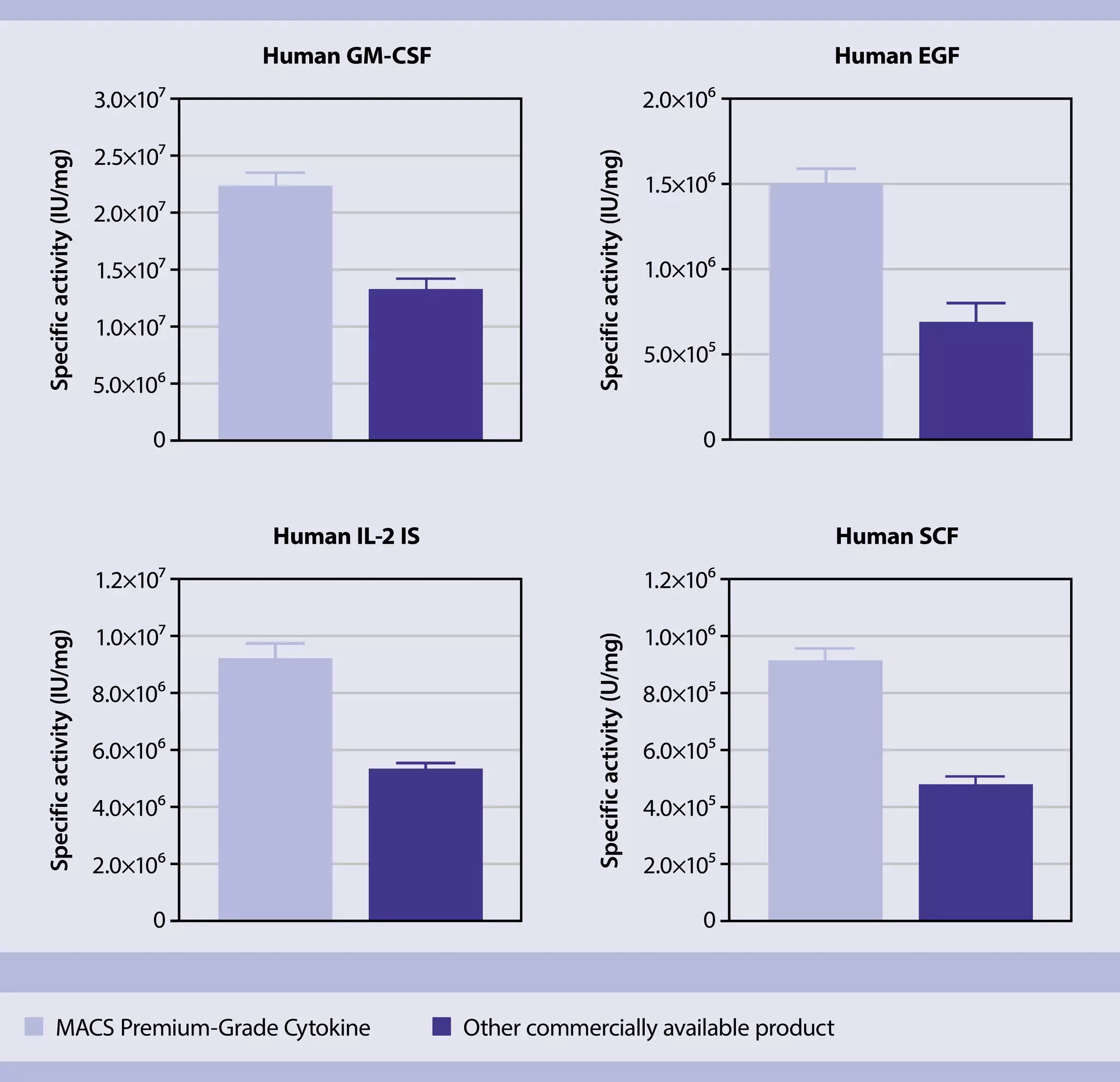
Using the same microgram quantities of cytokines from various sources or production batches can result in the addition of differing amounts of active protein. In these specific examples, dosing based on protein weight may lead to the inclusion of up to double (or half) the amount of active cytokine, thereby introducing variability into cell culture experiments. On the other hand, dosing based on biological activity helps minimize such variations and enhances the reproducibility of cell culture experiments.
Our recombinant cytokines and growth factors are available in three quality grades:
- Research grade
- Premium grade
- MACS GMP Grade
To streamline cytokine dosing based on biological activity, we offer lot-specific activity data with our MACS Premium-Grade Cytokines. This information enables consistent application of the same quantity of active cytokine, ensuring reproducible cell culture conditions without the need for extensive pre-testing. Moreover, utilizing MACS Premium-Grade Cytokines facilitates a smooth transition to clinical research with MACS GMP Cytokines.
At a glance: small molecules and cytokines to stimulate differentiation
Related content:
6.4 PSC Differentiation using organoid models
In recent years, the use of organoid models has emerged as a transformative approach in differentiation research, enabling scientists to recreate complex, three-dimensional structures that closely mimic human tissue architecture and function. Derived from PSCs, organoids provide a more physiologically relevant environment for cell differentiation than traditional two-dimensional cultures.
By utilizing specific growth factors, cytokines, and extracellular matrix components, researchers can guide PSCs through intricate differentiation pathways, forming miniaturized versions of organs such as the brain, liver, and intestine. This organoid-based approach enhances research accuracy, offering unprecedented insights that can improve translational research and therapeutic applications.
At a glance: Kit for dissociating the PSC-derived cells and organoids
| Use | Products |
| Neural organoids | Neural Tissue dissociation kit |
| PSC–derived cardiomyocytes | Multi Tissue Dissociation Kit 3 |
6.5 PSC differentiation using transcription factors
In addition to directed differentiation using small molecules and growth factors, mRNA transfection of critical transcription factors can be used to aid differentiation. Some neuronal and adipocytic mRNAs are available for the transfection of PSCs.
At a glance: reagents and transcription factors to stimulate differentiation
| Use | Products |
| For transfection | |
| Efficient mRNA delivery | StemMACS mRNA Transfection Kit |
| Positive control for mRNA transfections | StemMACS mCherry mRNA |
| Positive control for mRNA transfections | StemMACS eGFP mRNA |
| Positive control for mRNA transfections | StemMACS Nuclear eGFP mRNA |
| Positive control for mRNA transfections | StemMACS YPet mRNA |
| To induce differentiation | |
| Adipocyte differentiation | StemMACS Cebpb mRNA, human |
| Adipocyte differentiation | StemMACS Pparg mRNA, human |
| Neural cells differentiation | StemMACS NeuroD1 mRNA, human |
| Neural cells differentiation | StemMACS Brn2 mRNA, human |
| Neural cells differentiation | StemMACS NeuroG2 mRNA, human |
| Neural cells differentiation | StemMACS Myt1l mRNA, human |
| Neural cells differentiation | StemMACS Mash1 (Ascl1) mRNA, human |
| Dopaminergic neurons differentiation | StemMACS Lmx1a mRNA, human |
| Dopaminergic neurons differentiation | StemMACS Nurr1 (NR4a2) mRNA, human |
6.6 Isolation of PSC-derived cells during and after differentiation
Isolating PSC-derived cells post-differentiation is essential in scientific research, as it enables researchers to work with a homogeneous cell population, reducing contamination and variability in experimental outcomes. Additionally, isolated cells can undergo further characterization and functional assays, helping to deepen our understanding of their properties and behavior. This isolation process is also crucial for potential therapeutic applications, ensuring safe and precise administration of the intended cell types.
6.6.1 Isolation via MACS Technology
MACS (Magnetic-Activated Cell Sorting) technology is an advanced method for isolating nearly any cell type with precision. Using MACS MicroBeads, it enables the rapid isolation of cells from specific lineages at any stage of differentiation. Optimized kits are available for various PSC-derived cell types, providing a straightforward solution.
For unlisted cell types, the MACS Marker Screen, anti-human, can analyze 371 human markers to help identify suitable targets for isolation. Additionally, MACS MicroBeads support a wide range of indirect labeling strategies, enabling targeted cell isolation with flexibility.
At a glance: Kits and reagents for the separation of differentiated cells
| Target cell type | Starting material | Isolation strategy | Comments | Automation | Product |
| Ectoderm | |||||
| Neural crest | Single-cell suspension | Positive selection of target cells | Based on CD271 expression | Yes | Neural Crest Stem Cell MicroBeads, human |
| Neuronal progenitors | Single-cell suspension | Positive selection of target cells | Based on PSA-NCAM expression | Yes | Anti-PSA-NCAM MicroBead Kit, human and mouse |
| Glial progenitors | Single-cell suspension | Positive selection of target cells | Based on A2B5 expression | Yes | Anti-A2B5 MicroBead Kit, human and mouse |
| Immature oligodendrocyte | Single-cell suspension | Positive selection of target cells | Based on O4 expression | Yes | Anti-O4 MicroBeads, human, mouse, rat |
| Single-cell suspension | Positive selection of target cells | Based on GLAST (ACSA-1) expression | Yes | Anti-GLAST (ACSA-1) MicroBead Kit, human, mouse, rat | |
| Mesoderm | |||||
| Hematopoietic progenitors | Single-cell suspension | Positive selection of target cells | Based on CD34 expression | Yes | CD34 MicroBead Kit UltraPure, human |
| Cardiomyocytes | Single-cell suspension | Depletion of non-cardiomyocytes | Depletion cocktail | Yes | PSC-Derived Cardiomyocyte Isolation Kit, human |
| Endothelium | Single-cell suspension | Positive selection of target cells | Based on CD144 (VE-Cadherin) expression | Yes | CD144 (VE-Cadherin) MicroBeads, human |
| Endoderm | |||||
| Definitive endoderm | Single-cell suspension | Positive selection of target cells | Based on CD184 (CXCR4) expression | Yes | CD184 (CXCR4) MicroBead Kit, human |
At a glance: products for manual or automated cell isolation
| Use | Products |
| Automated cell separation systems | MACS Cell Separation Instruments |
| Manual cell separators | MACS Magnetic Separators |
| Columns for magnetic cell separation | MACS Columns |
| Buffers and solutions for cell isolation | MACS Buffers and Solutions |
MACS Handbook:
 Related products:
6.6.2 Isolation using flow cytometry cell sorting
Flow cytometry is another powerful technique for isolating PSC-derived cells, allowing for the analysis and sorting of individual cells based on specific surface markers. This method provides high-throughput capabilities and can simultaneously assess multiple parameters of each cell, ensuring precise identification and selection. Researchers can employ fluorescently labeled antibodies to target specific cell populations, enabling the isolation of homogeneous cell groups for downstream applications.
At a glance: Antibodies and instruments for cell isolation using flow cytometry
| Use | Products |
| Antibodies for flow cytometry | MACS Antibodies for flow cytometry |
| Cell sorter instruments | MACSQuant Tyto Cell Sorters family |
MACS Handbook:
Related products:
6.7 Characterization of the PSC-derived cells
Characterization of PSC-derived cells is crucial due to their heterogeneous nature, often comprising various subtypes within the desired cell lineage. Understanding the composition and properties of these subtypes is essential for ensuring the reproducibility and reliability of experimental outcomes, especially in applications such as regenerative medicine and disease modeling.
One of the primary methods for characterizing PSC-derived cells are flow cytometry and immunostaining. Flow cytometry enables high-throughput analysis of cell surface markers and intracellular proteins, allowing for the identification and quantification of specific cell populations within a heterogeneous culture. By using fluorescently labeled antibodies targeting lineage-specific antigens, flow cytometry provides valuable information about the composition and purity of PSC-derived cell populations. Similarly, immunostaining, combined with fluorescence microscopy or imaging techniques, allows for the visualization and spatial localization of proteins of interest within individual cells or tissues. By labeling cells with fluorescently tagged antibodies against specific markers, immunostaining enables the identification of cell types, assessment of differentiation status, and characterization of cellular morphology. Together, these complementary techniques provide comprehensive insights into the heterogeneity, phenotype, and functional properties of PSC-derived cells, facilitating their utilization in various biomedical applications with greater precision and confidence.
Our extensive portfolio of fully validated antibodies stands as a crucial asset for characterizing PSC-derived cells. These antibodies are adept at targeting various lineage-specific antigens, facilitating the identification and quantification of specific cell populations within heterogeneous cultures through techniques, such as, flow cytometry, microscopy, and functional assays. Leveraging our MACS Antibody Portfolio ensures high-performance consistency, with the majority of antibodies manufactured using our innovative recombinant antibody technology. Recombinant antibodies, derived from a defined set of genes, undergo a highly standardized production process, guaranteeing unrivaled lot-to-lot reproducibility and standardization. Their known gene sequence allows for easy modification, enhancing antibody properties and ultimately contributing to the reliability and precision of characterizing PSC-derived cells.
Our portfolio of proprietary antibodies, including REAfinity, REAlease®, and REAdye_lease™ reagents, are optimized for flow cytometry, flow sorting, and imaging applications, respectively. Each technology offers the benefit of high reproducibility and error-free analysis, with unique features suited for their respective applications.
At a glance: Kits and tested antibody panels for the characterization of differentiated cells
| Product category | PSC-derived cell types | Product/protocol |
| Flow cytometry-based antibody kit | Mesencephalic dopaminergic (mesDA) progenitors | PSC-mDA Neuron Analysis Kit, anti-human, REAfinity |
| Flow cytometry-based antibody panel | Mesencephalic dopaminergic (mesDA) progenitors | Miltenyi Biotec-tested panel 47 (MBTP 47) |
| Flow cytometry-based antibody panel | Cardiomyocytes | Miltenyi Biotec-tested panel 6 (MBTP 6) |
Related products:
You have previously chosen International as your location.
What do you want to do?