MHC multimer technology


1 Introduction to antigen-specific T cell biology 2 Analyzing antigen-specific T cells: MHC MACSimer technology 3 Designing a successful MHC MACSimer experiment 4 Expert tips and experiment optimization 4.1 Peptide pools vs. single peptides for T cell expansion 4.2 MHC allele–specific titration for optimal staining performance 4.3 Optimizing the efficiency of REAlease Technology on MHC MACSimers 4.4 Antibody clone compatibility 4.5 Increasing detection of ultra-rare T cells using the FASER Signal Amplification Kit 4.6 Increasing signal confidence with duplex staining using MHC MACSimers 4.7 Multiplexing for epitope screening
5 Troubleshooting guide
1 Introduction to antigen-specific T cell biology
1.1 Antigen-specific T cells and their relevance
Antigen-specific T cells are essential effectors of the adaptive immune system, responsible for recognizing and responding to antigens presented by aberrant or infected cells. Through their unique T cell receptors (TCRs), they are capable of discriminating between self and foreign antigens, enabling targeted immune responses against pathogens. Their specificity and ability to remember past infections underpin the effectiveness of immunological memory, which is the basis for vaccination strategies. Antigen-specific T cells are categorized into two subsets based on their coreceptor expression: CD8+ cytotoxic T cells and CD4+ helper T cells. CD8+ T cells are the main effectors of cell-mediated immunity, detecting and killing abnormal cells such as cancer or virus-infected cells. By contrast, CD4+ T cells are responsible for coordinating an efficient immune response against a specific threat or pathogen.
Recent advances in manipulating antigen-specific T cells hold promise for developing novel immunotherapies to treat cancer, infections, and autoimmune diseases. Antigen-specific therapeutic approaches include genetically engineered TCR T cells, designed to specifically target a patient’s cancer cells or to recognize susceptible patterns in pathogenic viruses not detected by the patient’s immune system.
1.2 Antigen recognition and MHC restriction
TCRs can recognize antigens only when these are presented by major histocompatibility complex (MHC) molecules, an immunological principle known as MHC restriction. MHC molecules are loaded with short peptide sequences, or T cell epitopes, derived from processed proteins inside the cell, and then present them to the extracellular space. The interaction of TCRs with MHC-bound peptides is highly specific and relies on the complementary fit between TCR and MHC complex. Every TCR is uniquely restricted to bind only one specific combination of peptide-MHC (pMHC) (fig. 1).
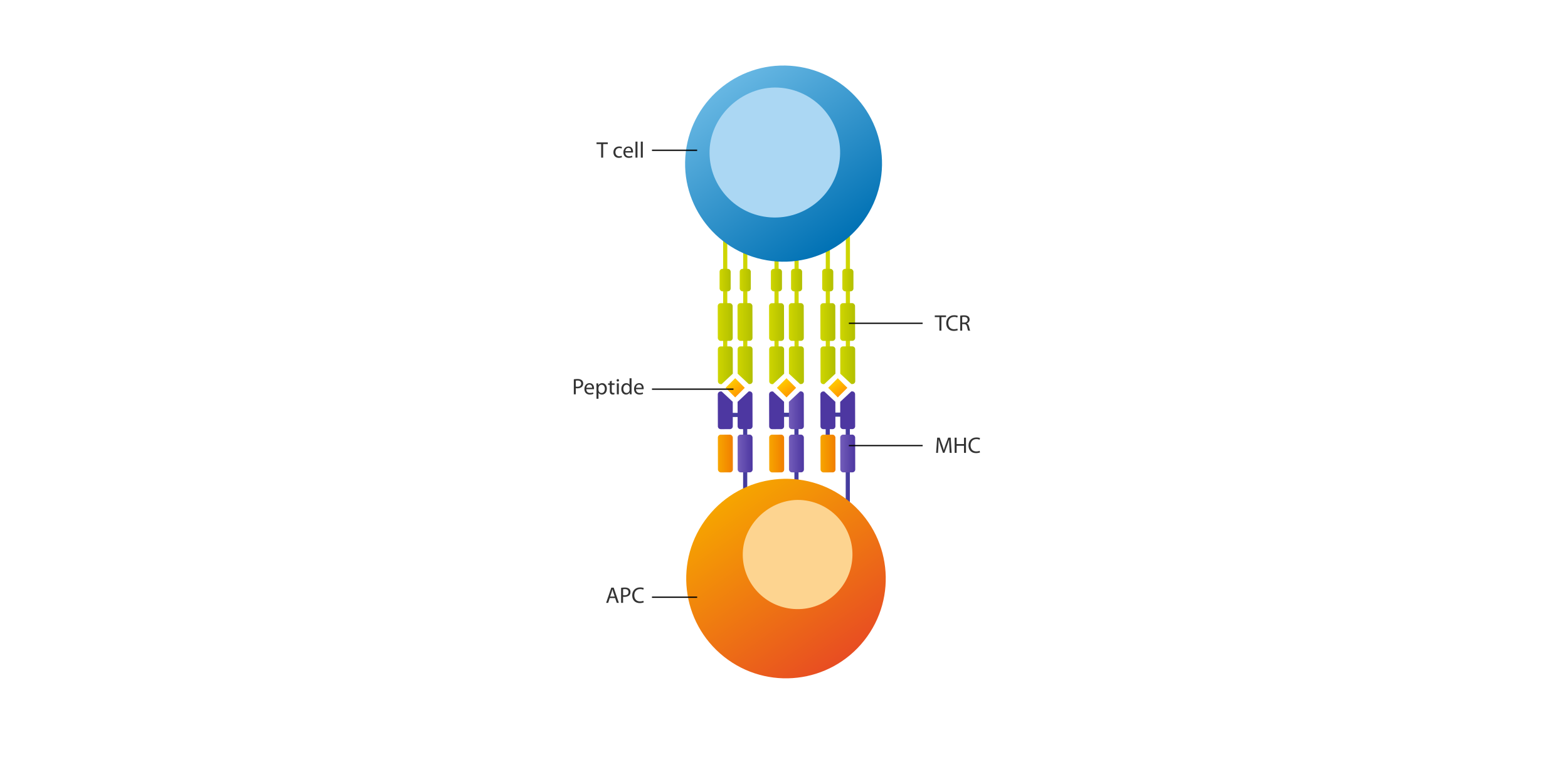


1.3 MHC and peptide classification
Two important classes of MHC molecules interact with antigen-specific T cells, MHC I and MHC II (table 1). CD8+ T cells recognize MHC class I molecules, expressed on virtually every nucleated cell. Peptides presented on MHC I mainly derive from intracellular proteins, enabling the immune system to monitor intracellular abnormalities like mutations and infections. By contrast, CD4+ T cells bind to MHC class II molecules, expressed only on professional antigen-presenting cells (APCs). MHC II typically presents peptides from extracellular proteins, uncovering foreign pathogens or allergens.
Table 1: Comparison of MHC class I and class II molecules.
| MHC class I | MHC class II | |
| Expressed on | All nucleated body cells. | Professional APCs, e.g., dendritic cells, macrophages, and B cells. |
| Length of bound epitope (amino acids) | 8–12 | 13–25 |
| Main peptide source* | Intracellular source, such as: - Self-peptides; - Mutated (neo-)antigens; - Peptides derived from replicating pathogens, such as viruses. | Extracellular source, such as: - Matrix peptides; - Bacterial peptides derived from extracellular infections; - Allergens. |
| Interacting T cell | CD8+ cytotoxic | CD4+ helper |
* Please note that cross-presentation of MHC I and II peptides is frequently observed.
1.4 MHC genetics and nomenclature
In humans, the MHC locus is known as the human leukocyte antigen (HLA) system, a highly polymorphic genomic region on chromosome 6. Each individual inherits a unique combination of HLA alleles that defines our immunological self (see table 2). This diversity is essential for immune defense and transplantation compatibility.
HLA genes are divided into three classes:
- Class I (e.g., HLA-A, HLA-B, HLA-C), presenting endogenous antigens to CD8⁺ T cells;
- Class II (e.g., HLA-DR, HLA-DQ, HLA-DP), presenting exogenous antigens to CD4⁺ T cells;
- Class III, which includes genes involved in immune function (e.g., components of the complement system) but do not encode classical MHC molecules.
Table 2: Overview of HLA terminology.
| Terminology | Meaning | Example |
| HLA genotype | The exact set of inherited HLA alleles, determined by genetic sequencing. This provides the precise nucleotide sequence of the HLA genes. | An individual inherits HLA-DRB1*03:01 from one parent and HLA-DRB1*04:01 from the other. Their HLA genotype is HLA-DRB1*03:01 / HLA-DRB1*04:01. |
| HLA phenotype | On an individual level: The set of HLA proteins expressed, determined by the inherited alleles. In a population: The proportion of individuals expressing a specific HLA protein variant. | Someone with the HLA phenotype HLA-A2 / HLA-B27 expresses these specific HLA proteins on their cells. |
| HLA allotype | A specific version of an HLA protein that comes from a particular gene variant. Allotypes reflect differences at the protein level, affecting peptide binding and immune responses. | HLA-B has different allotypes like HLA-B*27:05 and HLA-B*27:02, which have slight structural differences. |
| HLA serotype | A way to group HLA proteins based on their reactivity to specific antibodies (serotyping). Serotyping may not differentiate between allelic variants. | HLA-A2 is a serotype that includes multiple closely related HLA-A alleles. |
In mice, the MHC locus is termed histocompatibility-2 (H-2) complex and is located on chromosome 17. It includes class I (e.g., H-2K, H-2D, H-2L) and class II (e.g., H-2I-A and H-2I-E) molecules (table 3). Similar to humans, mouse MHC I interacts with CD8+ T cells, and mouse MHC II with CD4+ T cells, underscoring the evolutionary conservation of antigen presentation across species.
Table 3: MHC allotypes of frequently used laboratory mouse strains.
| MHC Class I | MHC Class II | |||||
| Mouse strain | MHC allotype | H-2K | H-2D | H-2L | H-2I-A | H-2I-E |
| C57BL/6 [B6] * | b | b | b | - | b | - |
| BaLB/cJ [C] | d | d | d | d | d | d |
| C3H/HeJ [C3] | k | k | k | - | k | k |
* Including the commonly used OT-I mice (C57BL/6-Tg(TcraTcrb)1100Mjb/J), expressing TCRs specific for ovalbumin residues 257–264 on the background of H-2Kb; and OT-2 mice (B6.Cg-Tg(TcraTcrb)425Cbn/J), expressing TCRs specific for ovalbumin residues 323–339 on the background of H-2I-Ab.
1.5 HLA frequency and association with disease
HLA allele frequencies significantly depend on population genetics, leading to great differences between geographic regions. Table 4 shows a global representation of the most frequent HLA variants based on aggregated data by the allele frequency net database (AFND). For example, HLA-A*0201 is one of the most frequent variants in North America and Europe, with phenotype frequencies of 40–50%. HLA-A*2402 represents another frequent variant, with phenotype frequencies of 20–40% in Northeast and Southeast Asia and 15–25% in North America.
Table 4: Globally prevalent HLA class I and II allotypes.
| HLA Class I | HLA Class II |
| HLA-A*01:01 | HLA-DPA1*01:03-DPB1*04:01 (DP401) |
| HLA-A*02:01 | HLA-DPA1*01:03-DPB1*04:02 (DP402) |
| HLA-A*03:01 | HLA-DQA1*01:01-DQB1*05:01 (DQ5.1) |
| HLA-A*11:01 | HLA-DQA1*01:02-DQB1*06:02 (DQ6.2) |
| HLA-A*24:02 | HLA-DQA1*02:01-DQB1*02:02 (DQ2.2) |
| HLA-B*07:02 | HLA-DQA1*05:01-DQB1*02:01 (DQ2.5) |
| HLA-B*08:01 | HLA-DQA1*05:05-DQB1*03:01 (DQ7.5) |
| HLA-B*35:01 | HLA-DRA*01:01-DRB1*04:01 (DR4a) |
| HLA-B*40:01 | HLA-DRA*01:01-DRB1*15:01 (DR2b) |
| HLA-B*44:02/B*44:03 | HLA-DRA*01:01-DRB1*07:01 (DR7a) |
| HLA-C*04:01 | HLA-DRA*01:01-DRB1*03:01 (DR3a) |
| HLA-C*07:01 | HLA-DRA*01:01-DRB1*13:01/13:02 (DR13a/b) |
*Aggregated data provided by AFND (effective Q1/2025).
In line with their enormous genetic diversity and ability to direct T cell responses, certain HLA variants are associated with human susceptibility to diseases (table 5). For instance, HLA-DRB1 variants are strongly associated with rheumatoid arthritis and multiple sclerosis, and with the outcome of infectious diseases like tuberculosis or malaria. Certain HLA-B variants are associated with immune control in human immunodeficiency virus-1 (HIV-1) infection.
Certain HLA-A and HLA-B variants have also been linked to higher susceptibility to cancer. Downregulation of HLA expression is one of the most important evasion mechanisms of tumor cells. While understanding how HLA is linked to disease is crucial, scientists still face major challenges in fully deciphering the complex genetic and environmental interactions that influence these associations.
Table 5: Known associations between HLA variants and human diseases.
| Disease | HLA variant | |
| Infection | Pulmonary tuberculosis | HLA-DQB1*0302 |
| Malaria | HLA-B53 | |
| Leprosy | HLA-DRB1*1501 | |
| HIV-1 infection | HLA-B*2705 | |
| Chronic hepatitis C infection | HLA-DQB1 | |
| Autoimmunity | Multiple sclerosis | HLA-DRB1*1501 |
| Psoriasis | HLA-C*06:02 | |
| Rheumatoid arthritis | HLA-DRB1*0401 | |
| Ankylosing spondylitis | HLA-B*27 | |
| Type 1 diabetes (T1D) | HLA-DRB1*0301; DQB1*0302 (DQ8), DQ2 | |
| Celiac disease | HLA-DQA1*0501-DQB1*0201 (DQ2) | |
| Neuropathies | Narcolepsy | HLA-DQB1*06:02 |
 Related content:
2 Analyzing antigen-specific T cells: MHC MACSimer technology
2.1 Principle of MHC MACSimer technology
For flow cytometry staining and sorting of antigen-specific T cells via their TCR, peptide-loaded MHC (pMHC) complexes can be used. When conjugated to a fluorochrome, they enable sensitive detection of rare T cell subsets even from a polyclonal population of T cells. This powerful method is well-established and applicable for basic research and TCR discovery, in-process/quality control in cell manufacturing, and post-infusion research in T cell therapy workflows.
A key challenge in detecting antigen-specific T cells lies in the low affinity of a single TCR for its cognate pMHC complex, typically about 10,000 times lower than the affinity of a conventional antibody for its antigen. To overcome this limitation, TCR avidity becomes a critical factor to detect specific TCRs with pMHC molecules.
Table 6 summarizes and compares TCR affinity, biochemical avidity, and functional T cell avidity, including their definitions, influencing factors, and typical measurement methods.
Table 6: TCR affinity and avidity.
| TCR affinity | TCR avidity (biochemical) | T cell avidity (functional) | |
| Definition | Interaction strength of one TCR and one pMHC complex | Interaction strength of multiple TCRs and coreceptors binding to multiple pMHC complexes | T cell responsiveness to a given concentration of a TCR ligand |
| Mode | Single interaction | Multiple molecule interactions | T cell activation by a natural or artificial antigen-presenting cell |
| Influencing factors | Molecular binding strength as defined by the equilibrium dissociation constant (KD) | Number of TCR and pMHC molecules, TCR clustering, and coreceptor binding | Biochemical TCR avidity, effector status, T helper subtype, and genetic factors |
| Measurement method | Determined by surface plasmon resonance (SPR) analysis of recombinant proteins (PMID: 27252176) | Estimated by mean fluorescent intensity (MFI) using MHC multimers (PMID: 23576742) | Concentration of a stimulus inducing 50% activation of antigen-specific T cells (PMID: 16818373) |
MACS Handbook:
 Related products:
2.1.1 Increasing TCR binding avidity
For flow cytometry–based detection of TCRs with pMHC molecules, TCR binding avidity is increased by multimerizing several identical pMHC molecules in one construct, enabling the binding of multiple TCRs on one single T cell simultaneously (“multivalency effect”, fig. 2). Conventional multimer technologies such as MHC tetramers use biotinylated pMHC monomers tetramerized on fluorescently tagged streptavidin.
In comparison, MHC MACSimers provide superior TCR avidity and stability, as they carry a precisely balanced number of covalently conjugated pMHCs and fluorochromes on a sterically flexible backbone. Learn more about MHC MACSimer technology, performance, and applications.

2.2 MHC MACSimer workflows and protocols
MHC MACSimers can be acquired as fully multimerized constructs, either preloaded with peptide (peptide-loaded MHC MACSimers), or in a loadable format (MHC MACSimer FleX Kits). For MHC MACSimer FleX Kit loading, peptide sequences can be chosen from the comprehensive MACSpep Single Peptide portfolio. After sorting of antigen-specific T cells, MHC MACSimer staining is reversible via REAlease® Technology, e.g., for sequential sorting or downstream assays.
Learn more about MHC MACSimer product formats and applications.
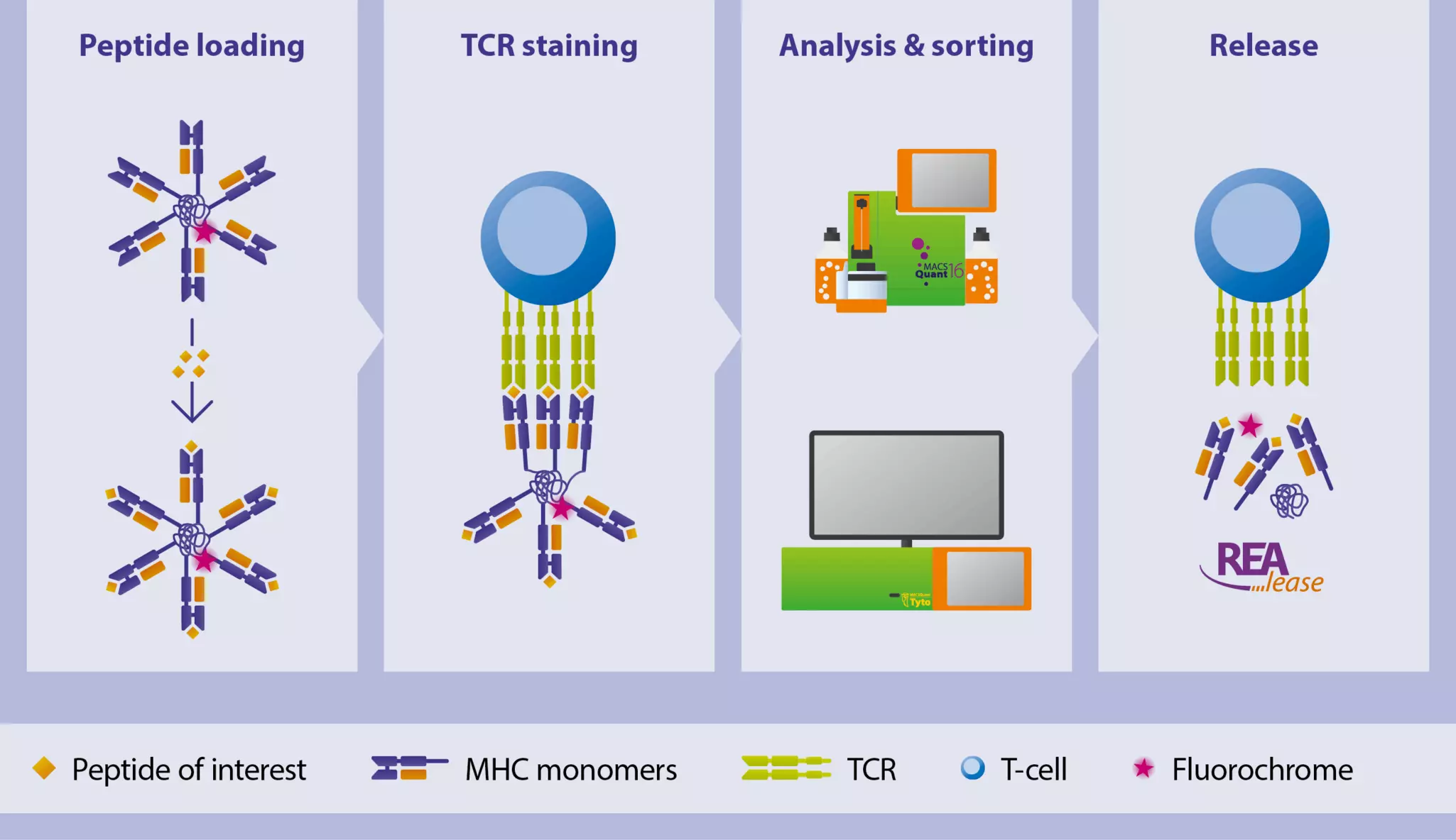
Related content:
Table 7: T cell analysis using MHC MACSimers: workflows and protocols.
| Detection and analyis |
| Detection and analysis of antigen-specific T cells using flow cytometry, e.g., for enumeration or phenotypical and functional characterization of T cells and their epitopes. |
| Preparation of MACSpep single peptides for peptide loading on MHC MACSimers MHC I MACSimer peptide loading and staining protocol MHC MACSimer multicolor panel for characterization of T cell maturation |
| Isolation and sorting |
| Isolation and sorting of antigen-specific T cells using flow sorting or MACS® Technology, e.g., for enrichment purposes or downstream assays (for expansion, cell culture, TCR sequencing, single-cell sequencing, protein biochemistry, and more). |
| Sorting of T cells using MHC MACSimers and the MACSQuant Tyto Cell Sorter Magnetic enrichment of T cells with MHC MACSimers and MACS MicroBeads |
| Stimulation and expansion |
| Stimulation of antigen-specific T cells in cell culture using MACSpep Single Peptides, e.g., for expansion against one specific epitope |
| Preparation of MACSpep single peptides for stimulation and expansion of antigen-specific T cells |
3 Designing a successful MHC MACSimer experiment
In order to ensure technical success, there are three important aspects that should be considered before designing an MHC MACSimer experiment: MHC compatibility, peptide selection, and sample preparation.
3.1 MHC allele compatibility
Ensuring compatibility between the MHC allele and the target T cells is essential, as TCRs recognize antigens only when presented by a matching MHC variant.
- The MHC variant of the MHC MACSimer reagent must match the variant of the intended cell sample. If the MHC variant of a sample is unknown, it can be assessed by different methods:
- Next-generation sequencing (NGS)
- HLA typing assays/ kits (qPCR–based)
- HLA serotyping using flow cytometry (anti-HLA antibodies, e.g., anti-HLA-A2)
- The MHC MACSimer must match the species of the intended cell sample, as MHC molecules do not cross-react with T cells from different species.
3.2 Peptide selection for loading onto MHC MACSimer FleX Kits
For successful and specific T cell detection with MHC MACSimer FleX Kits, careful peptide selection is critical. Only single peptides are suitable for loading, as peptide pools (e.g., PepTivator® Peptide Pools) containing multiple T cell epitopes compromise TCR binding avidity and are therefore not compatible.
For optimal results with MHC MACSimers, we recommend using our MACSpep Single Peptides portfolio, following the short instructions for MHC MACSimer peptide selection and loading. We offer a wide range of validated epitopes optimized for MHC MACSimer loading and supported by tailored protocols.
Alternatively, lyophilized single-peptide stocks from other sources may also be used, provided they meet key quality and handling requirements.
The following sections guide you through essential considerations for selecting a suitable peptide (3.2.1) and best practices when using peptides from external sources (3.2.2) to achieve optimal loading efficiency.
 Related PDFs:
3.2.1 Single peptide selection
There are three major considerations for selecting a suitable MHC peptide:
- Define your T cell epitope: Ensure the selected peptide is restricted to your MHC of interest, as each MHC variant naturally binds distinct peptide sequences. You can identify suitable epitopes using:
- one of our MACSpep Single Peptide epitopes;
- free online databases such as the Immune Epitope Database;
- well-characterized epitopes from scientific literature;
- previously identified immunogenic epitopes, e.g., from screening assays.
- Determine MHC binding affinity: The peptide must have sufficient molecular binding affinity to the MHC type of interest, or it cannot be loaded. Use online tools such as NetMHCpan to calculate the affinity.
- Ensure solubility*: The peptide must be soluble in aqueous solution. Use GRAVY calculators to assess the hydrophobicity of your peptide, and test solubility by dissolving a sample of the peptide in buffer before preparing the final MHC loading reaction (watch out for cloudiness and precipitation).
*Important note: Some peptides are highly hydrophobic due to their amino acid composition, which can make manual loading challenging or impractical. For these specific cases, we recommend using peptide-loaded MHC MACSimers . A list of these sequences is provided in the protocol for the preparation of MACSpep Single Peptides for peptide loading on MHC MACSimers.
3.2.2 Important recommendations when using single peptides from other sources
Lyophilized single-peptide stocks from other sources may be used as well. In such cases, it is essential to follow the guidelines below to achieve successful loading and performance:
- Dissolve peptides in accurately weighable amounts (≥1 µg). If precise weighing isn’t possible (e.g., no access to a microscale), use the entire lyophilized stock to ensure an exact peptide concentration.
- Determining peptide concentration in liquid phase is not recommended; instead, always calculate concentration based on the weight of the lyophilized peptide and the volume of the solvent used.
- Dissolve peptides in DMSO, as the solubility of peptides in water or buffer might be limited.
- Dissolve peptide to a stock concentration of 10 mmol/L or higher, as smaller dilution series from a higher concentrated stock are recommended for titration of the optimal peptide loading concentration.
- Rule out precipitation of the peptide in your dilution series (apparent by pellet formation and/or clouding). If precipitation is observed, consider increasing DMSO rate or decreasing peptide concentration.
- Dissolve peptide under sterile conditions if considering using it in cell culture applications.
- Peptides containing cysteines might need to be reduced with 10 mmol/L DTT for 30 minutes at room temperature before use (if stored at –20 °C a longer period might be necessary).
3.3 Sample source and preparation
MHC MACSimers are compatible with a wide range of sample types, including whole blood, PBMCs (isolated by density-gradient centrifugation), and preenriched or isolated T cells.
If magnetic preenrichment of T cells is required, we recommend using the negative isolation kits listed below. These kits deplete unwanted cell populations while leaving target T cells untouched and unlabeled, preserving TCR and coreceptor integrity for reliable posterior MHC MACSimer staining.
Important note: Avoid positive selection via CD4 or CD8 coreceptors, as direct labeling of these markers can interfere with TCR accessibility due to their close spatial proximity, potentially compromising MHC MACSimer staining.
Table 8: Recommended negative selection kits for untouched (label-free) cell isolation.
| Kits for magnetic preenrichment of T cells | Comment |
| StraightFrom® Whole Blood PBMC Isolation Kit, human | |
| MACSprep™ PBMC Isolation Kit, human | |
| MACSexpress® Whole Blood Pan T Cell Isolation Kit, human | |
| Pan T Cell Isolation Kit II, mouse | |
| Pan T Cell Isolation Kit, human | |
| CD8+ T Cell Isolation Kit, human | MHC Class I only |
| CD4+ T Cell Isolation Kit, human | MHC Class II only |
Related products:
3.3.1 How to prepare a cell sample for MHC MACSimer staining
T cell characteristics and origin significantly influence MHC MACSimer staining efficiency. The relative frequency of TCR-expressing cells in a sample, as well as the expression levels of surface TCRs, are decisive factors for the technical success of the staining. To ensure reliable and specific staining of antigen-specific T cells, consider the following factors when preparing your cell sample:
- Viability: T cells should be fresh or properly cryopreserved to maintain viability and TCR integrity. Viability should be tested before staining.
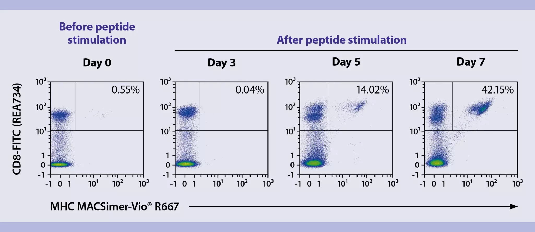
- TCR internalization: In response to peptide–based stimulation, antigen-specific T cells internalize their TCRs, making them temporarily undetectable by MHC MACSimers. Allow a resting phase of 5–7 days post-stimulation to enable TCR re-expression on the cell surface (see chapter 4.1., fig. 4).
- Fixation compatibility: Fixation protocols may affect TCR conformation and multimer binding. Test your fixation method for every specific application, e.g., by comparing staining results from fixed and unfixed samples to ensure compatibility with your workflow.
- Target cell frequency: The frequency of target cells in your sample should exceed the detection threshold of MHC MACSimer technology for reliable results. When analyzing ultra-rare T cells, increase the number of cells stained and events acquired by flow cytometry. Include proper negative controls to exclude the background signal.
4 Expert tips and experiment optimization
4.1 Peptide pools vs. single peptides for T cell expansion
Antigen-specific T cells can be expanded using either peptide pools (e.g., PepTivator Peptide Pools) or defined single-peptide epitopes (e.g., MACSpep Single Peptides), applied before or after MHC MACSimer staining or isolation. Table 9 outlines key differences between both approaches to help you select the most suitable method for your experiment.
Importantly, T cells typically internalize their TCRs following peptide stimulation. To allow for effective MHC MACSimer staining, expanded cells should undergo a resting phase of 5–7 days in culture to enable TCR re-expression (fig. 4).
Table 9: Functional differences between peptide pools and single peptides in T cell assays.
| PepTivator Peptide Pools | MACSpep Single Peptides | |
| Format | Peptide pool | Single peptides |
| Peptide length | 15-mers with 11 amino-acid overlaps, covering full protein sequences. | Specific T cell epitopes with a defined length (8–12-mer for MHC class I, 13–25-mer for MHC class II). |
| Sequence coverage | Covering multiple peptide sequences. | Single defined epitope, restricted to bind either an MHC I or MHC II molecule. |
| Main applications | Quick and robust activation and expansion of antigen-specific CD4+ and CD8+ T cells for functional assays (e.g., cytokine secretion assays, proliferation, T cell enrichment). Validated for use with rapid ARTE protocol for T cell enrichment and analysis. | Epitope-specific stimulation and expansion of T cell populations in cell culture, e.g., before MHC multimer staining. |
| T cell expansion properties | Broad expansion: stimulates diverse T cell populations (multiple peptide-MHC combinations). | Narrow expansion: stimulates only one T cell specificity (one peptide-MHC combination). |
| T cell tracking | Tracks multiple T cell populations with different TCR specificities. | Tracks a single epitope-specific T cell population. |
4.1.1 Epitope-specific expansion protocol using MACSpep Single Peptides
The following protocol provides a general framework for expanding antigen-specific T cells from PBMCs using MACSpep Single Peptides. Please note that expansion efficiency can vary significantly depending on donor, HLA type, and T cell epitope. Optimization may be required to achieve optimal results.
Cell culture conditions: To expand antigen-specific T cells, such as human cytomegalovirus (CMV) pp65-reactive CD8⁺ T cells, PBMCs should be cultured at a density of 1×10⁷ cells/mL in TexMACS™ Medium supplemented with 5% human serum albumin (or an alternative suitable culture medium).
Day 0: Add the single peptide of choice (e.g., MACSpep CMV pp65, NLVPMVATV) to a final concentration of 1 nmol/mL. Incubate cells at 37 °C and 5% CO₂.
Day 2: Replace the culture medium with fresh medium containing 50 U/mL of Interleukin-2 (IL-2).
From day 3: Monitor cells daily to ensure nutrient levels remain sufficient. Based on cell density and expansion kinetics, fresh medium may be added every 2–3 days. Additional cytokines can be added as needed, depending on the experimental setup and application.
4.2 MHC allele–specific titration for optimal staining performance
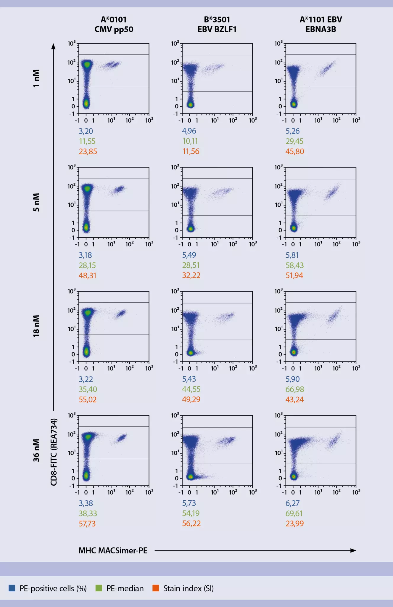
The optimal staining performance of MHC MACSimers can vary depending on the specific peptide–MHC (pMHC) complex and the corresponding TCR avidity. While the standard staining protocol recommends a concentration of 36–60 nM (as given by standard staining protocol), certain MHC alleles may require adjustment to avoid suboptimal or non-specific binding. For example, HLA-B*3501 and HLA-A*1101 can exhibit non-specific over-titration effects at 36 nM (fig. 5). Therefore, we recommend empirically determining the optimal concentration for each MHC allele through titration.
4.3 Optimizing the efficiency of REAlease Technology on MHC MACSimers
The efficiency of REAlease Technology on MHC MACSimers after flow staining and sorting depends on several factors. To achieve optimal results, please consider the following recommendations:
- Using Anti-PE MACS MicroBeads for magnetic enrichment of MHC MACSimer+ T cells blocks the REAlease reagent. Therefore, REAlease Technology is effective only in the context of flow cytometry.
- Not all MHC MACSimer variants are releasable. Check the product data sheet to confirm whether the “release feature” is available. Releasability is defined as achieving at least a 90% reduction in mean fluorescence intensity (MFI) following the REAlease protocol, which corresponds to freeing 90% or more of TCRs from staining.
- REAlease efficiency is highest with Vio R667–labeled MHC MACSimers, due to the dye’s biochemical and molecular properties. For complete optical release, we recommend using Vio R667-labeled reagents.
- Avoid incubating MHC MACSimers at temperatures above 2–8 °C if you plan to release the staining afterward. Incubation at room temperature may impair release due to accelerated TCR internalization.
- Expanded or stimulated T cells often show increased TCR internalization, which can reduce the efficiency of the release.
Related products:
4.4 Antibody clone compatibility
MHC MACSimers are compatible with multicolor flow cytometry. For costaining of CD8+ or CD4+ antigen-specific T cells with MHC MACSimers, the following standard antibody panel is recommended to achieve optimal performance and clear signal resolution (table 10).
Table 10: Standard panel for antigen-specific T cell analysis in combination with MHC MACSimers.
| Specificity | Clone | Gating | Fluorochrome | Detection channel (nm) | Titer |
| ViobilityTM 405/520 Fixable Dye | - | Live/dead discrimination | 405/520 | 525/50 (violet) | 1:100 |
| CD14 | REA599 | Detection of macrophages and monocytes | VioGreen™ | 525/50 (violet) | 1:50 |
| CD20* | LT20 | Detection of B cells and dendritic cells | VioGreen | 525/50 (violet) | 1:50 |
| *Or instead CD19 | REA675 | Detection of B cells and dendritic cells | VioGreen | 525/50 (violet) | 1:50 |
| CD3 | REA613 | Detection of T cells | PE-Vio 770 | 785/62 (blue) | 1:50 |
| CD4 | REA623 | Detection of helper T cells | APC-Vio 770 | 785/62 (red) | 1:100 |
| CD8 | REA734 | Detection of cytotoxic T cells | Vio Bright V600 | 615/20 (violet) | 1:100 |
| MHC class I or II MACSimer-PE | - | TCR staining | PE | 585/40 (blue) | |
| MHC class I or II MACSimer-Vio R667 | - | TCR staining | Vio R667 | 667/30 (red) |
Importantly, some CD8 and CD4 antibody clones can interfere with pMHC–TCR binding, reducing MHC MACSimer staining efficiency. This interference occurs because CD8 and CD4 coreceptors are positioned very close to the TCR and play an essential role in stabilizing the pMHC–TCR interaction during T cell activation. As a result, some antibody clones targeting these coreceptors can sterically hinder or disrupt this binding, leading to reduced staining efficiency of MHC MACSimers. Table 11 lists antibody clones that have been thoroughly tested and verified to be compatible with MHC MACSimers, providing accurate and reliable detection of antigen-specific T cells.
Table 11: Validated antibody clones for coreceptor staining with MHC MACSimers.
| Antibody | Clone |
| CD8 Antibody, anti-human, REAfinity | REA734 |
| CD8a Antibody, anti-mouse, REAfinity | REA601 |
| CD8a Antibody, anti-mouse, REAfinity | 53-6.7 |
| CD4 Antibody, anti-human, REAfinity | REA623 |
Related products:
4.5 Increasing detection of ultra-rare T cells using the FASER Signal Amplification Kit
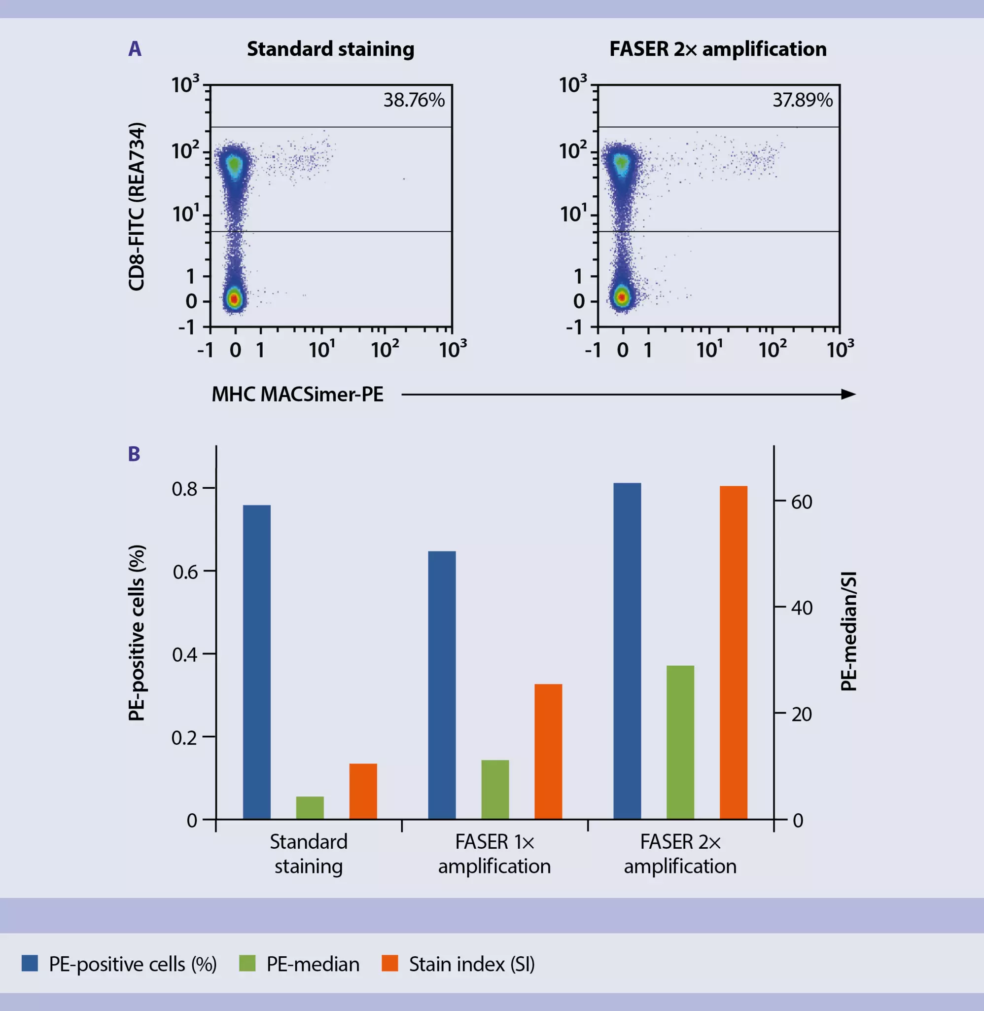
While MHC MACSimers provide high sensitivity, detecting ultra-rare T cell populations may require further signal enhancement. The fluorescence amplification by sequential employment of reagents (FASER) kit boosts low fluorescence signals to improve detection accuracy.
The FASER-PE kit works by sequentially adding a PE-specific activator followed by a fluorochrome-conjugated enhancer. The activator binds to the PE-labeled MHC MACSimer, and the enhancer attaches to the activator, amplifying the fluorescence intensity. This amplification cycle can be repeated multiple times to significantly increase signal intensity (fig. 6).
Please note that REAlease Technology, which allows antibody removal after staining, is incompatible with FASER-amplified samples.
4.6 Increasing signal confidence with duplex staining using MHC MACSimers
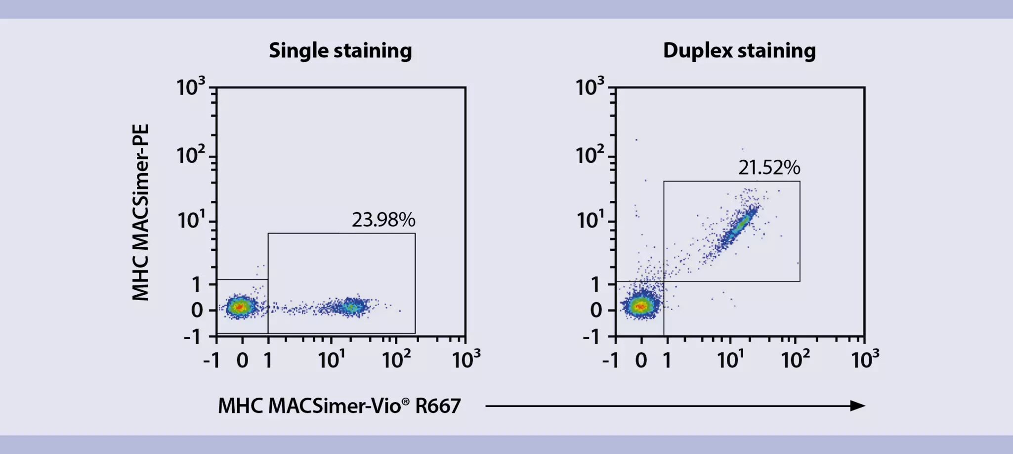
Staining cells with two MHC MACSimers of identical specificity but conjugated to different fluorochromes, a technique known as duplexing, offers two key advantages for the detection of antigen-specific T cells:
- Increased signal confidence: Using two differently labeled MHC MACSimers targeting the same antigen-specific TCR reduces the risk of false-positive events caused by background noise or gating errors.
- Robust validation of TCR-pMHC specificity: Using two different conjugates with the same TCR specificity can confirm that the interaction is biologically specific and not due to fluorophore- or batch-related artifacts. Only double-positive events indicate antigen-specific T cells. A specific duplex signal typically appears as a diagonal population, reflecting the affinity of both reagents for the same TCR (fig. 7).
Related products:
4.7 Multiplexing for epitope screening
For large-scale epitope screening, multiplexing can be beneficial, especially when working with limited cell material. This approach allows simultaneous testing of multiple MHC allele variants and/or peptide sequences within a single sample. MHC MACSimers are compatible with multiplexing; however, careful experimental design is essential to avoid potential pitfalls:
- Multiplexing with peptide-loaded MHC MACSimers: Preloaded MHC MACSimers of different MHC variants and/or peptide sequences can be combined to stain one sample simultaneously. Notably, when selecting the same fluorochrome for multiple MHC variants and/or peptide sequences, it is not possible to assign the resulting fluorescent signal to one of these variants. If this distinction is desired, we recommend choosing different fluorochrome conjugates.
- Multiplexing with MHC MACSimer FleX Kits: When multiplexing different peptide sequences restricted to one or more MHC allele variants using the MHC MACSimer FleX Kit, each peptide loading reaction must be carried out separately for each specificity. Before combining the loaded MHC MACSimers in one sample, each must be individually purified, e.g., via spin column purification. This step is essential, as cross-loading of peptides due to overlapping binding affinities cannot be ruled out, even between different MHC variants.
5 Troubleshooting guide
If your T cell staining shows no signal or unusually high background when using MHC MACSimers, please refer to the questions in table 12 as a diagnostic tool to identify potential issues. This troubleshooting guide outlines key factors that may influence staining quality, from sample preparation to data analysis, and is designed to support you in optimizing your workflow.
If you’ve gone through the list and still can’t identify a potential source of error, don’t hesitate to reach out to our technical support team – we’re here to help.
Table 12: MHC MACSimer troubleshooting guide
| 1. Sample preparation, staining, MHC compatibility | |
| Are your cells healthy and expressing surface TCRs? | Cells should be viable and healthy before staining. If they've been expanded or stimulated, they may have internalized TCRs. In this case, a resting phase of 5 to 7 days is recommended before staining. |
| Does your T cell sample express the MHC allotype that matches the MHC MACSimers? | MHC MACSimers must match the MHC allotype your cells are restricted to. Binding won’t occur if there’s a mismatch, even within the same species. |
| How was your T cell epitope selected, and do your T cells still reflect the intended specificity? | Ensure that your epitope is well-defined, ideally registered in an immune epitope database (e.g., IEDB) or described in peer-reviewed literature. If you're working with expanded T cells or T cells transduced with a specific TCR, confirm that the population still reflects the intended specificity. Be aware that enrichment methods, especially those using peptide pools, can sometimes skew the TCR repertoire toward off-target pMHC combinations. |
| Did recent stimulation or expansion cause TCR internalization, reducing multimer binding? | Expanded/stimulated T cells internalize their TCRs and need 5 to 7 days resting in culture to re-express their surface TCRs and enable proper TCR staining. |
| Is your antibody panel compatible with MHC MACSimers? | Some antibody clones used for CD8/CD4 coreceptor staining may interfere with MHC multimer binding. Compatibility with MHC multimers must be tested. |
| Is your fixation method compatible with MHC MACSimers? | Fixation can compromise TCR integrity. Always assess the compatibility of your fixation method for your specific application, e.g., by comparing staining results from fixed and non-fixed samples. |
| Is the frequency of antigen-specific T cells in your sample sufficient for detection? | Rare populations may fall below the detection threshold. Detection sensitivity depends on the MHC allele, peptide, and type of T cell sample. For ultra-rare T cells, increase the number of stained cells and acquired events, and use appropriate negative controls to exclude background. With optimized protocols, MHC MACSimers have detected as few as 0.02% antigen-specific cells within the CD3⁺CD8⁺ population of PBMCs. |
| Was the number of PBMCs used adequate for reliable detection? | Low cell input can lead to increased background noise. For PBMCs, we recommend a minimum concentration of 2×10⁷ cells/mL (equivalent to 1×10⁶ cells in 55 µL for the standard MHC MACSimer protocol). Note that optimal cell numbers may differ for other T cell sample types, such as TCR-transduced lines or expanded T cells. |
| 2. MHC MACSimer handling and staining conditions | |
| Have the MHC MACSimers been stored and handled appropriately? | MHC Class I MACSimers must be stored at 2–8 °C protected from light. Please refer to the vial label for the expiration date. The shelf-life of the MHC MACSimer FleX Reagent after peptide loading varies significantly depending on the specific pMHC combination. As a result, a general post-loading expiration period cannot be provided. |
| Have the MHC MACSimers been centrifuged before staining? | A colorful pellet after centrifugation at 3300×g for 5 minutes indicates precipitation of the pMHC complex, which can interfere with staining performance. |
| Was the staining time and concentration optimized for the experiment? | TCR-binding affinity and avidity of every pMHC combination vary depending on allele, peptide sequence, and T cell sample. Titration of the optimal staining concentration before experimental setup is essential to technical success. |
| Were there washing steps included between staining steps? | Washing between the fluorescent staining steps and before measurement reduces background noise and improves signal resolution. |
| Have the staining conditions been optimized? | We recommend optimizing MHC MACSimer staining conditions for every application. Longer incubation times (20 minutes instead of 10 minutes) or higher staining temperatures (room temperature instead of 2–8 °C) can improve staining performance for certain types of T cells. |
| 3. Experimental controls | |
| Were the necessary positive and negative controls included? | Controls are essential for the accurate detection of target cell populations. Include both types when possible to guide analysis and gating. See chapter 4 of the MHC MACSimer staining protocol for more details on recommended staining control strategies. |
| 4. Flow cytometry setup and analysis | |
| Is the flow cytometer calibrated? | Proper instrument calibration is a prerequisite to technical success. |
| Have compensation controls been set up? | Use single-stained controls and verify settings for each fluorophore used. Check that fluorescence spillover is appropriately corrected. Inadequate compensation can lead to missed detection of rare T cell populations or misinterpretation of background signals. |
| Has my antibody panel been optimized? | Professional panel design is essential when working with rare antigen-specific T cells. Refer to our panel building resources and the panel builder tool for help. |
| Are gating strategies in place to isolate the T cell population? | Accurate gating improves data quality. We recommend the following strategy: 1) Exclusion of singlets and selection of live lymphocytes; 2) Exclusion of B cells and monocytes; 3) Positive selection of CD3+ T cells and CD8+ or CD4+ T cells; 4) Within the T cell population, MHC MACSimers should be gated using the appropriate gating controls. |
| Has the fluorescence minus one (FMO) control been used? | FMO controls help define gating boundaries and reduce the risk of false positives, especially for rare populations. |
To further optimize your MHC MACSimer workflows, we recommend consulting the following scientific resources:
- Wooldridge L. et al. (2009) Immunology 126(2):147-64. | Read article
- Chang J. (2021) Mol Cells 44(5):328-334. | Read article
- Dolton G., Tungatt K. et al. (2015) Immunology 146(1):11-22. | Read article
You have previously chosen International as your location.
What do you want to do?