

Synonyms: IFNg, IFG, IFI
EXTENDED VALIDATION
- Clone:
- 45-15
- Type of antibody:
- Primary antibodies
- Isotype:
- mouse IgG1κ
- Applications:
- ICFC, FA Glossary
- Select a conjugate:
- Select a size:


Other sizes
Related products for IFN-γ Antibody, anti-human


Intracellular flow cytometry applications
APC
APC-Vio 770
FITC
PE
PE-Vio 770
Recommended dilution:
1:50
Remark:
The antibody is suited for staining of formaldehyde-fixed cells.
Data source:
Miltenyi Biotec
Conjugate:
APC
Used products:
130-114-016, 130-113-490
Description:
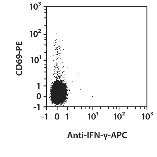
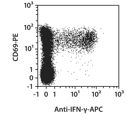
Human peripheral blood mononuclear cells (PBMCs) were either left unstimulated (left images) or stimulated with SEB for six hours. After two hours, brefeldin A was added for stimulated and unstimulated cells. The cells were fixed, permeabilized, and intracellularly stained with Anti-IFN-γ antibodies as well as with CD4 antibodies and CD69 antibodies. Cells were then analyzed by flow cytometry using the MACSQuant® Analyzer. The Tandem Signal Enhancer has been used to increase binding specificity of tandem-dye–conjugated antibodies. CD4+ lymphocytes were pre-gated for the analysis. Cell debris were excluded from the analysis based on scatter signals.
 view details
 view details
Functional assay applications
pure-functional grade
Data source:
Miltenyi Biotec
Type of functional assay:
promotion of Tʜ2 cell and Tʜ17 cell polarization
Conjugate:
pure-functional grade
Used products:
130-095-743
Extended validation for IFN-γ Antibody, anti-human
Specificity
In order to compare the epitope specificity of an antibody, the clone being used is compared with other known clones recognizing the same antigen in a competition assay.
Other clones
Overlap in epitope recognition with 45-15
25723.11
partial
B27
low
4S.B3
low
W19227A
low
REA1343
high
Cells were incubated with an excess of purified unconjugated IFN-γ (45-15) antibody followed by staining with fluorochrome-conjugated antibodies from other known clones targeting the same antigen. Fluorescence intensity was then used to assess epitope overlap: a minimal or absent signal indicated high epitope overlap, an intermediate signal suggested partial overlap, and a strong signal reflected low overlap.
Sensitivity
Selected fluorochrome conjugated antibodies from Miltenyi Biotec were compared to commercially available hybridoma clones in flow cytometry analysis.
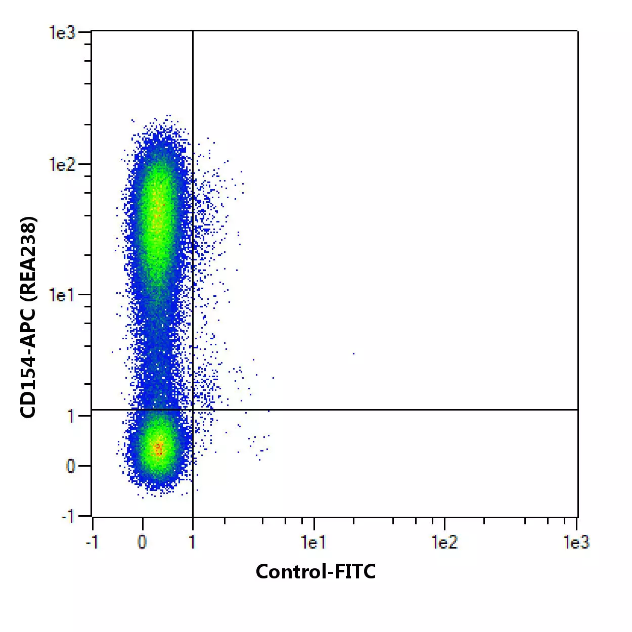
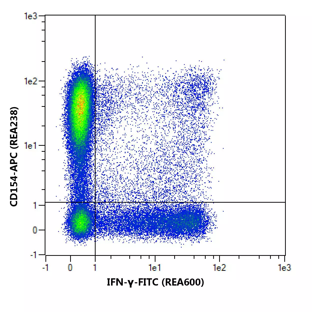
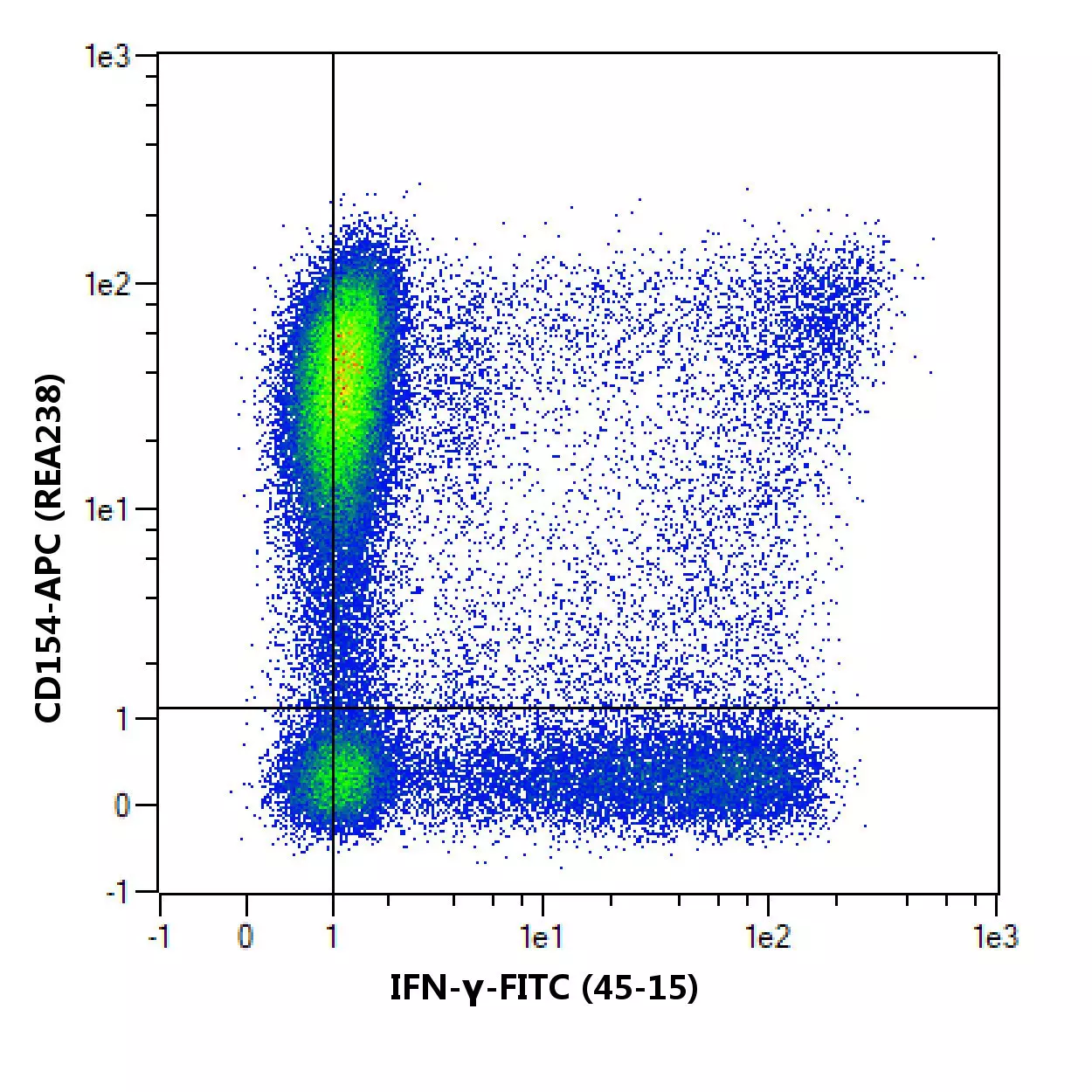
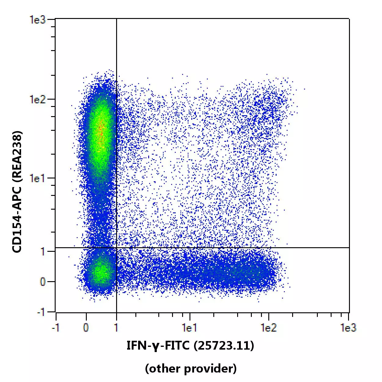
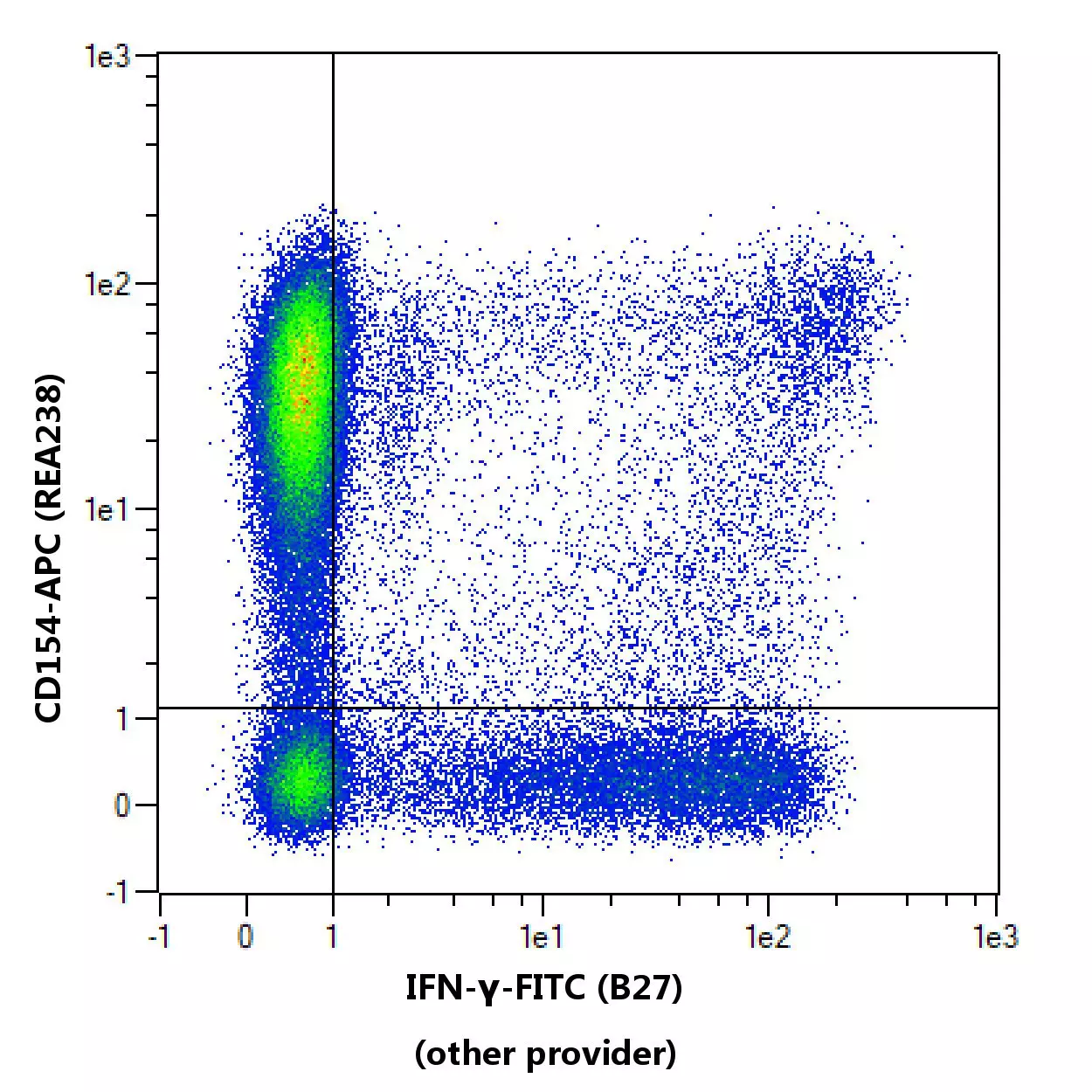

Flow cytometric comparison of different clones for IFN-y. Human peripheral blood mononuclear cells (PBMCs) were stimulated with 20 ng/mL Phorbol 12-myristate 13-acetate (PMA) and 1 µg/ml Ionomycin for 1 hour, followed by an incubation with 1µg/mL Brefeldin A for 4 hours. Cells were stained with a suitable counterstaining and then fixed and permeabilized followed by a staining with IFN-y antibodies. As a control, IFN-y antibody staining was omitted and cells were measured in the same channels. Flow cytometry was performed with the MACSQuant® Analyzer. CD3+ cells were pregated for the analysis. Cell debris and cell doublets were excluded from the analysis based on scatter signals. No FcR Blocking Reagent was used. The recommended titers of respective antibodies from different suppliers were used.
Specifications for IFN-γ Antibody, anti-human
Overview
Interferon gamma (IFN-γ) is produced by cells that are involved in inflammatory immune responses. IFN-γ is predominantly released by memory and effector CD4+ and CD8+ T cells as well as by NK cells upon activation. Quantitative analysis of IFN-γ–producing cells can provide important information on the course of immune responses. Anti-IFN‑γ antibodies are designed for intracellular staining of IFN‑γ–producing cells. Cells can be stimulated for IFN-γ–production, e.g. by polyclonal stimulation with mitogens. For induction of IFN‑γ–production by antigen-specific T cells, cells are restimulated with respective antigen. IFN‑γ can be accumulated in the cells by addition of secretion inhibitors like brefeldin A. After fixation and permeabilization of the cell sample, IFN‑γ–producing cells can be stained intracellularly with Anti-IFN‑γ antibodies. Staining of surface markers allows simultaneous flow cytometric analysis of subsets and activation status of the IFN-γ–producing cells.
IFNg, IFG, IFI
Detailed product information
Technical specifications
Clone
45-15
Clonality
monoclonal
Isotype
mouse IgG1κ
Isotype control
Isotype Control Antibody, mouse IgG1
Host
mouse
Type of antibody
Primary antibodies
Species reactivity
human, non-human primate
Cross-reactivity
rhesus monkey (Macaca mulatta), cynomolgus monkey (Macaca fascicularis)
Antigen
IFN-γ
Molecular mass of antigen [kDa]
16
Entrez Gene ID
RRID
AB_2751116, AB_10830868, AB_2733719, AB_2733589, AB_2733080, AB_2751183, AB_2751182, AB_2733718, AB_2733588, AB_2751117, AB_2733079
Resources for IFN-γ Antibody, anti-human
Documents and Protocols
App notes/customer reports
Certificates
To access a Certificate of Analysis (CoA) follow the link below and enter the specific lot of your product.
Should you wish to request additional documentation, such as a Safety Data Sheet (SDS) in other languages than English or Swedish, an SDS-replacement document, or a Certificate of Origin (CoO), please direct your inquiry to your local technical support team.
 Powered by Bioz
You have previously chosen International as your location.
What do you want to do?