MICS technology workflow
Pancreatic cancer
Lung cancer
Brain tissue
Multiple sclerosis
Content
- MICS technology workflow
- Pancreatic cancer
- Lung cancer
- Brain tissue
- Multiple sclerosis
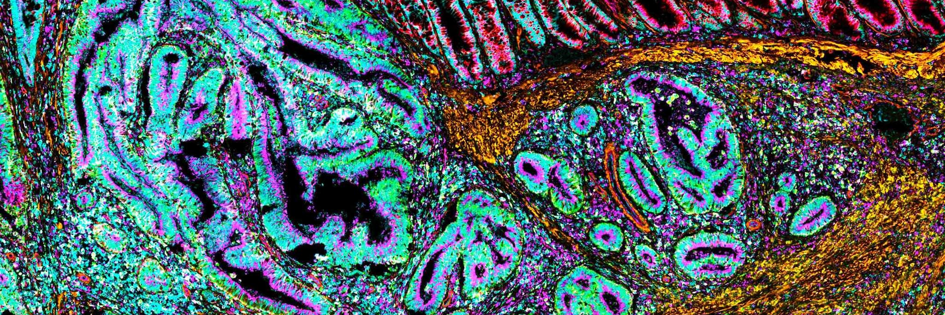

High-plex spatial proteomics brings a powerful new dimension to high-plex protein analysis by preserving the physical context of biological systems. Instead of measuring proteins in bulk, spatial approaches reveal where proteins are expressed within tissues, how cell types interact, and how microenvironments shape biological function.
By combining high-plex detection with spatial resolution, researchers can characterize complex tissue architecture, identify rare or transitional cell states, and uncover spatially organized signaling networks that would otherwise remain hidden. This integrated view of protein abundance and location is especially valuable in areas such as tumor microenvironment profiling, immune response mapping, and understanding disease heterogeneity. Ultimately, spatial proteomics accelerates discovery by providing a richly detailed, context-aware picture of biology that is far more informative than protein measurements alone.
Discover how MACSima® Imaging Cyclic Staining (MICS) turns spatial proteomics into deeper tissue insight, helping you reveal cellular interactions, complex microenvironments, and the spatial patterns that drive discovery.
MICS technology workflow
Pancreatic cancer
Lung cancer
Brain tissue
Multiple sclerosis
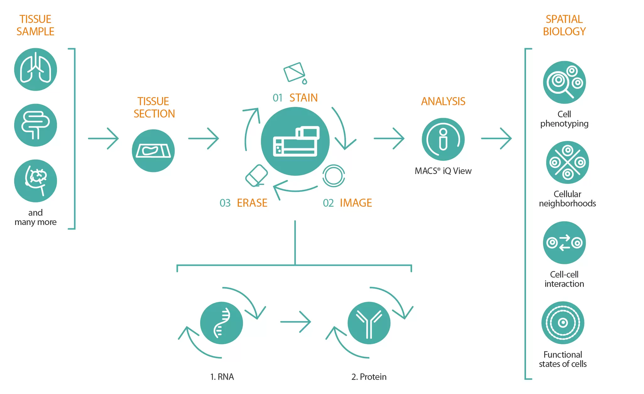
The MACSima Platform analyzes a wide range of sample types, from lung and breast to colon and beyond. Powered by MICS technology, an innovative, high-resolution multiplex imaging method, it relies on an iterative, fully automated staining cycle with three steps:
1. simultaneous staining of the sample with multiple primary conjugated antibodies or RNA probes,
2. image acquisition using widefield microscopy, including automated image processing, and
2. gentle removal of the fluorescence signals.
This three-step process is repeated as often as necessary, allowing a single sample to be analyzed for dozens of RNA molecules and hundreds of proteins. In addition, a newly developed image processing pipeline that reliably removes artifacts enables subcellular resolution of protein and RNA localization. The resulting high-dimensional datasets can then be analyzed with MACS iQ View to address questions related to cell phenotyping, cellular interactions and neighborhoods, and the functional states of different cell types.
Pancreatic cancer remains one of the deadliest malignancies, with five-year survival rates showing little improvement. Most patients are diagnosed at an advanced, unresectable stage, and only ~20% are eligible for surgery – the sole potentially curative option. Even then, median survival ranges from just over two years in resectable cases to less than six months in cases with metastasis. These poor outcomes highlight the need for new therapies. CAR T cell approaches, successful in hematologic cancers, are now being explored for solid tumors such as pancreatic cancer. At the same time, emerging insights into pancreatic ductal cell biology and high-plex spatial technologies are revealing previously unrecognized ductal heterogeneity, offering new clues to disease mechanisms and therapeutic targets.
The MACSima Platform enables high-resolution, automated analysis of hundreds of proteins at the single-cell level. Explore the study on CAR T therapy for pancreatic cancer, which validated tumor-specific targets and clarified biological mechanisms in preclinical models.
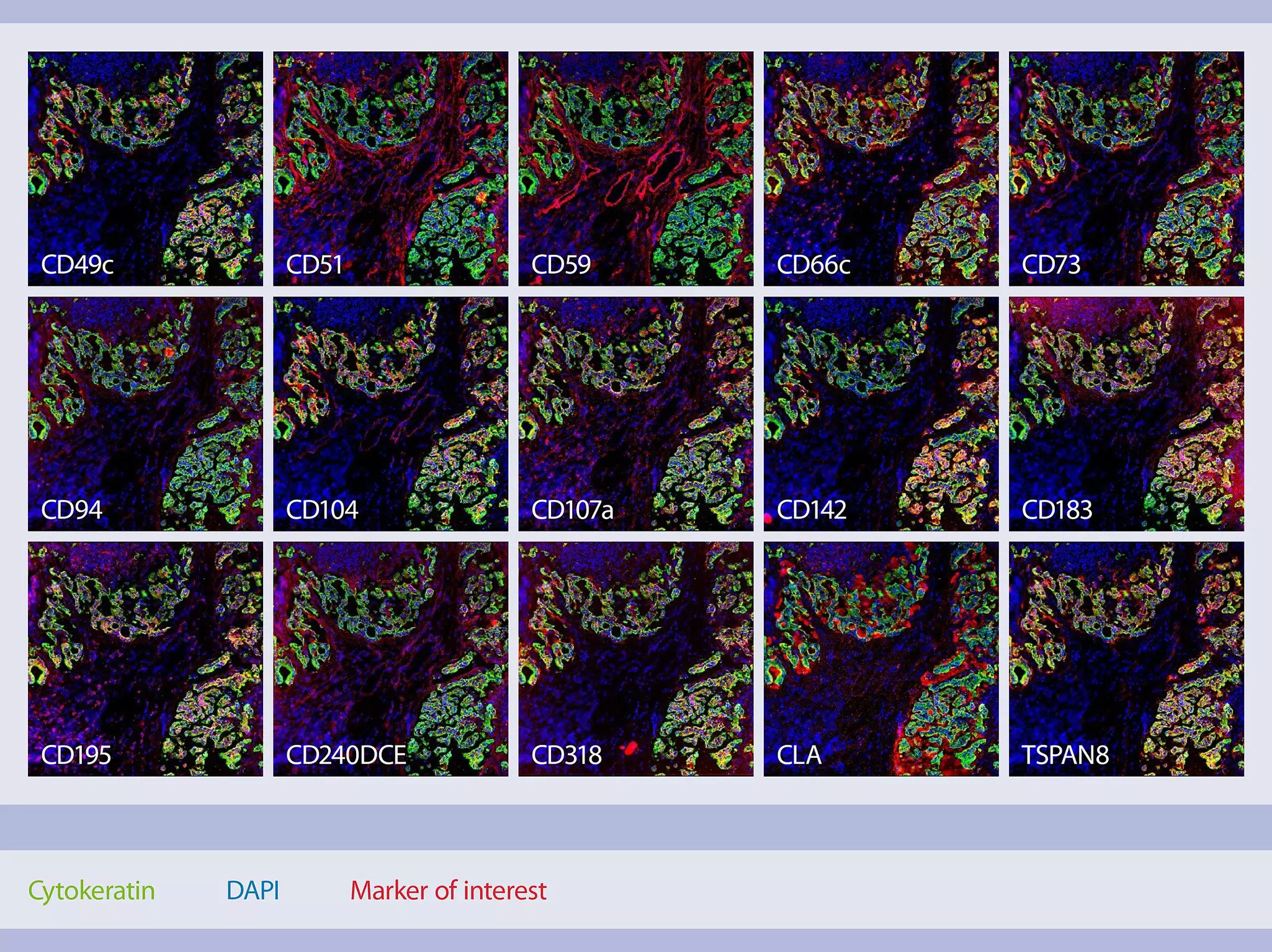
Identification of CD318, TSPAN8, and CD66c as target candidates for CAR T cell-based immunotherapy of pancreatic adenocarcinoma
Schäfer, D. et al. (2021) Nat. Commun. 12: 1453 | Read the article
Optimization of pancreatic cell subset classification in multiplex immunofluorescence
Coleman, et al. (2024) UF J. Undergrad. Res. 26: UF Journal of Undergraduate Research | Read the article
A single-cell atlas of the murine pancreatic ductal tree identifies novel cell populations with potential implications in pancreas regeneration and exocrine pathogenesis
Fernandez, et al. (2024) Gastroenterology | Read the article
Lung cancer remains one of the deadliest malignancies worldwide, causing approximately 1.8 million deaths each year and continuing to impose a major global health burden. Despite advances in immunotherapy, the absence of robust and easily applicable biomarkers to predict responses to immune checkpoint inhibitors hampers effective patient selection, as traditional measures such as PD-L1 expression show limited predictive value. Given the lung’s constant exposure to environmental antigens and its ability to form ectopic germinal centers that locally shape B cell responses, understanding the complex immunological landscape of the lung and its tumor microenvironment is essential for improving therapeutic strategies and identifying more reliable biomarkers.
Explore how high-plex imaging can help identify tumor-associated antigens in lung adenocarcinoma and squamous cell carcinoma by enabling the screening of 155 markers on single tissue sections.
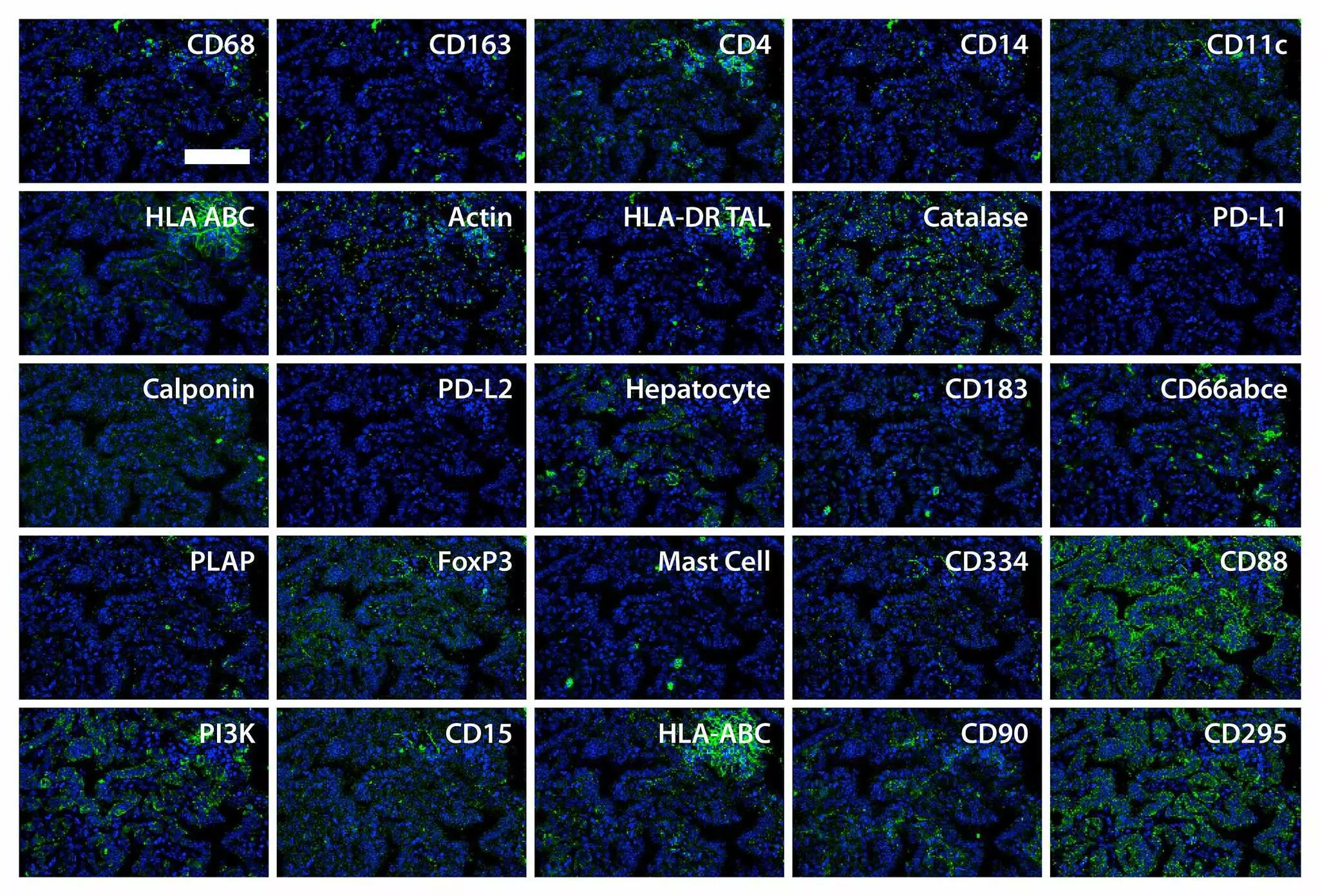


Lung B cells in ectopic germinal centers undergo affinity maturation
Guillaume, S.M. et al. (2024) PNAS 12(14) | Read the article
Platelet PD-L1 reflects collective intratumoral PD-L1 expression and predicts immunotherapy response in non-small cell lung cancer
Hinterleitner, C. et al. (2021) Nat. Commun. 12: 7005 | Read the article
Pitfalls in the characterization of circulating and tissue-resident human γδ T cells
Beucke, N. et al. (2020) J. Leukoc. Biol. 107(6): 1097–1105 | Read the article
Alzheimer’s disease is driven by complex interactions among diverse cell types, each with distinct marker signatures. Early cognitive decline is preceded by changes in neurons, microglia, and astrocytes, resulting in neuroinflammation, vascular alterations, necroptosis, and the accumulation of amyloid‑beta plaques and tau tangles. Intracellular hyperphosphorylated tau (pTau) is a key pathological hallmark and a promising biomarker. Traditional immunofluorescence, limited to around five markers, captures only a fraction of this complexity. View our application note to explore how highly multiplexed tissue imaging with the MACSima Platform enables spatially resolved, single-cell analysis of hundreds of markers in a single tissue sample, overcoming the limitations of traditional methods.
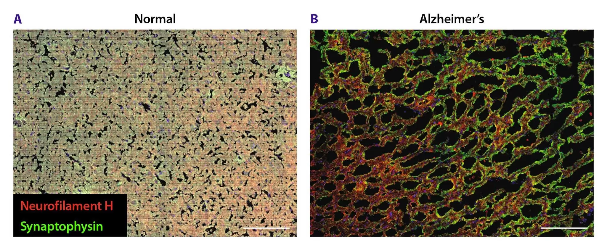
In this application note, discover how the MACSima Platform enables simultaneous analysis of multiple neural markers and, combined with MACS iQ View analysis, allows precise comparison of healthy and Alzheimer’s-affected hippocampal tissue. This workflow lets researchers quantify reduced synaptic density and increased phosphorylated tau, providing a powerful multiplexed spatial biology approach for studying neurological diseases. To explore the research in more detail, read the full study in this application note.
Multiple sclerosis (MS) is an autoimmune disease of the central nervous system that causes neuroinflammation, neurodegeneration, and loss of myelin. Immune cells attack oligodendrocytes and their myelin sheaths, forming characteristic MS lesions, while reactive microglia and astrocytes further drive inflammation.
Because many central and peripheral cell types contribute to MS, detailed characterization of these cells and their spatial organization is essential. Conventional microscopy is limited by the small number of markers it can detect simultaneously.
To address this, we used the MACSima Platform with its MICS technology, which performs fully automated iterative staining cycles to reveal the complex cellular composition and organization in MS brain tissue.
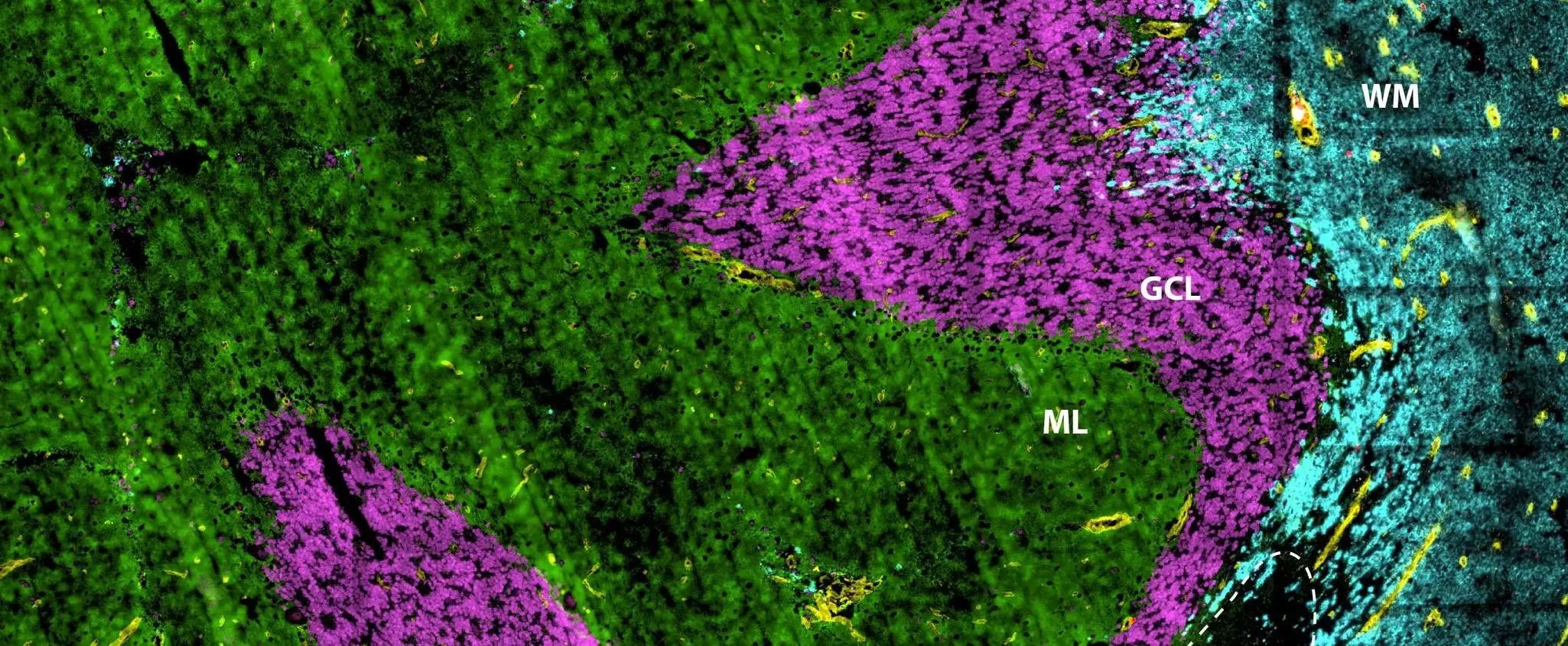
In this application note, we present how a spatial biology approach based on the MACSima Platform and its unique MICS technology was used to explore the cellular composition and organization of postmortem brain tissue from MS patients. High-quality, single-cell staining with 87 antibody conjugates enables unbiased qualitative analysis to distinguish healthy and lesion areas in spatial context. Quantitative marker expression analysis, for example via heatmaps, can further support the discovery of new biomarkers and drug targets. Read the full application note to find out more about this unbiased epitope analysis.
AAV vectors trigger DNA damage response-dependent pro-inflammatory signaling in human iPSC-derived CNS models and mouse brain
Costa-Verdera, H. et al. (2025), Nat Commun., 16, 3694. | Read the article
Brain tumoroids: Treatment prediction and drug development for brain tumors
Souberan, A. et al. (2024), Neuro Oncol., 27(2), 415–429. | Read the article
Hypoxic-ischemic brain injury in neonatal mice sequentially recruits neutrophils with dichotomous phenotype and function
Richter, M. et al. (2025), Nat Commun., 16, 9696. | Read the article
You have previously chosen International as your location.
What do you want to do?