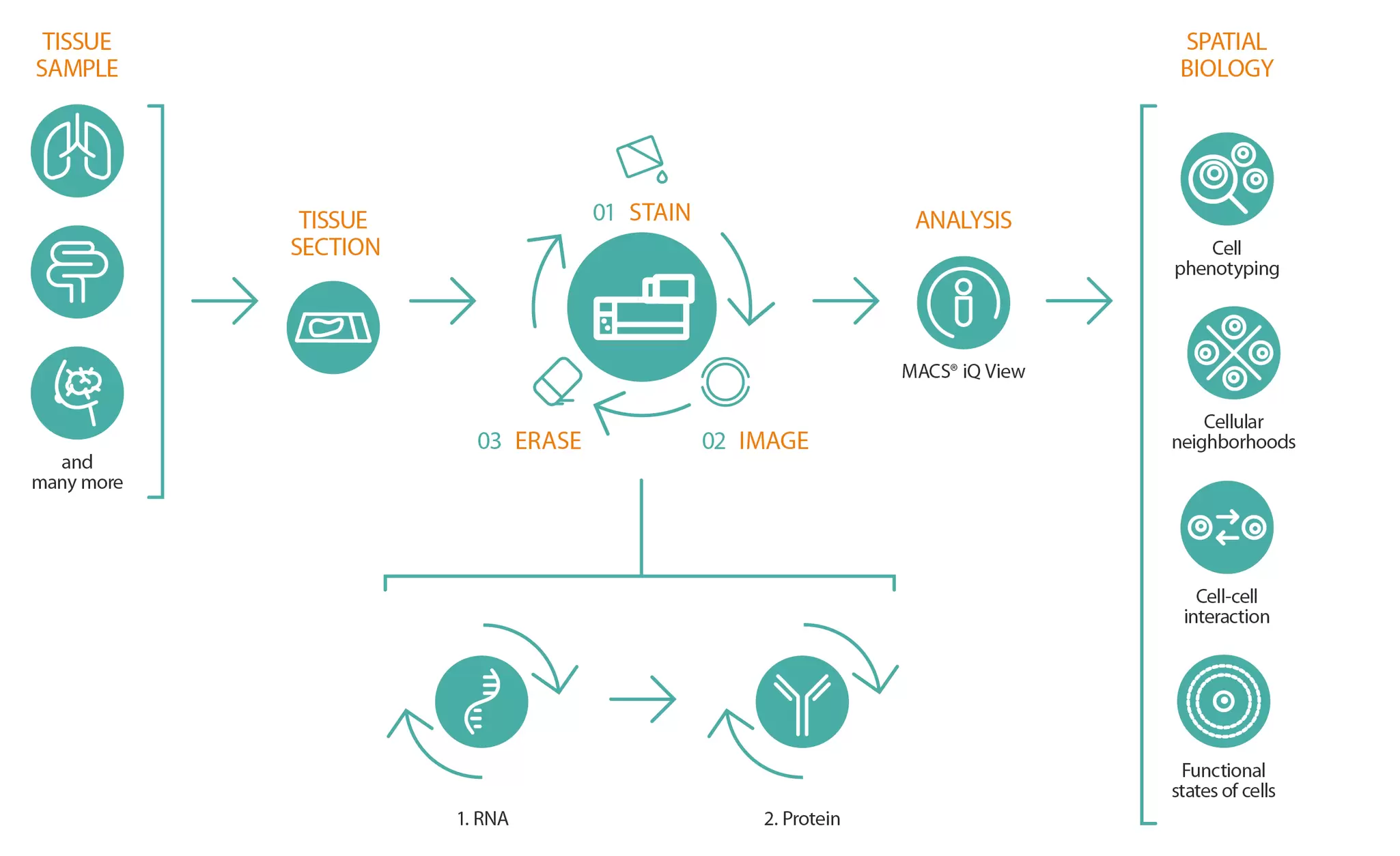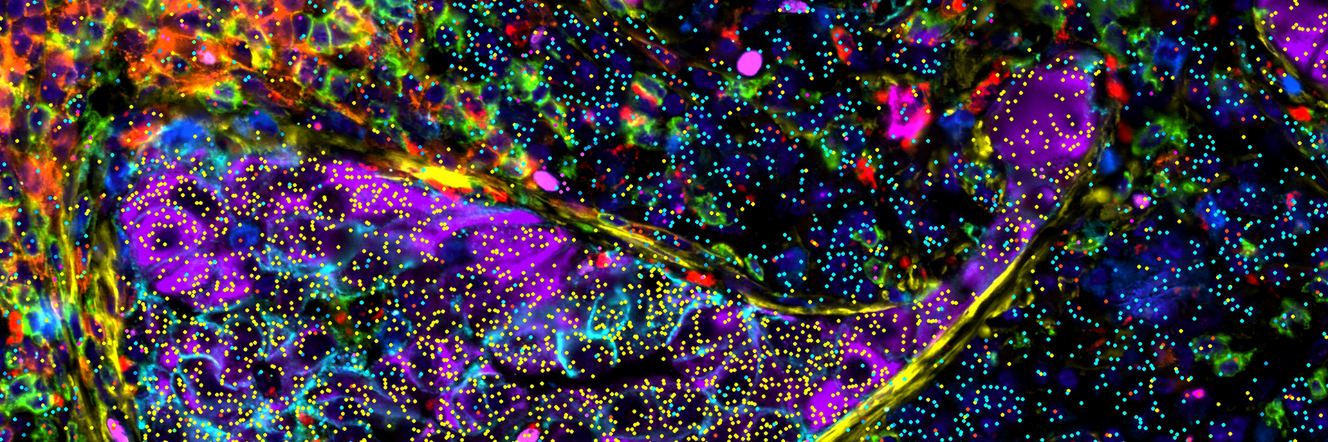
Same-section multiomics analysis (protein and RNA).
Logo

High-plex spatial multiomics research

High-plex spatial multiomics integrates RNA and protein detection within intact tissues, providing a powerful view of biology that surpasses traditional approaches. While proteins reflect the functional state of a cell, RNA reveals early transcriptional programs and regulatory activity that often precede detectable protein changes.

By combining both layers, spatial multiomics delivers a more complete and sensitive picture of cellular states, detecting subtle or early biological shifts that remain hidden when analyzing proteins alone. Ultimately, spatial multiomics offers a comprehensive, high-resolution perspective of biological systems, accelerating insights in both basic research and translational applications.

Explore how the MACSima® Platform with Imaging Cyclic Staining (MICS) technology adds this multiomic depth to spatial biology, uncovering key cellular interactions, fibroblast diversity, and immune landscapes that shape the tumor microenvironment (TME).

Discover the MICS workflow and its application in TME analysis

MICS technology workflow

Illustration of the MACSima imaging cyclic staining (MICS) workflow.
MICS technology utilizes cycles of staining, imaging, and signal erasure to enable high-plex detection of both RNA and proteins within the same sample.

The MACSima Platform analyzes a wide range of sample types, from lung and breast to colon and beyond. Powered by MICS technology, an innovative, high-resolution multiplex imaging method, it relies on an iterative, fully automated staining cycle with three steps:

1. simultaneous staining of the sample with multiple primary conjugated antibodies or RNA probes,

2. image acquisition using widefield microscopy, including automated image processing, and

3. gentle removal of the fluorescence signals.

This three-step process is repeated as often as necessary, allowing a single sample to be analyzed for dozens of RNA molecules and hundreds of proteins. In addition, a newly developed image processing pipeline that reliably removes artifacts enables subcellular resolution of protein and RNA localization. The resulting high-dimensional datasets can then be analyzed with MACS iQ View to address questions related to cell phenotyping, cellular interactions and neighborhoods, and the functional states of different cell types.

Cancer-associated fibroblasts in the TME

Colorectal cancer (CRC) contains a complex TME comprising immune cells, stromal components, and cancer cells whose interactions drive tumor progression. Cancer-associated fibroblasts (CAFs) have emerged as key players, influencing tumor growth, invasion, and immune modulation.

Multiomic profiling using MICS technology was performed on the same CRC tissue sections to resolve CAF subpopulations and their spatial context. A custom 48-plex RNA panel combined with a 40-plex antibody panel enabled in-depth characterization of CAF subsets and immune activation states. Formalin-fixed, paraffin-embedded (FFPE) CRC samples underwent pathologist-guided region selection, followed by RNA detection and antibody-based protein analysis on the same section.

The analysis identified four spatially distinct CAF subpopulations. Neighborhood and proximity analyses revealed that some CAF subsets promoted tumor growth, while others, such as antigen-presenting CAFs (apCAFs), localized near and interacted with T cells. These findings enhance understanding of CAF heterogeneity and immune–stromal interactions in CRC and may help guide future therapeutic strategies.

Multiomic characterization of the TME with MICS technology

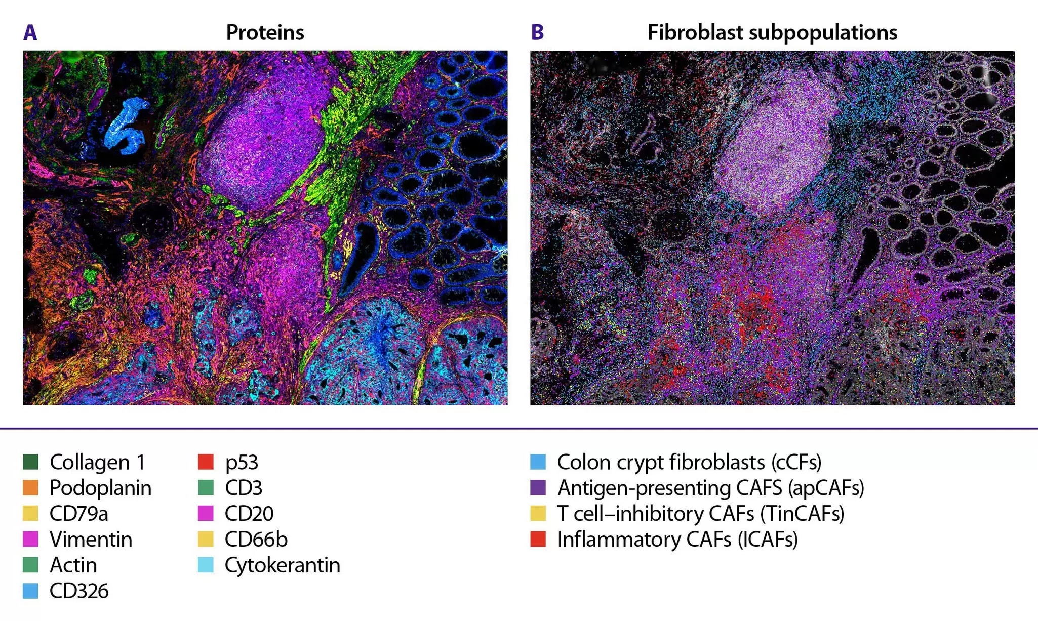
Multiomics characterization of the tumor microenvironment with MICS technology - colorectal cancer.

To characterize the tumor microenvironment (TME) at single-cell resolution, MICS technology was applied for integrated RNA and protein mapping in colorectal cancer (CRC) tissue. High-resolution imaging revealed interactions among immune, stromal, and tumor cells and uncovered previously undescribed CAF diversity. Four fibroblast subpopulations were defined based on hallmark gene expression and spatial distribution, including a novel population termed T cell–inhibitory CAFs (TinCAFs). Colon crypt fibroblasts (cCFs, blue) were evenly distributed and enriched in actin-positive regions, while antigen-presenting CAFs (apCAFs, purple) localized broadly, especially in immune-dense areas. In contrast, inflammatory CAFs (iCAFs, red) and the novel TinCAFs (yellow) appeared more sparsely and in specific niches.

Immune and tumor cell population analysis

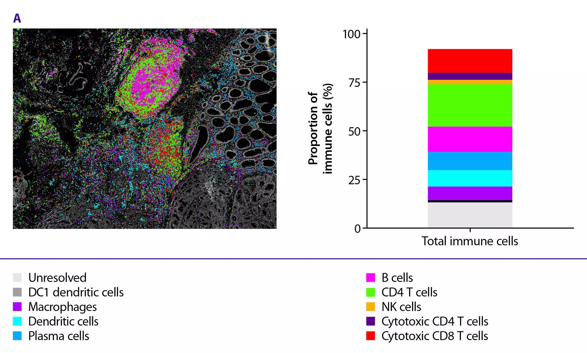
To further elucidate immune–tumor interactions within the TME, immune cells were classified based on protein and gene expression. Spatial profiling revealed a prominent lymphocyte presence at the tumor edge and within a tertiary lymphoid structure (TLS) distal to the tumor. We then assessed immune activation states, identifying cytotoxic lymphocytes that accounted for 19% of all immune cells, with 36% of these activated, as defined by two hallmark genes. Activated cytotoxic lymphocytes were most abundant at the tumor edge and in the TLS. In addition, interferon (IFN)-induced lymphocytes, marked by two additional hallmark genes, represented 5% of immune cells and showed the highest density at the tumor edge.

Multiomic characterization of the tumor microenvironment with MICS - tumor distance map.

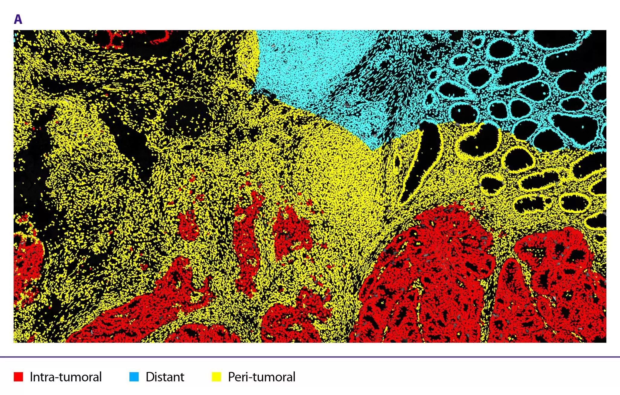
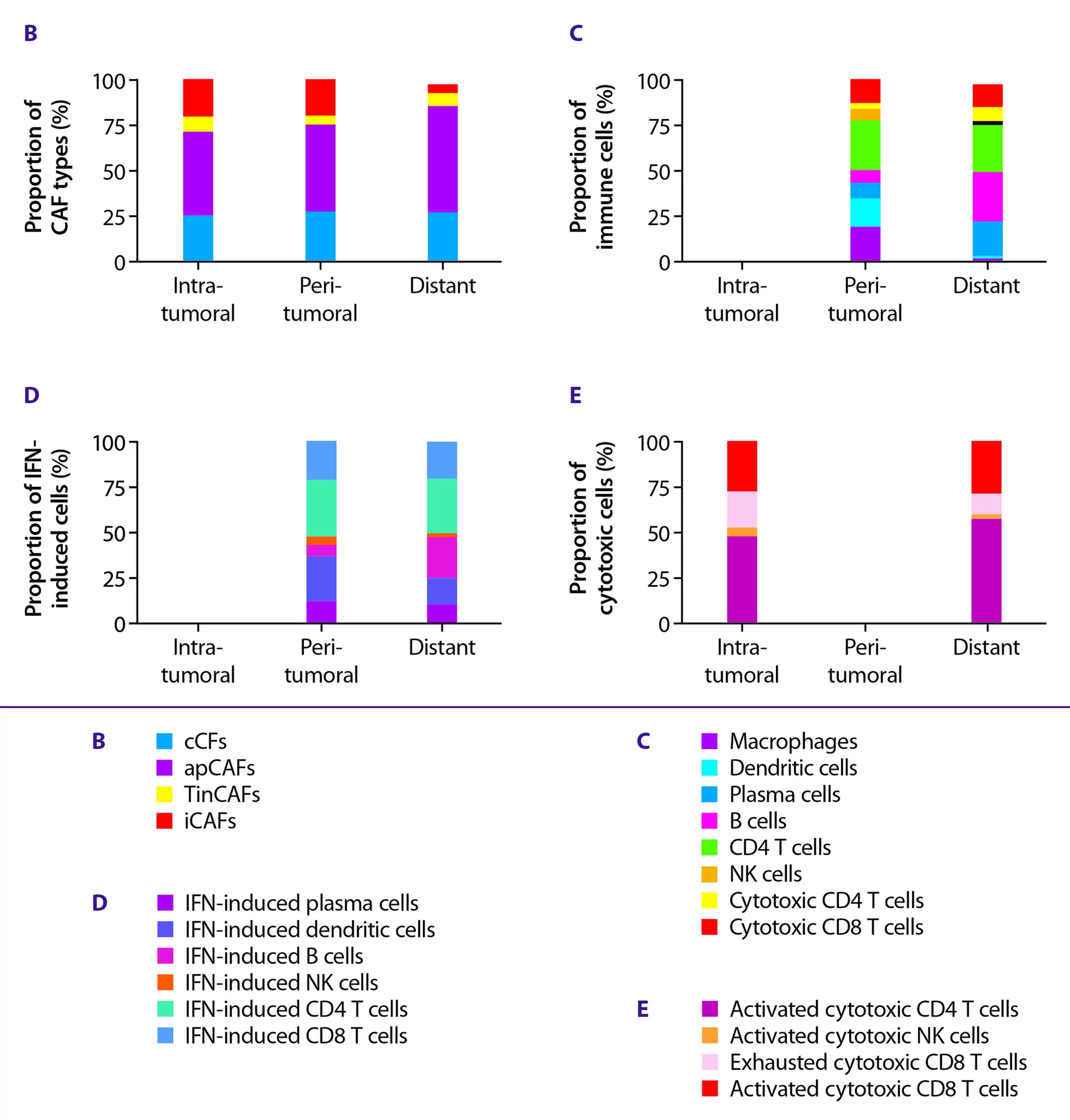
The spatial distribution of CAFs and immune cells was analyzed by defining intra-tumoral, peri-tumoral, and distant areas relative to the tumor. iCAFs and TinCAFs showed the highest concentrations in intra- and peri-tumoral regions. cCFs were evenly distributed regardless of proximity to the tumor, while apCAFs were more concentrated in the peri-tumoral and distant regions.

Multiomics characterization of the tumor microenvironment with MICS technology - tumor distance map analysis.

For our analysis, we established exclusion gates between immune and cancer cells, so immune analyses focused on the peri-tumoral and distant regions. Both cytotoxic lymphocytes and CD4 T cells were present in similar quantities in these areas, whereas macrophages and dendritic cells showed the highest concentrations in the peri-tumoral region. B cells were concentrated in the distant region due to the presence of a TLS. IFN-induced and activated cytotoxic NK cells were most concentrated in the peri-tumoral area. The highest number of exhausted CD8 T cells was also observed in the peri-tumoral region.

Illustration showing that TinCAFs inhibit T cells.

TinCAFs and activated lymphocytes did not colocalize. TinCAFs were present in intra- and peri-tumoral regions but were absent in areas with a high density of activated lymphocytes. We hypothesize that TinCAFs are immunosuppressive and act to inhibit T cells via the immune-interacting function of the hallmark genes for this population.

Conclusion and outlook

MICS technology generates highly specific and quantitative protein signatures and gene expression profiles for investigating TME dynamics. The heterogeneity of fibroblasts and immune cells in colorectal cancer was assessed, and a novel immunosuppressive CAF subpopulation was identified. Due to the immune-interacting genes that define this novel CAF population, TinCAFs show promise as a potential target for future immunotherapies. These findings indicate that same-section spatial tumor profiling has the potential to improve biomarker identification, tumor cell phenotyping, and clinical prognoses for patients.

Characterization of a novel cancer-associated fibroblast population with immune-suppressive function

Speaker: David Agorku, PhD, Group Leader Cancer Research at Miltenyi Biotec

Abstract: David Agorku presents how the team uses multiomic profiling of CAFs in colorectal cancer to identify potential subtypes, spatial relationships, and signaling interactions between CAFs and other cell types within the TME. Learn how they combined single-cell RNA sequencing results with spatially resolved protein and RNA detection to discover a TinCAF subtype that may play a key role in the immune-suppressive TME of colorectal cancer.

Related publications

Deciphering functional tumor-immune crosstalk through highly multiplexed imaging and deep visual proteomics
Zheng, X. et al. (2025), Mol Cell., 85(5), 1008–1023. | Read the article

Unveiling spatial complexity in solid tumor immune microenvironments through multiplexed imaging
Scheuermann, S. et al. (2024), Front. Immunol., 15. | Read the article

Colorectal cancer-associated fibroblasts inhibit effector T cells via NECTIN2 signaling
Agorku, D. et al. (2024), Cancer Lett., 595, 216985. | Read the article

Related antibodies

AntigenCloneFluorochrome
CD3REA1151APC
CD4REA1307PE
CD8REA1024PE
FoxP3REA1253PE
CD20REA1087FITC
CD56REAL1142FITC
CD68REA1306PE
CD326REA1311PE
Cytokeratin LMWREAL648FITC
CD66bREA306FITC
p53REA1132PE
CD79aREA1168APC
VimentinREAL1008APC
Actin (Smooth Muscle)REAL650FITC
PodoplaninREA446APC
Collagen IREAL958APC
Ki 67REAL1047FITC
CD31REA1312PE

You have previously chosen International as your location.
What do you want to do?