Immunophenotyping of CAR T cell transduction efficiency and viability using flow cytometry
MBTP 43 = Miltenyi Biotec-tested panel 43
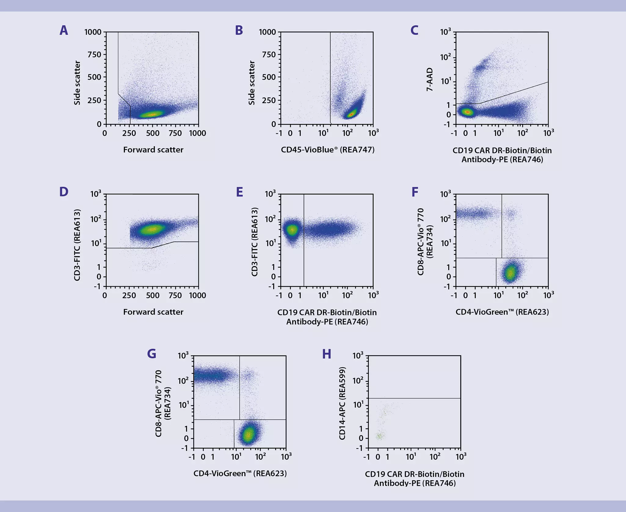
This application protocol describes the analysis of the surface expression of chimeric antigen receptors (CARs) on T cells after transduction. To demonstrate the state-of-the-art panel, CAR T cells were generated within 12 days using the CliniMACS Prodigy® T Cell Transduction process. The data was analyzed using Miltenyi Biotec’s automated data acquisition and analysis Express Mode program “CAR T Cell Express Mode Package”.
- Volumes given below are for up to 106 nucleated cells. When working with fewer than 106 cells, use the same volumes as indicated. When working with higher cell numbers, scale up all reagent volumes and total volumes accordingly (e.g. for 2 × 10⁶ nucleated cells, use twice the volume of all indicated reagent volumes and total volumes).
- Cell number was determined.
- Cells were centrifuged at 300 × g for 10 minutes. Supernatant was aspirated completely.
▲Note: If using a PE-conjugated CAR Detection Reagent, directly proceed with step 7. - Up to 106 nucleated cells were resuspended in 100 µL of PEB buffer.
- CD19 CAR Detection Reagent, human, Biotin was added according to the dilution mentioned in the respective data sheet.
▲Note: Alternative CAR Detection Reagents can be used. - Cells were resuspended and incubated for 10 minutes in the dark at room temperature (+19 to +25 °C).
- Cells were washed by adding 1−2 mL of PEB buffer and centrifuged at 300 × g for 10 minutes. Supernatant was aspirated completely.
- A staining cocktail with all antibodies was prepared according to the dilutions mentioned in the respective antibody data sheets. To detect biotinylated CAR Detection Reagents, Biotin Antibody, PE, REAfinity was used. All reagents were diluted in PEB buffer.
- 100 µL staining cocktail was added to the cells.
- Cells were resuspended and incubated for 10 minutes in the dark at room temperature (+19 to +25 °C).
- Cells were washed by adding 1−2 mL of PEB buffer and centrifuged at 300 × g for 10 minutes. Supernatant was aspirated completely.
- Cells were resuspended in 100 µL of PEB buffer and 1 µL of 7-AAD and incubated for 5 minutes in the dark at room temperature (+19 to +25 °C) right before flow cytometric analysis using the MACSQuant® 10 Analyzer.
16 products found
|
For research use only
FC, MICS, IF, IHC
100 tests in 200 µL
(excl. tax)
For research use only
FC
100 tests in 200 µL
(excl. tax)
For research use only
FC
100 tests in 200 µL
(excl. tax)
For research use only
1 mL
(excl. tax)
For research use only
FC
100 tests in 200 µL
(excl. tax)
For research use only
FC, MICS, IF, IHC
100 tests in 200 µL
(excl. tax)
For research use only
FC
100 tests in 200 µL
(excl. tax)
For research use only
FC
30 tests in 60 µL
(excl. tax)
For research use only
FC
30 tests in 60 µL
(excl. tax)
For research use only
FC
30 tests in 60 µL
(excl. tax)
For research use only
FC
30 tests in 60 µL
(excl. tax)
For research use only
FC
30 tests in 60 µL
(excl. tax)
CE–IVD
for 100 tests
(excl. tax)
For ex vivo cell processing only
2× 5 mL
(excl. tax)
For ex vivo cell processing only
2× 5 mL
(excl. tax)
For ex vivo cell processing only
5 mL
(excl. tax)
You have previously chosen International as your location.
What do you want to do?