Antibody research for cancer treatment
Advance your antibody research with these optimized solutions for B cell isolation, sequencing, and functional testing.
Monoclonal antibody development
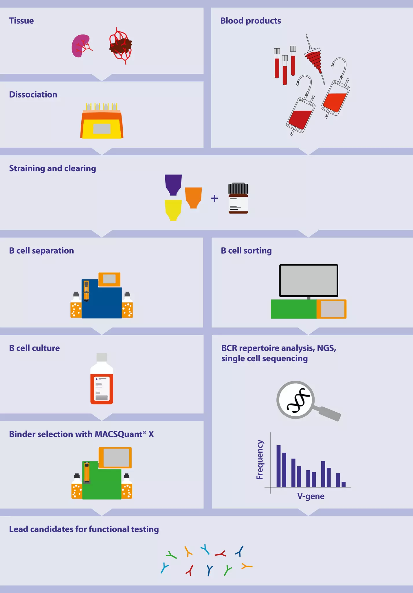
At Miltenyi, we offer a complete workflow for your antibody discovery research.
Whether you start from tissue samples, which need to be dissociated first, or from blood products – We offer optimized solutions to clear your samples for the following B cell application steps. Your workflow might be based on B cell sorting followed by BCR sequencing. Or you might perform magnetic B cell separation, followed by B cell culture and flow cytometry–based binder selection. We support your preferred workflow, as well as subsequential functional testing of lead candidates.
Therapeutic antibody research: Discovery to functional assays
In this webinar we share insights into our complete, comprehensive workflow for therapeutic antibody research. Join us as we discuss our solutions and products available for antibody discovery, functionality analysis and functional assays.
B cell sorting with the MACSQuant® Tyto® for BCR and single B cell sequencing
The MACSQuant® Tyto® Cell Sorter is revolutionizing cell sorting. Our patented microchip-based technology opens up new possibilities with high-speed multiparameter cell sorting in the safety of a fully enclosed and sterile cartridge system, the MACSQuant Tyto Cartridge. The closed cartridge system prevents aerosol and droplet formation, allowing highest safety for the operator. Furthermore, intuitive instrument handling make cell sorting suitable for any lab professional.
The unique features are beneficial for single B cell sequencing and BCR sequencing. The technology enables sorting of rare cell populations with high precision. Cell enrichment of rare target populations improves reads, excludes dead cells and debris avoiding DNA/RNA artefacts and decreases costs.
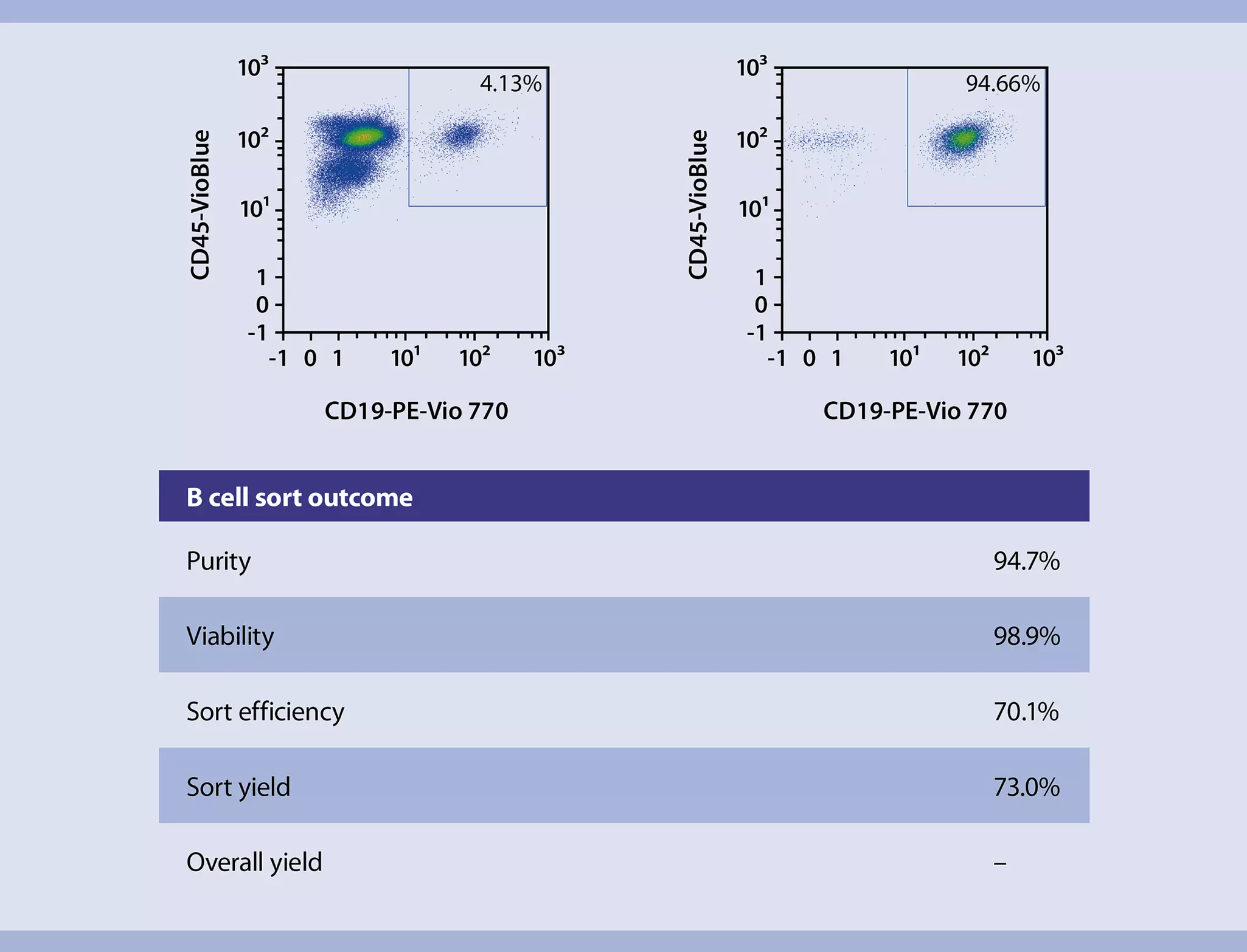
Sorting of highly pure B cells from PBMCs
The MACSQuant Tyto Cell Sorter enables gentle sorting of CD19+ B cells from PBMCs, resulting in a highly pure and viable target fraction.
Magnetic separation and cultivation of B cells to identify therapeutic monoclonal antibody candidates

Cell separation directly from blood products
Developing antibody-based therapies can be labor intensive and time consuming. With our StraightFrom® MicroBead Kits, your workflow is streamlined and saves valuable hands-on time bringing you closer to your new discovery. StraightFrom MicroBeads allow magnetic cell separation directly from human blood products, such as whole blood, buffy coat, leukoreduction system chambers (LRSCs) and leukopaks. CD19+ and CD138+ B cells can be purified using the autoMACS® Pro Separator, the MultiMACS™ Cell24 Separator Plus, or manual separators.
Isolation of human CD19+ cells from whole blood
The StraightFrom LRSC CD19 MicroBead Kits enables isolation of highly pure and viable B cells directly from blood products.
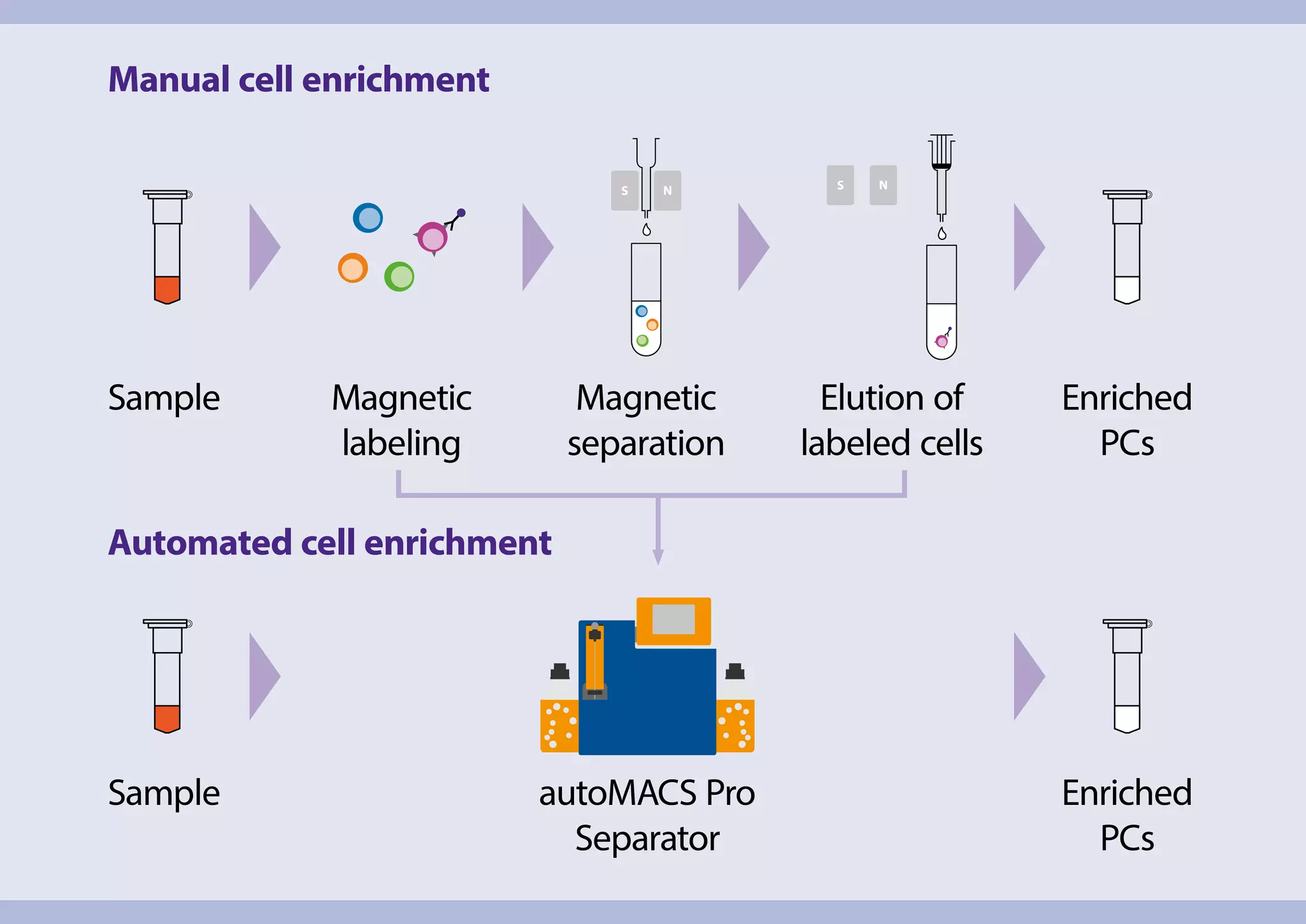
Maximum timesaving with fully automated cell labeling and separation
Automated cell enrichment using the autoMACS® NEO Separator eliminates time-consuming, hands-on steps usually performed by the operator, including labeling, separation, and washing. After separation, the enriched cells can be directly collected for further analysis. A stable separation of B cells or even Plasma cells can be achieved. This robustness is important as it enables standardized workflows without operational bias for daily lab routines. The system can be used with a variety of surface marker-specific MicroBeads available for other cell types and can be used for other applications, e.g., characterization of B cells (CD19+ or CD20+) or B cell subsets such as Plasma cell (CD138+).
Manual versus automated pre-enrichment workflows
Enrichment of PCs directly from whole blood using autoMACS NEO Separator saves hands-on time and minimizes the procedure compared to manual enrichment.
Video
Direct cell isolation from blood or bone marrow on the autoMACS® NEO Separator

Standardized culture of primary human B cells
Ex vivo cultivation is a suitable choice to increase B cells in numbers, especially antigen-specific B cells which are very low infrequency. Studying their antibody production, as well as specificity, is a very important step to develop antibody-based drugs. Our B Cell Expansion Kit, human, can streamline your workflow and simplify the process. The formulation offers the perfect solution to expand and activate human B cells from various sources. This enables expansion of B cells that are fully functional and ready for any downstream application.
Reliable activation and expansion of B cells is a clear requirement for effective downstream experiments. The B Cell Expansion Kit, human is optimized for primary B cell expansion in vitro.
Standardized B cell expansion
Within only 14 days, the B Cell Expansion Kit, human enables a 10-fold expansion of B cells. Furthermore, the kit offers a standardized system for reproducible conditions.
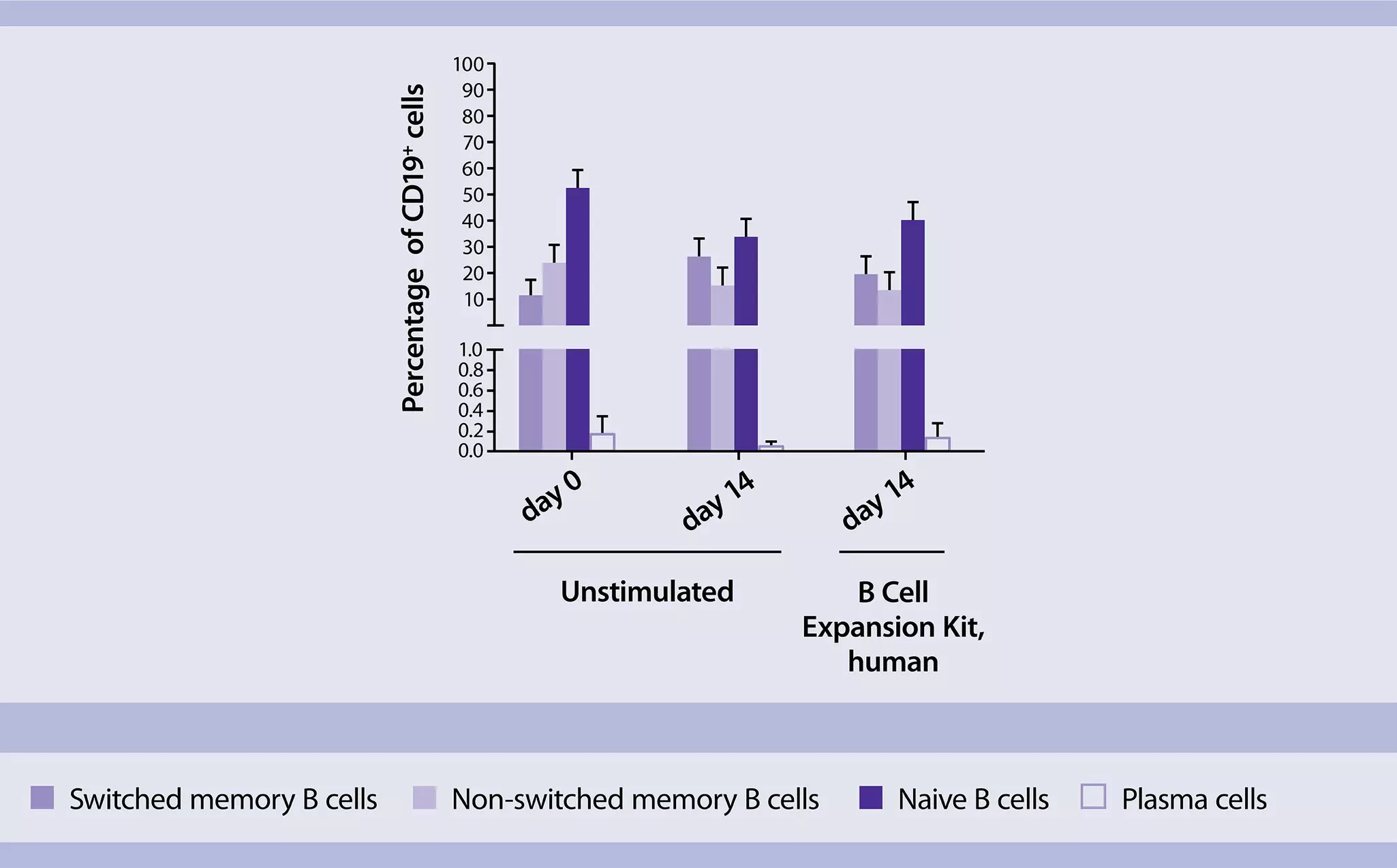
Frequency of B cell subsets after 14 day culture
Expanded B cells display an activated phenotype with a distribution of memory and plasma cells similar to the original fraction. The amount of naive B cells decreased under all culture conditions on day 14. Likewise, reports describe that naive B cells have a shorter survival rate.
Maintained B cell phenotype
The phenotype of the original cell fraction (plasma cells, naive, and memory B cells) are maintained when expanding B cells with the B Cell Expansion Kit, human. The distribution of different phenotypes is similar between expanded and untreated cells, with a reduction of naive B cells after 14 days and a similar amount of memory cells in both setups.

Analyze antigen specificity and find your lead candidate with automated flow cytometry
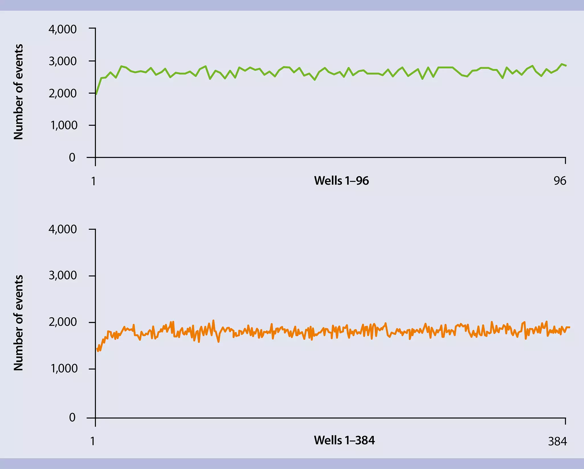
The most important step in antibody discovery research is the identification of lead candidates. To identify antigen-specific antibodies in tissue culture supernatants, flow cytometry analysis can be used. With the MACSQuant® X Flow Cytometer, these assays can be performed in a fast, standardized, and automated manner. This generates high-content information, which is both reproducible and reliable. The MACSQuant X Instrument is equipped with a syringe needle capable of volumetric pipetting, thereby enabling absolute counting from sample volumes starting at 5 µL. The instrument is fully automated in its calibration and compensation procedures, on top it’s pre-set templates, and instrument settings significantly reduce experiment setup and optimization time.
Reliable high-throughput analysis
The MACSQuant X Flow Cytometer enables reliable sample analysis even for high-throughput analyses like lead candidate identification. The instrument can analyze a 384-well plate in less than 60 minutes and a 96-well plate in less than 15 minutes.
Functional screening of lead candidates
Testing the mode of action or the functionality of the antibody lead candidate is one of the most vital steps in the therapeutic antibody development process. It is important to assess effects the therapeutic antibody candidates have on body, and how they interact with the human immune system. Cell-based assays have been widely used, not only to discover and develop therapeutic antibodies, but also for immunogenicity testing for neutralizing anti-drug antibodies and in the manufacturing QC process.
The most important cell-based assays are:
ADCC assay:
- Automated isolation of CD56+ natural killer cells and antibody-dependent cellular cytotoxicity (application note)
MLR:
- MLR: MACS® Technology facilitates mixed lymphocyte reaction (application note)
- MLR: Antigen-specific restimulation of CD8+ T cells by cross-presenting dendritic cells (application note)
CDC assay:
- A complete macrophage suppression assay workflow making exclusive use of Miltenyi Biotec products (application note)
- A simple and fast method for enrichment of lymphocyte subsets for complement-dependent cytotoxicity assays (scientific poster)
Viability assays:
- Reliable, hands-free, and high-throughput cell viability assays on the MACSQuant® Flow Cytometer (Application note)
- Viability dye guide
Immunogenicity testing:
- Video: Accelerating drug development by early unwanted immunogenicity testing
- Immunogenicity assays in drug discovery
Toxicity assay:
Cytokine Secretion Assay:
Webinar
Automation meets cell separation: primary cells for any drug discovery assay
Primary cell separation and isolation are essential for drug discovery. Conclusive assays can benefit from flexible, automated instruments and reagents.
Target discovery
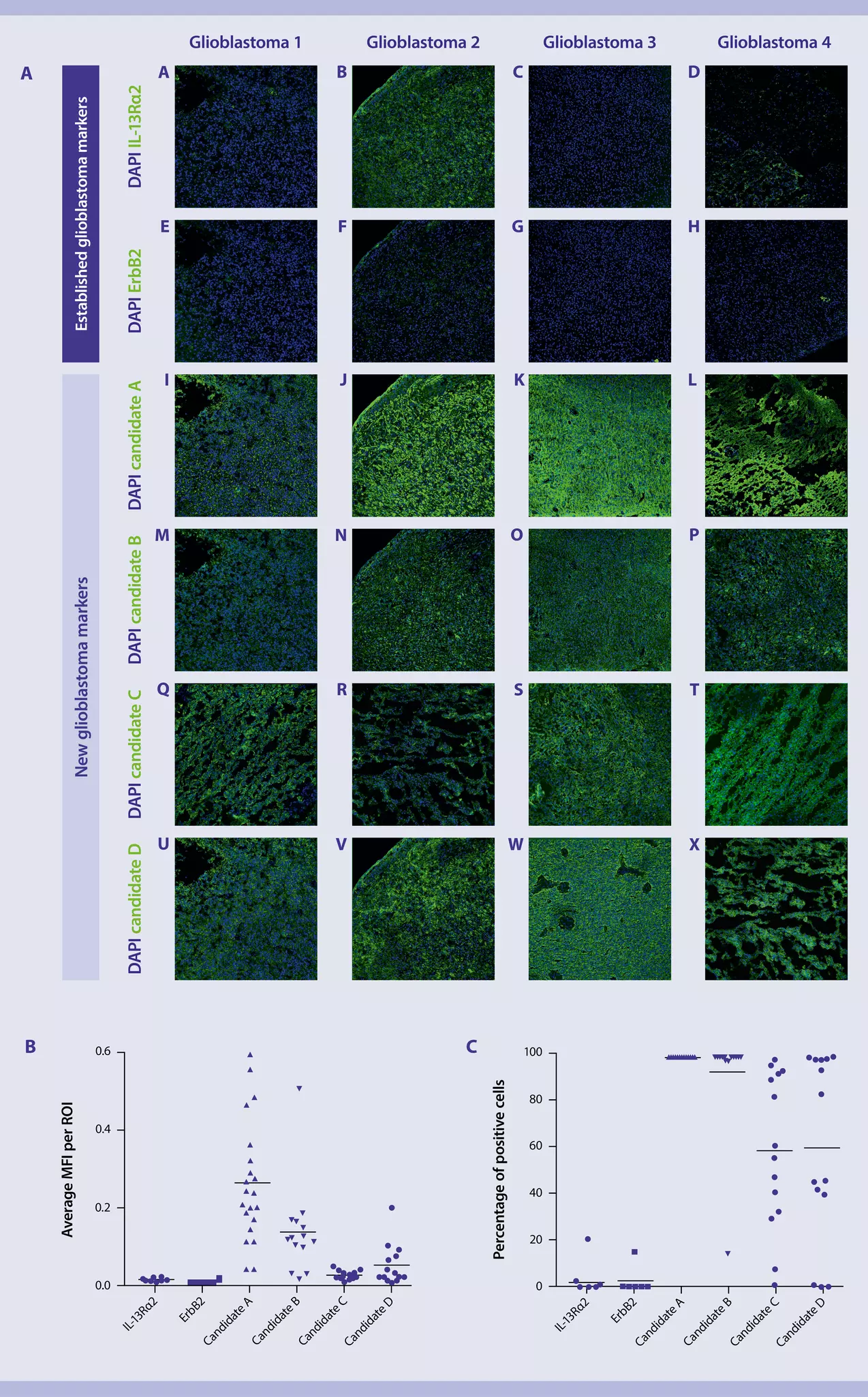
Identification of novel molecular targets for immunotherapies, as well as the analysis and diagnosis of cancers, is a crucial step in cancer research. It is vital for developing monoclonal antibody-based drugs, such as immune checkpoint antibodies, but also for other immunotherapies such as CAR T cell therapy.
However, these approaches are often hampered by a lack of technologies that allow for sensitive and comprehensive multiplexed protein analysis. The MACSima™ Imaging Platform enables high-content, cyclic fluorescence–based imaging of individual biological samples in a fully automated fashion.
In this example, the MACSima Imaging Platform was applied for identification of new glioblastoma-specific markers. Screening of 96 antibodies led to the identification of new marker candidates.
Step-by-step protocols for antibody research in cancer treatment
Get acquainted with our protocols for your workflows in antibody research.
Search:
6 results
You have previously chosen International as your location.
What do you want to do?