Recombinant human TGF-β1 is used for the cultivation of embryonic stem cells and induced pluripotent stem cells, but can also promote Th17 and Treg differentiation of T cells. The transforming growth factor (TGF) beta 1 is associated with diverse processes including chondrogenesis, wound healing, embryogenesis, proliferation, and apoptosis.


Related products for Human TGF-β1
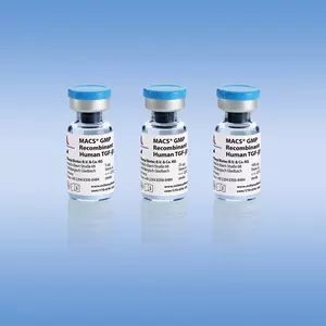
Recombinant human TGF-β1 is used for the cultivation of embryonic stem cells and induced pluripotent stem cells, but can also promote Th17 and Treg differentiation of T cells. The transforming growth factor (TGF) beta 1 is associated with diverse processes including chondrogenesis, wound healing, embryogenesis, proliferation, and apoptosis.
Human TGF-β1 can be used for a variety of applications, including:
- In vitro differentiation of naive CD4+ T cells towards TH17 cells.
- In vitro generation of FoxP3+ inducible regulatory T cells (iTregs).
- Embryonic stem cell differentiation models, for example, for vasculogenesis and angiogenesis.
- In vitro chondrogenesis of mesenchymal progenitor cells and redifferentiation of expanded chondrocytes.
Biological activity
Transforming growth factor β1 (TGF-β1) belongs to a family of homologous, disulfide-linked, homodimeric proteins. These highly pleiotropic cytokines inhibit proliferation of most cells, but can promote the growth of mesenchymal cells and enhance extracellular matrix formation. The pivotal function of TGF-β1 in the immune system is to mediate immunosuppression and maintain tolerance by regulating lymphocyte proliferation, differentiation, and survival. In addition, TGF-β1 controls inflammatory responses through chemotactic attraction and activation of inflammatory cells and fibroblasts. TGF-β1 is produced by many cell types, but is reported to be most abundant in mammalian platelets and bone. All three TGF-β members are synthesized as an homodimeric precursor of 390 residues, which is intracellularly processed by proteolysis into a 112 aa form. The resulting N-terminal latency-associated peptide (LAP) remains non-covalently associated with the TGF-β dimer, and the complex binds to another protein called Latent TGF-β-Binding Protein (LTBP), forming a larger complex called Large Latent Complex (LLC). The LLC is secreted into the extracellular matrix, and prevents the binding of TGF-β to its specific cell surface receptor. Several extracellular factors such as matrix metalloproteases, low pH, reactive oxigen species and thrombospondin-1 can induce release of the active mature TGF-β dimer from the inactive complex. This sophisticated mechanism of activation is important for a fine-tuning of TGF-β signaling. Human TGF-β1 is a recombinant homodimer corresponding to the fully mature form of TGF-β1 without LAP. The amino acid sequence of human TGF-β1 shares 99% identity with TGF-β1 from mouse and rat, therefore human TGF-b1 is commonly used also for mouse cell culture.
Premium-grade cytokines offer the convenience of high and well-defined biological activities and allow exact unit dosing for demanding applications. The biological activity is determined after lyophilization and reconstitution, and normalized to WHO/NIBSC standards whenever available. In general, endotoxin levels are <0.01 ng/μg (<0.1 EU/μg), and purities are >97%. Lot-specific activities are stated in the Certificate of Analysis (www.miltenyibiotec.com/certificates).
Human TGF-β1 activity assay. The biological activity of Human TGF-β1, premium grade was determined by inhibition assay using IL-5-induced TF-1 cells.
SDS-PAGE of Human TGF-β1, premium grade under reducing (R) and non-reducing (NR) conditions.
Mass spectrometry analysis (ESI-MS) of Human TGF-β1, premium grade. The peak corresponds to the calculated molecular mass of 25572 Da.
To access a Certificate of Analysis (CoA) follow the link below and enter the specific lot of your product.
Should you wish to request additional documentation, such as a Safety Data Sheet (SDS) in other languages than English or Swedish, an SDS-replacement document, or a Certificate of Origin (CoO), please direct your inquiry to your local technical support team.
- Ferreras, C. et al. (2019) Chondral Differentiation of Induced Pluripotent Stem Cells Without Progression Into the Endochondral Pathway. Front Cell Dev Biol. 7: 270
- Pfalzgraff, A. et al. (2016) Synthetic antimicrobial and LPS-neutralising peptides suppress inflammatory and immune responses in skin cells and promote keratinocyte migration. Sci Rep 6: 31577
- Neybecker, P. et al. (2020) Respective stemness and chondrogenic potential of mesenchymal stem cells isolated from human bone marrow, synovial membrane, and synovial fluid. Stem Cell Res Ther. 11 (1): 316
- Ricciardi, S. et al. (2018) The Translational Machinery of Human CD4 + T Cells Is Poised for Activation and Controls the Switch from Quiescence to Metabolic Remodeling. Cell Metab. 28 (6): 895-906
- Souza-Fonseca-Guimaraes, F. et al. (2012) NK cell tolerance to TLR agonists mediated by regulatory T cells after polymicrobial sepsis. J Immunol 188 (12): 5850-5858
- Riedel, L. et al. (2018) microRNA-29b mediates fibrotic induction of human xylosyltransferase-I in human dermal fibroblasts via the Sp1 pathway. Sci Rep 8 (1): 17779
- Leleu, D. et al. (2020) Elevated Circulatory Levels of Microparticles Are Associated to Lung Fibrosis and Vasculopathy During Systemic Sclerosis. Front Immunol 11: 532177
- Bosch, J. et al. (2013) Comparing the gene expression profile of stromal cells from human cord blood and bone marrow: lack of the typical "bone" signature in cord blood cells. Stem Cells Int. 2013: 631984
- Neybecker, P. et al. (2018) In vitro and in vivo potentialities for cartilage repair from human advanced knee osteoarthritis synovial fluid-derived mesenchymal stem cells. Stem Cell Res Ther. 9 (1): 329
- Chiurchiù, V. et al. (2016) Proresolving lipid mediators resolvin D1, resolvin D2, and maresin 1 are critical in modulating T cell responses. Sci Transl Med 8 (353): 353ra111
- Nguyen, T. L. et al. (2011) Antigen-specific TGF-β-induced regulatory T cells secrete chemokines, regulate T cell trafficking, and suppress ongoing autoimmunity. J Immunol 187 (4): 1745-1753
- Randall, L. A. et al. (1993) A novel, sensitive bioassay for transforming growth factor β. J. Immunol. Methods 164: 61-67
You have previously chosen International as your location.
What do you want to do?