Recombinant human Flt3-Ligand can be used to generate and expand hematopoietic stem cells or to differentiate the hematopoietic progenitors. In the myeloid lineage this cytokine is crucial for dendritic cells and in the lymphoid lineage it its involved in B and NK cell differentiation. Human Flt3-Ligand recombinant protein is developed for use in cell culture, differentiation studies, and functional assays.


Related products for Human Flt3-Ligand
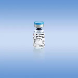



Recombinant human Flt3-Ligand can be used to generate and expand hematopoietic stem cells or to differentiate the hematopoietic progenitors. In the myeloid lineage this cytokine is crucial for dendritic cells and in the lymphoid lineage it its involved in B and NK cell differentiation. Human Flt3-Ligand recombinant protein is developed for use in cell culture, differentiation studies, and functional assays.
Human Flt3-Ligand can be used for a variety of applications, including:
- In vitro expansion of CD34+ hematopoietic progenitor cells.
- Differentiation of ES-derived cells towards the hematopoietic lineage.
- In vitro generation of Langerhans cells, dendritic cells, or eosinophils from CD34+ cells.
Biological activity
Fms-related tyrosin kinase 3 ligand (Flt3-Ligand) is a growth factor that regulates early hematopoiesis. Flt3-Ligand belongs to a small family of α-helical cytokines and promotes in synergy with other growth factors like G-CSF, GM-CSF, SCF, and IL-3 the proliferation and differentiation of primitive hematopoietic stem cells. Early B cell lineage differentiation as well as expansion of monocytes and immature dendritic cells is stimulated. Flt3-Ligand is expressed by T lymphocytes and bone marrow stromal fibroblasts as a membrane-bound and a soluble isoform. Both isoforms signal through the tyrosine kinase receptor Flt3/Flk-2, which is restricted to cells of hematopoietic origin. Human Flt3-Ligand is also active on mouse cells.
Research-grade cytokines are suitable for a wide variety of cell culture applications. They are sterile-filtered prior to lyophilization. Generally, endotoxin levels are <0.1 ng/μg (<1 EU/μg), and purities are >95%. The biological activity is tested in appropriate bioassays.
Premium-grade cytokines offer the convenience of high and well-defined biological activities and allow exact unit dosing for demanding applications. The biological activity is determined after lyophilization and reconstitution, and normalized to WHO/NIBSC standards whenever available. In general, endotoxin levels are <0.01 ng/μg (<0.1 EU/μg), and purities are >97%. Lot-specific activities are stated in the Certificate of Analysis (www. miltenyibiotec.com/certificates).
Human Flt3-Ligand activity assay. The biological activity of Human Flt3-Ligand is determined by proliferation assay using OCI-AML5 cells. Activity of Human Flt3-Ligand, premium grade, (red line) was compared to another commercially available product (black line).
SDS-PAGE of Human Flt3-Ligand, premium grade under reducing (R) and non-reducing (NR) conditions.
Human Flt3-Ligand biological activity. Activity of Human Flt3-Ligand, premium grade, (red bar) was compared to another commercially available product (black bar).
Mass spectrometry analysis (ESI-MS) of Human Flt3-Ligand, premium grade. The peak corresponds to the calculated molecular mass of 17934 Da.
To access a Certificate of Analysis (CoA) follow the link below and enter the specific lot of your product.
Should you wish to request additional documentation, such as a Safety Data Sheet (SDS) in other languages than English or Swedish, an SDS-replacement document, or a Certificate of Origin (CoO), please direct your inquiry to your local technical support team.
- Laurenti, E. et al. (2015) CDK6 levels regulate quiescence exit in human hematopoietic stem cells. Cell Stem Cell 16 (3): 302-313
- Tang-Huau, T.-L. et al. (2018) Human in Vivo-Generated Monocyte-Derived Dendritic Cells and Macrophages Cross-Present Antigens Through a Vacuolar Pathway. Nat Commun 9 (1): 2570
- Meyer, C. and Drexler, H. G. (1999) FLT3 ligand inhibits apoptosis and promotes survival of myeloid leukemia cell lines. Leuk Lymphoma 32: 577-581
- Herrera, L. et al. (2017) OP9 Feeder Cells Are Superior to M2-10B4 Cells for the Generation of Mature and Functional Natural Killer Cells from Umbilical Cord Hematopoietic Progenitors. Front Immunol 8: 755
- Narla, A. et al. (2011) Dexamethasone and lenalidomide have distinct functional effects on erythropoiesis. Blood 118 (8): 2296-2304
- Boyette, L. B. et al. (2017) Phenotype, function, and differentiation potential of human monocyte subsets. PLoS One 12 (4): e0176460
- Chae, H.-D. et al. (2020) RSK inhibitor BI-D1870 inhibits acute myeloid leukemia cell proliferation by targeting mitotic exit. Oncotarget. 11 (25): 2387-2403
- Li, Y. et al. (2017) Targeted Disruption of TCF12 Reveals HEB as Essential in Human Mesodermal Specification and Hematopoiesis. Stem Cell Reports 9 (3): 779-795
- Brault, J. et al. (2014) Optimized generation of functional neutrophils and macrophages from patient-specific induced pluripotent stem cells: ex vivo models of X0-linked, AR220- and AR470- chronic granulomatous diseases. Biores Open Access 3 (6): 311-326
- Garcia-Perez, L. et al. (2020) Successful Preclinical Development of Gene Therapy for Recombinase-Activating Gene-1-Deficient SCID. Mol Ther Methods Clin Dev 17: 666-682
- Dussiau, C. et al. (2015) Targeting IRAK1 in T-cell acute lymphoblastic leukemia. Oncotarget. 6: 18956-18965
- Chae, H.-D. et al. (2017) Niclosamide suppresses acute myeloid leukemia cell proliferation through inhibition of CREB-dependent signaling pathways. Oncotarget. 9 (4): 4301-4317
- Velardi, E. et al. (2014) Sex steroid blockade enhances thymopoiesis by modulating Notch signaling. J. Exp. Med. 211 (12): 2341-2349
You have previously chosen International as your location.
What do you want to do?