Spatial biology


1 Introduction 2 Spatial omics 3 Applications in spatial biology 4 Spatial biology workflow 5 Current trends and developments in spatial biology 5.1 Integration of multiomics technologies in spatial biology 5.2 3D spatial imaging of organ tissue architecture 5.3 Computational challenges in spatial omics data analysis 5.4 Efficient workflows and time-to-data in spatial biology 5.5 Clinical and translational adoption and implementation of spatial biology
6 Overview / Conclusion
1 Introduction
1.1 What is spatial biology?
Spatial biology is a rapidly developing field with the goal of understanding biological processes within their spatial context. It takes a holistic approach by looking at how molecules like proteins or RNA are organized and interact within tissues. This provides deep insights into complex disease mechanisms, drug responses, and the development of personalized medicine.
In recent years, a breakthrough in spatial biology has been seen, merging classic methods like immunohistochemistry with high-throughput techniques like transcriptomics and proteomics. While RNA analysis is well established, high-plex proteomics is emerging quickly. This is important, as proteins drive biological processes and offer tissue architecture insights missing in RNA data. Examples include:
- Understanding how cancer cells interact with their surroundings
- Studying how the immune system functions in different tissues
- Developing new drugs that target specific cell types and interactions
Until recently, spatial biology relied on single omics approaches, like transcriptomics or high-plex proteomics. New platforms enable multiomics analysis, ranging from high-plex transcriptomics with limited proteins to high-plex protein panels with focused RNA. These advancements have the potential to revolutionize both scientific research and clinical practice.
 Related PDFs:
 Related content:
2 Spatial omics
Two of the major techniques used in spatial biology are spatial transcriptomics and spatial proteomics. Both techniques provide valuable information about the biological processes occurring within a tissue sample, but they differ in the type of biomolecule they measure and the strengths and limitations they possess. Table 1 provides a comparison of spatial proteomics and spatial transcriptomics.
Table 1: Comparison of spatial omics techniques
| Spatial proteomics | Spatial transcriptomics | |
| Target read-out | Expression of functionally relevant proteins | Gene expression on RNA level |
| Benefits | Detection of the functional status of a cell Detection of protein isoforms Detection of post-translational modifications giving insight into protein status/activity Identification of novel protein targets and signaling pathways for drug development Flexible sample preparation and long-term storage without significant degradation, giving potential for retrospective analysis | High throughput Analysis of thousands of genes (up to the whole transcriptome) Suitable for discovery-driven research Quantitative Independent of antibody availability |
| Challenges | Availability of high-quality antibodies for specific proteins No coverage of the whole proteome | No full correlation between cellular RNA landscape and cell state/function Limited RNA integrity in stored samples |
| Goals | Understanding the functional state of a cell and protein expression/activity Development of targeted therapies | Identification of new genes involved in a process Study of gene expression patterns |
The future of spatial biology might lie in combining both techniques – spatial multiomics. This allows researchers to gain a comprehensive picture of gene and protein expression within a tissue section, providing the most detailed understanding of cellular processes.
2.1 Spatial proteomics approaches
Spatial proteomics leverages the power of antibodies to unlock a new dimension in protein analysis. Unlike methods like flow cytometry or single-cell sequencing, which homogenize samples, spatial proteomics preserves crucial spatial information. This allows researchers to directly visualize protein expression patterns within cells and tissues, offering a deeper understanding of cellular function and interaction.
Spatial proteomics goes beyond simple protein abundance. It can detect intricate post-translational modifications (PTMs) and protein isoforms, providing valuable insights into protein activity and regulation. This capability makes spatial proteomics a powerful tool for drug discovery, aiding in the identification of novel therapeutic targets and the characterization of signaling pathways involved in disease development.
While antibody availability and multiplexing capabilities were initial limitations, advancements like automated cyclic immunofluorescence are rapidly expanding the scope of analysis. These technologies allow researchers to probe hundreds of protein markers within a single sample, providing a comprehensive picture of the cellular proteome in its native context.
There are two main approaches in spatial proteomics (fig. 1):
- Imaging-based: This approach relies on repeated cycles of staining, imaging, and erasing the signal. Three main variations exist:
- Directly labeled antibodies: Fluorescently tagged antibodies directly bind to the target protein. After imaging, the signal is erased (bleached or enzymatically removed) for the next round of staining with different antibodies.
- Secondary antibody labeling: Here, a non-fluorescent primary antibody binds the target protein, followed by a fluorescently labeled secondary antibody for detection.
- Oligo-conjugate labeling: Antibodies are linked to unique DNA sequences (oligos). The sample is then exposed to complementary DNA strands attached to fluorescent dyes, enabling the detection of specific proteins.
- Sequencing-based: Antibodies are conjugated with specific DNA sequences. After binding to the target protein, these oligos are cleaved and sequenced, revealing the identities of the targeted proteins.
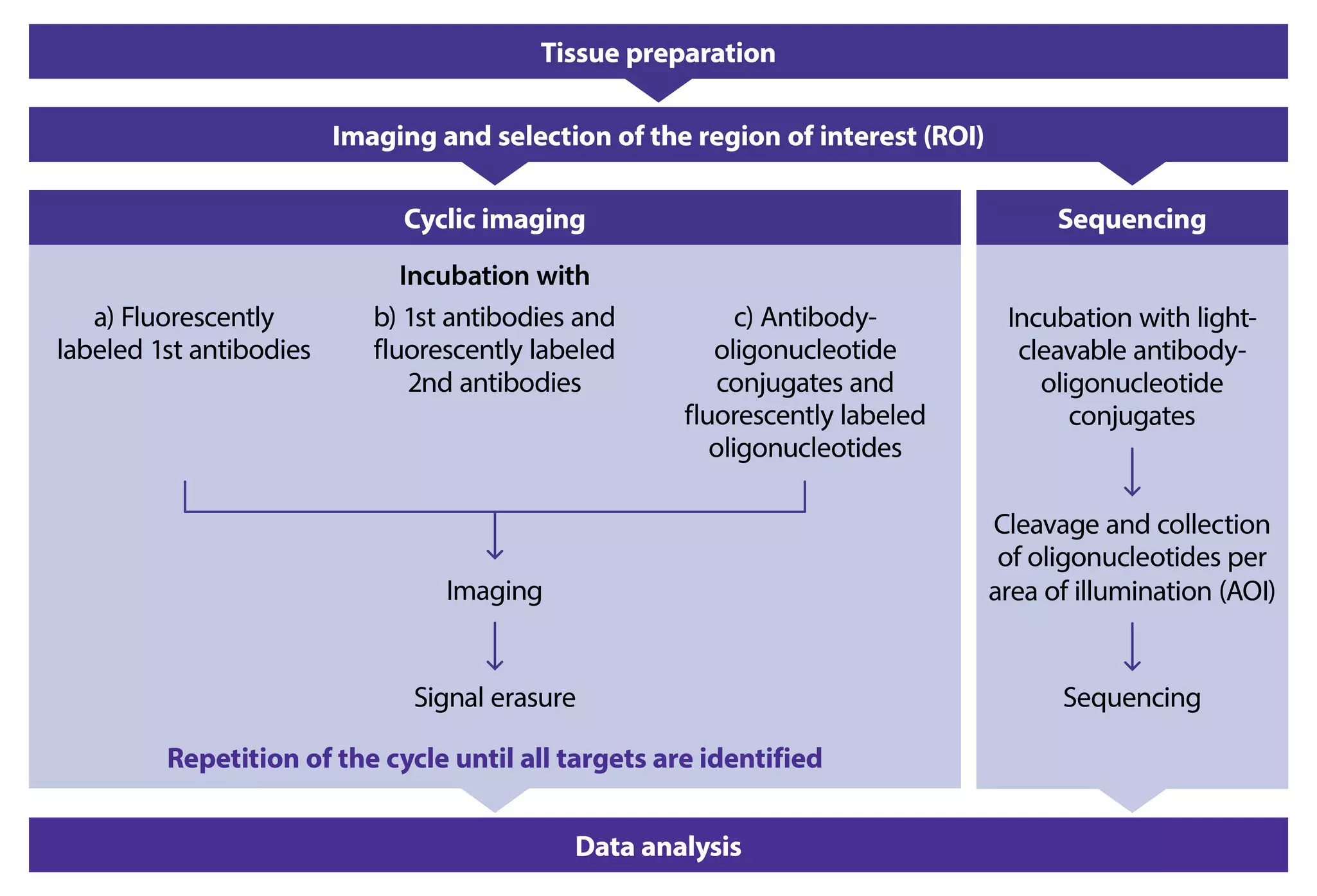
Both imaging- and sequencing-based methods have limitations in the number of detectable proteins within a tissue. Imaging-based methods offer direct visualization and insights into protein expression at the single-cell level. In contrast, sequencing-based methods provide indirect insights into protein expression, as they require an additional sequencing step and subsequent data analysis. Furthermore, sequencing-based methods cannot achieve single-cell resolution due to the necessity of pooling cells for sequencing readout.
2.1.1 Multiplex imaging as a crucial tool within the spatial biology toolkit
Spatial biology can be viewed as advanced imaging through multiplex techniques. These techniques enable simultaneous detection of multiple targets in a single sample, surpassing traditional microscopy. Several techniques are used in multiplex imaging, each with its own advantages and limitations (table 2).
Table 2: Overview of multiplex imaging techniques used in spatial biology
| Principle | Advantages | Challenges | |
| Multiplexed immunofluorescence (MxIF) | Labeling of multiple protein targets with different fluorophores. | Highly sensitive and specific, allowing detailed single-cell mapping. | Overlapping emission spectra. Very limited number of proteins per samples ( max. 5) due to overlapping emission spectra. |
| Cyclic immunofluorescence (CycIF) | Repeated cycles of staining, imaging, and signal removal. | High levels of multiplexing. Adaptable to different types of samples. Multiomics-capable (proteins and RNA). | Specialized software required. Automation needed. |
| Multiplexed ion beam imaging (MIBI) | Staining with unique metal-tagged antibodies. Dislodging and identification of metal tags via secondary ion mass spectrometry. | High sensitivity. No spectral overlap Subcellular resolution. | Complex and time-consuming process. Specialized equipment and expertise needed. Sample gets destroyed during the process. Limitation in number of metals. |
| Co-detection by indexing (CODEX) | Staining with DNA barcodes attached to antibodies. Subsequent cyclic staining and imaging using fluorescent reporter molecules. | High levels of multiplexing. High spatial resolution. | Complex data analysis requiring advanced computational tools and expertise. Barcode cross-reactivity. Steric hindrance as all antibodies are added at once. |
2.2 Spatial transcriptomics approaches
Spatial transcriptomics builds on established methodologies like smFISH and single-cell sequencing by incorporating high multiplexing and spatial dimensions. This innovation enables the exploration of interactions between cell types and cellular states, allowing for a deeper characterization of cells and an understanding of how surrounding cells influence their behavior – such as identifying immune cell populations within tumors. While initially limited in target identification and resolution, the technology has advanced to the point where hundreds of RNAs can be analyzed at the single-cell level. This high level of multiplexing facilitates a more comprehensive understanding of cellular states and holds promise for future advancements, including the identification of mutations and SNPs. Such insights can contribute to a better understanding of why different samples or individuals respond differently to the same stimuli.
There are two primary approaches to identifying transcripts in tissues: imaging-based and sequencing-based methods (fig. 2).
- Imaging-based spatial transcriptomics approaches rely on fluorescently labeled probes designed to bind specific RNA molecules. These methods are further divided into:
- In situ hybridization (ISH): Probes directly target RNA in the tissue. However, traditional ISH is limited in the number of detectable transcripts. Newer techniques like barcoded ISH use multiple probes per RNA, requiring additional imaging cycles to decode the identity of each transcript.
- Rolling circle amplification (RCA): A padlock probe binds to the target RNA, followed by amplification of the bound sequence into a larger structure. This allows for easier detection by fluorescent probes but adds another step to the process.
- Sequencing-based spatial transcriptomics approaches offer two main strategies:
- Targeted approaches: Probes designed for specific RNAs are applied to the tissue. The bound probes are then sequenced, allowing quantification of specific RNA molecules.
- Unbiased approaches (3' capture): The entire 3' end of all RNA molecules in the tissue is captured by complementary oligonucleotides on a surface. This is followed by standard library preparation and sequencing. This approach offers single-cell resolution in some technologies but with varying spatial resolution depending on the specific method.

Both imaging and sequencing-based approaches offer valuable tools for spatial transcriptomics. Imaging approaches provide visualization but are limited in the number of detectable transcripts. Sequencing-based approaches, particularly unbiased capture methods, offer a broader view of the transcriptome with higher resolution, making them ideal for whole transcriptome analysis.
3 Applications in spatial biology
Spatial biology offers a comprehensive view of how cellular composition, interaction, and overall tissue architecture[1] drive tissue function and disease mechanisms. This is essential for advancing our knowledge in various areas of biology and medicine, leading to more effective diagnostic tools and therapeutic strategies and revolutionizing a variety of research areas by providing detailed insights into the spatial organization of tissues.
3.1 Cancer research and oncology
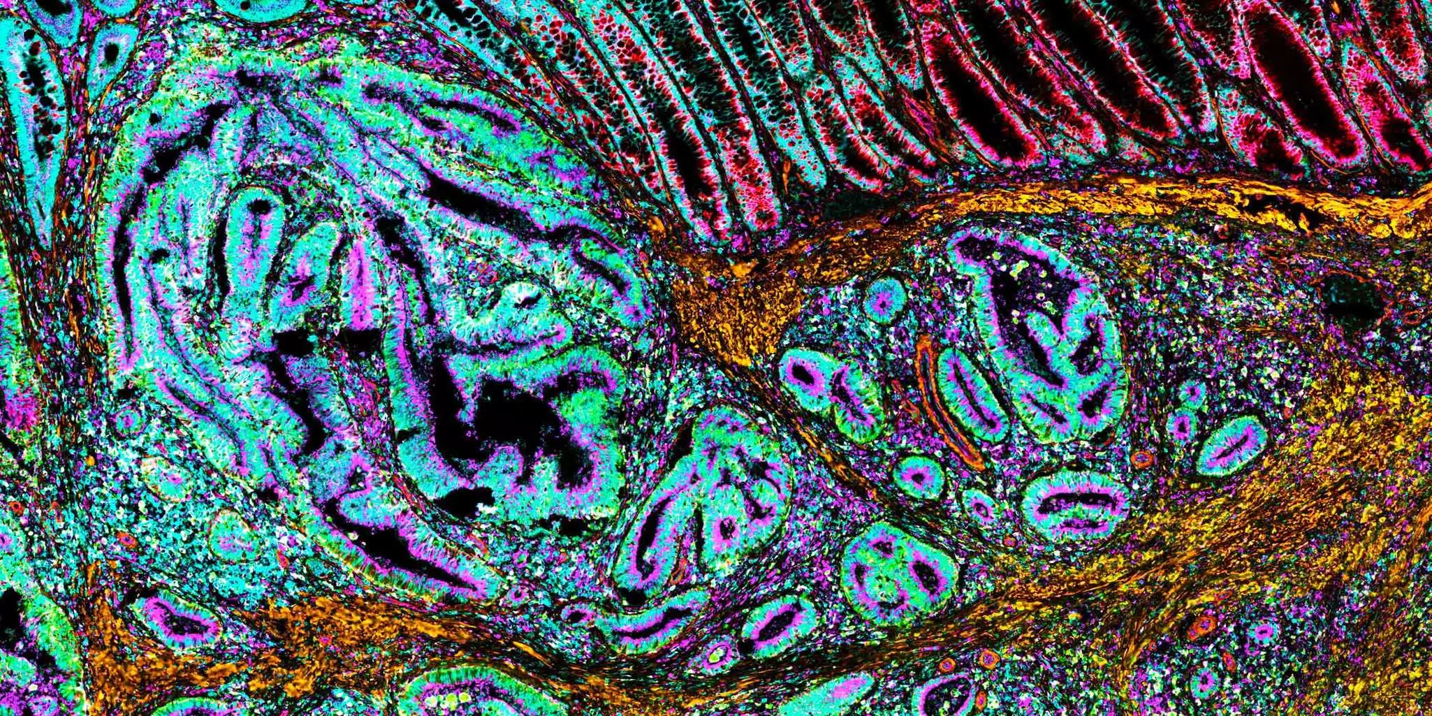
Spatial biology helps in mapping the tumor microenvironment[2], revealing how cancer cells interact with surrounding stromal and immune cells. This information is vital for understanding tumor progression and metastasis. By identifying spatial patterns of protein expression in tumor tissues, spatial biology can facilitate biomarker discovery[3] for cancer diagnosis, prognosis, and treatment response[4].
3.2 Immunology
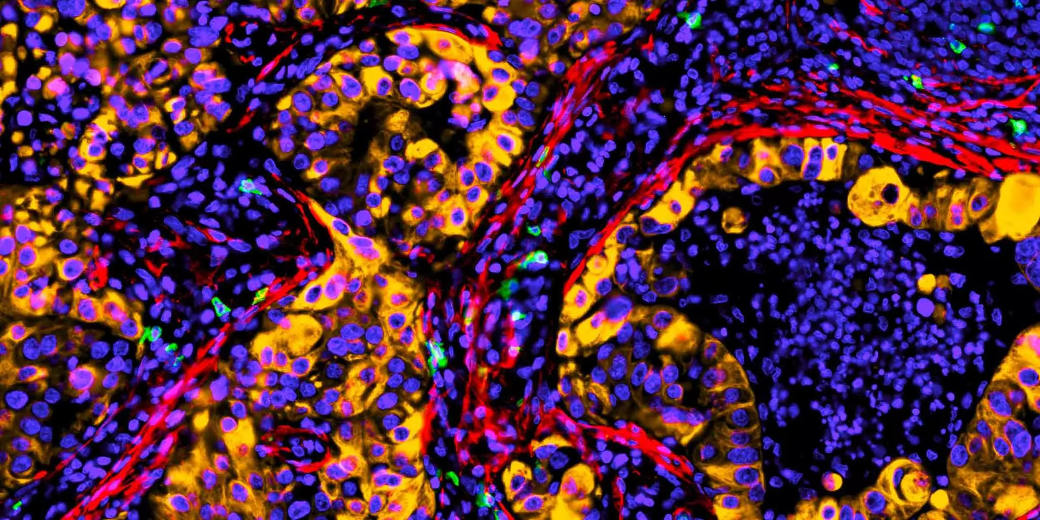
Mapping immune cell distribution during inflammation helps understand immune system activation and response within tissues[4], enabling identification of immunotherapy targets. Spatial information can also reveal pathogen invasion and interaction with host tissues, crucial for developing targeted treatments for infectious diseases.
3.3 Neuroscience
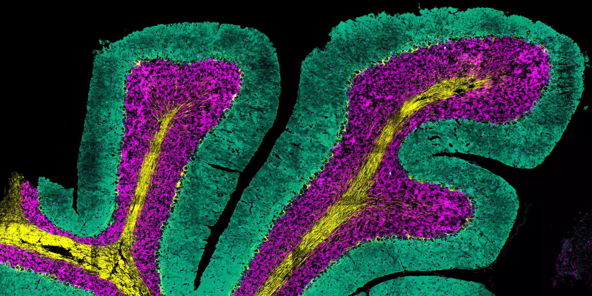
Spatial biology techniques excel in mapping the distribution of various neuronal cell types, immune cells, and aggregates within the brain. This detailed information is instrumental in diagnosing and furthering our understanding of brain function and pathologies associated with neuroinflammation and Alzheimer's disease. Brain samples are often rare and precious. High-plex imaging capabilities of spatial biology allow researchers to gather a wealth of data from a single sample, maximizing information gain.
3.4 Developmental biology
Spatial biology empowers researchers to study cellular differentiation and tissue formation during development. This knowledge can unlock a deeper understanding of congenital abnormalities, developmental disorders, and the spatial organization of cells during organogenesis. These insights can then drive advancements in regenerative medicine and tissue engineering.
4 Spatial biology workflow
This chapter provides a comprehensive overview of the spatial biology workflow, discussing general principles and considerations. For each step, we also delve into the specific capabilities and advantages of the MACSima® Platform in that particular context.
4.1 Sample preparation
4.1.1 General considerations for sample preparation
The foundation of any spatial biology experiment lies in meticulous sample preparation.
While specific protocols may vary across technologies, all spatial proteomics approaches share a core principle: specific antibody binding to a target protein, followed by signal detection. After formulating your research question and identifying the key protein markers for visualization, consider the following sample-related factors:
Table 3: General considerations for sample preparation in spatial proteomics
| What to consider when preparing samples in spatial proteomics | |
| Species compatibility | Ensure the chosen antibodies recognize proteins from the sample species. Protein sequences can differ between organisms, potentially affecting antibody binding. |
| Sample processing | Fixation methods like in formalin-fixed paraffin-embedded (FFPE) samples can crosslink proteins, altering their structure and masking antibody binding sites (epitope masking). Epitope retrieval techniques can counteract this but may not be compatible with all antibodies. |
| Minimizing degradation | Minimize the time between sample collection and fixation to preserve the epitopes you want to detect. |
| Automated platforms | Most spatial proteomic platforms utilize automated staining and washing within dedicated reaction chambers. These chambers may come in disposable formats compatible with standard slides or require instrument-specific flow chambers. Choose the appropriate sample carrier solution that accommodates your sample type (tissue vs. cultured/extracted cells). |
4.1.2 Sample preparation for the MACSima Platform
The MACSima Platform offers flexibility in accommodating various sample types for spatial analysis. Miltenyi Biotec provides protocols for preparing these various sample formats, including RNA protocols for same-section multiomics. All the protocols can be customized to suit your specific research needs. Here's an overview of supported fixation methods:
Table 4: Fixation methods supported by the MACSima Platform
| Tissue samples | |
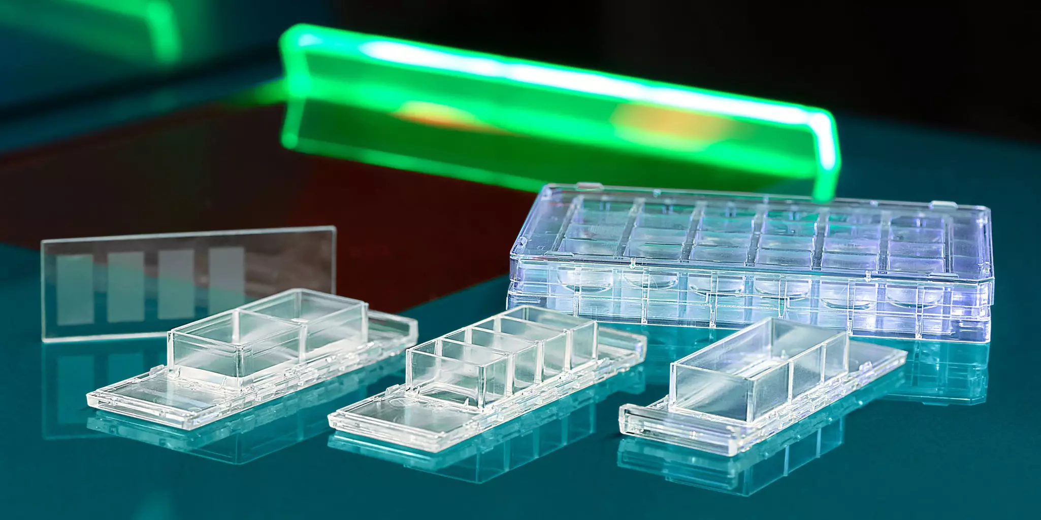
| Formalin-fixed paraffin-embedded (FFPE) tissue sections Frozen tissue sections PFA- or acetone-fixed frozen tissue sections | The tissue samples can be presented on standard microscope slides. For high-resolution imaging, consider using MACSwell HighRes Slides with their 170 μm thin glass window. The standard and high resolution slides are compatible with specifically designed MACSwell Imaging Frames that create a reaction chamber for antibody solutions. Additionally, MACSwell Imaging Frames support tissue microarrays (TMAs), collections of tiny tissue cores arranged on a single slide. |
| Cell culture | |
| Adherent | The platform offers preassembled MACSwell 24 Imaging Plates specifically designed for adherent cells. |
| Single-cell suspensions | For these cells, MACSwell 24 Micro Imaging Plates are ideal. These plates feature hundreds of thousands of tiny microcavities, each holding a single cell. You can simply pipette your cells onto these plates and insert them into the MACSima System for analysis. |
 Related products:
4.2 Selecting targets and setting up panels
4.2.1 General considerations when selecting antibody clones and setting up antibody panels
Once you've chosen the species, fixation method, and sample processing protocol (including epitope retrieval), the next step is finding antibodies to visualize your desired markers. Especially for large panels (over 100 markers), preliminary staining experiments can be very time consuming. Here's how to streamline this process:
Table 5: Selecting antibodies for a spatial proteomics experiment
| How to select antibodies | |
| Literature search | Begin with a thorough literature search. Publications describing similar experiments using the same species and fixation method can offer valuable insights into the antibody clones used for specific markers. |
| Preliminary testing | If no prior research aligns with your specific needs, consider testing antibodies against the desired epitopes that haven't been previously tested for your application. Ideally, use samples identical to your experiment and test different antibody dilutions to find the optimal concentration. |
With individual antibodies chosen, it's time to assemble them into an "experimental panel". This refers to a sequence of staining steps designed to visualize all markers in a single sample. Here are some key considerations for panel building:
Table 6: Building antibody panels for a spatial proteomics experiments
| What to consider when building antibody panels | |
| Antibody conjugation | Purchase or manufacture antibodies conjugated to oligonucleotides (short DNA sequences) for detection with complementary dye-labeled oligos. Select appropriate secondary antibodies if using primary antibodies without fluorophores. Conjugate primary antibodies to fluorophores for direct visualization. Commercially available solutions often exist for common targets. However, custom conjugation of existing antibodies might be necessary for novel or specific markers. |
| Signal interferences | Consider potential interference between markers when assembling the panel. Protein abundance can vary widely, and highly abundant proteins may obscure signals from less abundant proteins. If signal interference occurs, try adjusting antibody dilutions or the order of staining within the panel. If signal removal between cycles is insufficient (e.g., FITC signal persists in later cycles), optimize the signal removal settings. |
| Spectral separation | Fluorescence-based readouts require a higher degree of spectral separation compared to techniques like flow cytometry. Most platforms can only handle 3-4 markers per cycle. Even with separation, spectral overlap can occur: a highly expressed protein with a FITC conjugate can bleed over into the channel detecting a less abundant protein with a PE conjugate. |
| Resources for panel design | The Human Protein Atlas (https://www.proteinatlas.org/) provides a valuable reference for expected staining patterns of various markers. OMAP (Antibody Mapping Project) offers community-tested and shared antibody panels. Predesigned vendor-provided panels for specific applications. However, ensure compatibility with your sample type. |
Optimizing the antibody panel often requires iterative adjustments. This includes tweaking antibody concentrations, staining parameters, and the staining sequence. The complexity arises from protein expression levels, which can vary greatly depending on the sample. However, by utilizing these strategies and resources, you can effectively build robust panels for your spatial proteomics experiments.
4.2.2 General considerations for combining RNA and protein analysis in spatial biology for a multiomics approach
Spatial proteomics offers valuable insights into protein distribution within tissues. However, it has limitations, including:
- Limited protein coverage: Not all proteins are expressed at detectable levels or may be masked by technical limitations.
- Quantitative accuracy challenges: Measuring absolute protein abundance can be difficult.
Spatial RNA-protein co-detection combines spatial proteomics with RNA detection, providing complementary information to address these limitations. The benefits and goals of combining RNA and protein data in a multiomics approach are summarized in tables 7 and 8.
Table 7: Benefits and goals of a multiomics approach in spatial biology
| Benefits of spatial RNA-protein co-detection | |
| Comprehensive understanding | Combining data provides a more detailed picture of biological processes. |
| Enhanced protein coverage | RNA data reveals gene activity, even when protein levels are low or undetectable. |
| Improved quantitative accuracy | RNA data provides an additional measurement layer for protein expression. |
| Higher spatial resolution | RNA data helps interpret protein localization within a cellular context. |
| Mitigating technical challenges | Combined data offers a more robust picture, reducing the impact of technical and sample preparation issues. |
| Simplified data analysis | RNA data can streamline analysis and interpretation of protein data. |
| Functional insights | RNA data offers insights into cellular states and dynamic changes. |
| Goals of spatial RNA-protein co-detection | |
| Cross-validation of upstream screening methods | Spatially resolved multiomics data validates initial findings from bulk RNA sequencing or single-cell RNA sequencing by adding spatial context. This helps identify true patterns and avoid artifacts, crucial for confirming potential therapeutic targets or biomarkers. |
| Detecting secreted factors | Spatial RNA analysis is particularly valuable for studying the distribution of signaling molecules like chemokines and cytokines within tissues. These molecules are challenging to detect at the protein level due to low abundance and short half-lives. Spatial RNA methods offer a more reliable and sensitive approach. |
| Analyzing RNA-protein co-expression | Analyzing RNA and protein interactions within cells provides insights into regulatory relationships. This approach helps identify: • Cell type–specific gene expression • Cellular sources of secreted proteins • Cell type–specific interactions This information aids in identifying disease pathways, potential drug targets, and cell type–specific functions. Additionally, analyzing co-expression in a spatial context reveals tissue architecture and multicellular organization, guiding therapeutic development. Identifying co-expression changes in specific regions (e.g. tumor margins) helps pinpoint disease-associated alterations. |
| Replacing unavailable or poorly performing antibodies | Spatial RNA hybridization methods offer an alternative to antibodies for protein detection. RNA probes specifically target RNA sequences, providing high specificity and avoiding potential antibody cross-reactivity. Additionally, researchers can easily synthesize RNA probes for any target, allowing for rapid customization and detection of non-coding RNAs, fusion genes, or splice variants. |
| Studying signaling pathways | The correlation between mRNA and protein levels can provide clues about post-translational regulation. High levels of RNA alongside abundant protein might suggest active gene expression and a functioning pathway. Conversely, low RNA with little protein might indicate a silenced pathway or a bottleneck in protein production. This spatial information provides a more nuanced view of pathway activity compared to bulk analysis. |
4.2.3 Defining antibody and RNA panels for the MACSima Platform
The MACSima Platform offers users flexibility in designing panels for protein and RNA analysis within a single experiment.
The available options for antibody panels can be seen in table 8.
Table 8: Three available antibody options for the MACSima Platform
| Liquid antibodies | Dried antibodies | |
| Single antibody vials | The platform offers a selection of over 600 pretested recombinant primary conjugated antibodies optimized for either FFPE or frozen human or mouse samples. For a quick start, researchers can choose from over 2,000 preconjugated antibodies. Users can also integrate their own or third-party antibodies for maximum flexibility. | |
| Predefined antibody panels | REAplex Panels These modular panel kits address specific research questions by providing a selection of ready-to-use antibody conjugates (e.g., 20-plex panels). This option is ideal for researchers with predefined targets of interest. | REAscreen™ Panels These ultrahigh-plex, plug-and-play panels are designed for deep phenotyping (analysis of hundreds of markers) from valuable samples. Offered in a convenient 96-well format, this approach saves time by eliminating laborious panel design while still allowing the addition of custom or third-party antibodies. |
| Customized antibody panels | REAscreen Design Panels For researchers with specific research questions, this option allows for custom panels tailored to their application in a plug-and-play 96-well format. |
Dried REAscreen Antibody Panels save researchers time by eliminating pipetting errors and reducing handling mistakes, leading to more reliable and reproducible results.
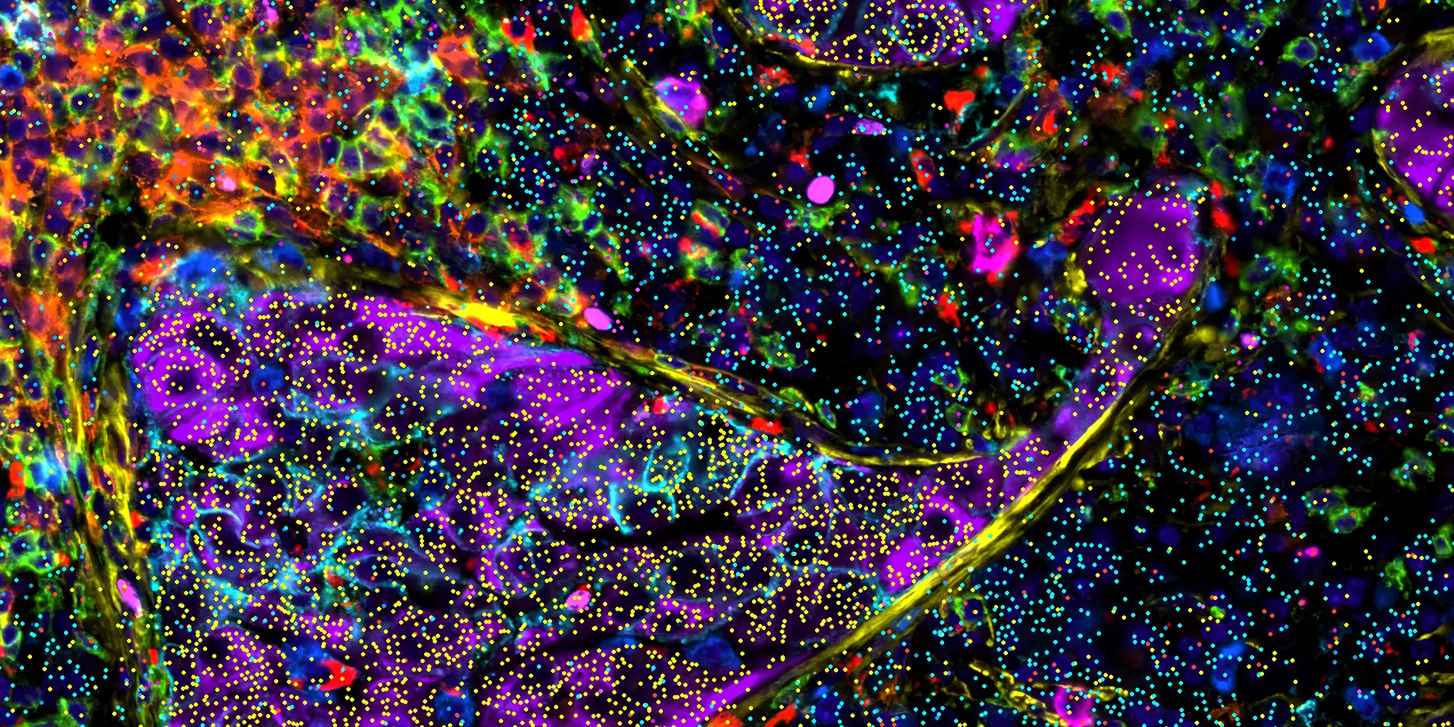
For same-section multiomics analysis[5], users can seamlessly integrate RNA panels with their protein analysis experiment.
Table 9: Available RNA panel options for the MACSima Platform
| RNA detection probes | |
| Single RNA vials | RNAsky Detection Probes are designed to target virtually any RNA of interest with high sensitivity and specificity. Researchers can design their own custom RNAsky Panels. |
| Predefined RNA panels | The 24-plex RNAsky IO Explore Panel focuses on immuno-oncology markers. |
 Related videos:
4.3 Data acquisition
4.3.1 General considerations regarding automation of data acquisition systems
Spatial biology experiments, particularly those involving multiplexed analysis (CycIF), often require complex staining protocols with numerous cycles of staining and washing. Manual execution of these protocols can be time-consuming, labor-intensive, and prone to errors due to human variability in pipetting and handling. Automated liquid handling systems address these challenges by offering several advantages:
- High precision and accuracy: Automated systems ensure consistent and precise reagent volumes across samples, which is critical for reproducible staining results in techniques like CycIF.
- Reduced processing time: Automation significantly speeds up cyclic staining processes, allowing researchers to analyze more samples in a shorter timeframe.
- Enhanced reproducibility: By automating staining, washing, and other steps, researchers achieve consistent and reproducible results across multiple samples, leading to more reliable data.
- Improved workflow efficiency: Automation frees up researchers' time from tedious manual tasks, allowing them to focus on data analysis and interpretation, accelerating scientific discovery.
- Increased accessibility: Manual multiplex imaging often requires specialized skills and expertise, limiting its accessibility to highly trained personnel. Automation can potentially broaden access to these technologies by reducing reliance on manual expertise.
- Improved safety and ergonomics: Automation minimizes manual handling of potentially hazardous reagents, reducing exposure risks for laboratory personnel. Additionally, it reduces the physical strain associated with repetitive tasks, improving overall lab safety and ergonomics.
In recognition of these benefits, most spatial biology platforms incorporate either semi-automated or fully automated systems for data acquisition.
4.3.2 High quality vs. speed in multiplex imaging
In multiplex imaging, high-quality images are paramount. They provide the resolution and detail needed for accurate analysis. Detailed and high-resolution images enable more precise quantification of molecular markers, which is essential for reliable biomarker discovery and validation. Poor-quality images can lead to misinterpretations and hinder reproducibility, a cornerstone of research. Furthermore, advanced analysis techniques like machine learning and artificial intelligence also rely heavily on high-quality input data.
While speed is important for efficiency, it should not compromise image quality. Slower techniques often involve multiple controlled steps to remove non-specific binding and ensure consistent protocols across samples. These steps, although time-consuming, are crucial for maintaining data integrity and generating reliable results that justify the longer runtime.
4.3.3 High-throughput and high-content approaches in spatial biology
Two approaches in spatial biology are high-throughput and high-content analyses. Each serves distinct but complementary roles.
Table 10: High-throughput and high-content approaches in spatial biology
| What is it? | Benefits | Challenges | |
| High-throughput spatial biology | High-throughput analysis is characterized by its ability to screen a vast number of samples or spatial datasets. High-throughput techniques rely heavily on automation and computational tools, enabling researchers to process and analyze spatial data from numerous samples efficiently. | High-throughput spatial biology is beneficial for hypothesis generation or screening for spatial biomarkers on a large scale. | High-throughput approaches generate extensive datasets that require advanced bioinformatics for analysis and proper data management to store large data. |
| High-content spatial biology | High-content analysis delves deeper, aiming to extract detailed, multiparametric spatial information from a smaller set of samples. | High-content spatial biology seeks to understand the intricate interactions and functional implications of spatial organization within cells and tissues. The resulting datasets are rich and complex, offering insights into the mechanisms underlying spatial patterns and the functional significance of spatial arrangements. | High-content approaches generate complex datasets that require dedicated analysis tools. |
The complexity and cost of dedicated slide scanning devices and the hardware and software required for high-throughput and high-content imaging can complicate implementation and accessibility for some research laboratories. Additionally, as the throughput and multiplexing capabilities increase, so does the complexity of the data generated. Extracting meaningful information from these large datasets requires advanced tools for cell identification, classification, and spatial analysis. Despite the advancements, there are still technological limitations that restrict the efficiency and effectiveness of high-throughput multiplex imaging in certain contexts, necessitating ongoing research and development to overcome these hurdles.
Integrating high-throughput and high-content approaches in spatial biology research offers a holistic view of biological complexity, paving the way for groundbreaking discoveries in understanding disease progression, developmental biology, and cellular function. As technology advances, the synergy between these approaches will undoubtedly deepen, further enhancing our ability to decipher the spatial codes that govern life at the molecular level. These methodologies leverage automation and advanced imaging technologies to process and analyze thousands of samples simultaneously, providing comprehensive insights into complex biological systems.
4.3.4 Data acquisition with the MACSima System: MACSima Imaging Cyclic Staining (MICS)
The MACSima System employs a fully automated approach for staining and imaging, simplifying the experimental workflow. Users can load samples and antibody panels, define regions of interest, and initiate the experiment with minimal hands-on time. The system can process up to four slides in parallel. To optimize image acquisition, the system performs an initial overview scan to identify regions of interest for detailed analysis.
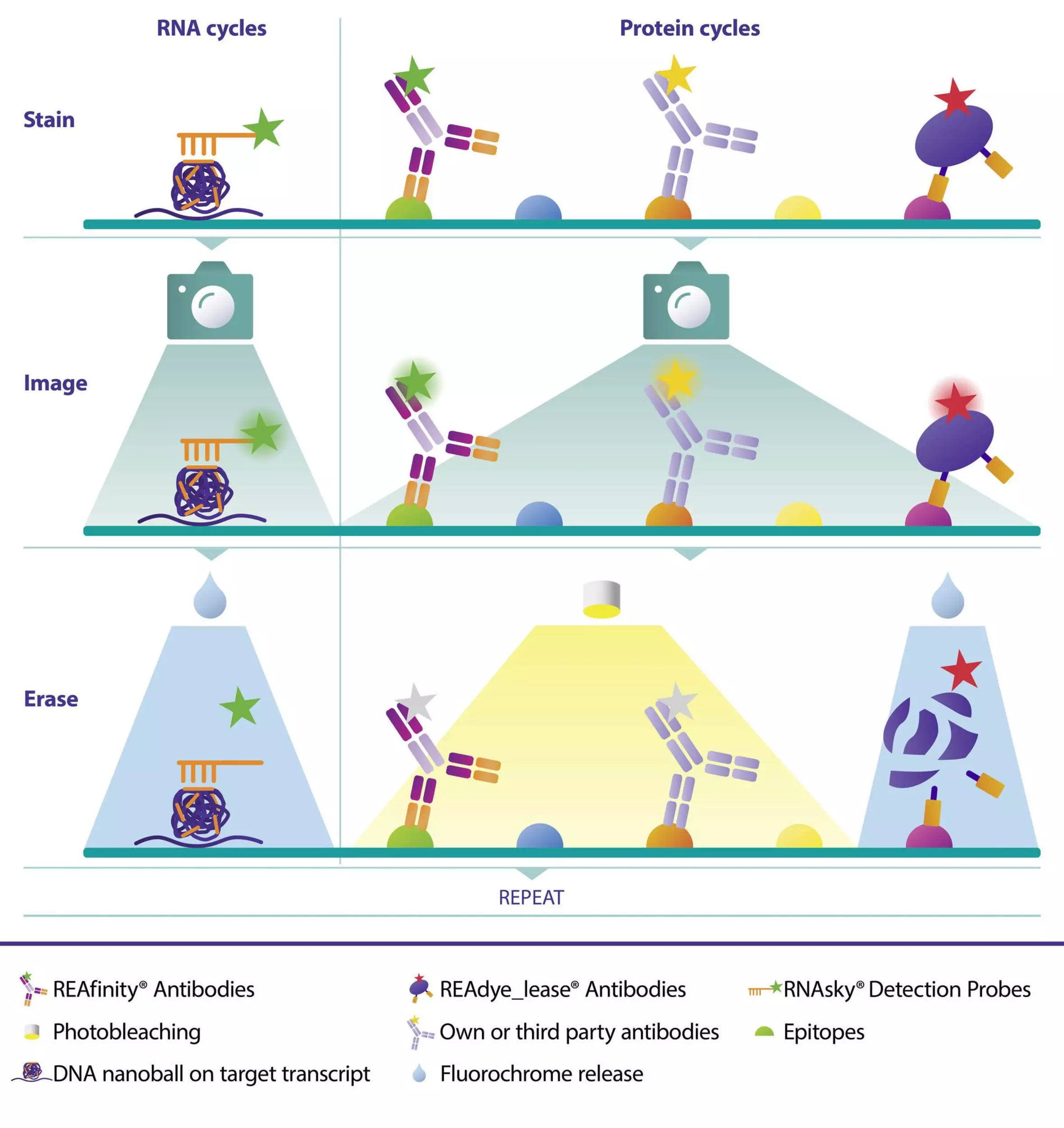
The MACSima Platform incorporates MICS technology, an iterative process involving staining, imaging, and signal erasure (fig. 9). This enables the simultaneous analysis of numerous protein and RNA markers on the same tissue section without compromising sample integrity. The MICS process consists of three steps:
- Staining: The sample is incubated with fluorochrome-conjugated antibodies (for proteins) or RNAsky Detection Probes (for RNA).
- Imaging: The stained sample is imaged using a widefield fluorescent microscope.
- Erasure: The fluorescence signal is removed without damaging the sample, allowing for subsequent staining cycles. For signal erasure, the MICS technology supports two different mechanisms:
- Photobleaching (REAdye_lease®, REAlease®, and REAfinity® Antibodies): Fluorescent signals are erased from any antibodies conjugated to photosensitive fluorochromes such as FITC, PE, or APC.
- Fluorochrome release: Fluorochromes are detached from antibody complexes (REAdye_lease, REAlease Antibodies) and RNAsky® Detection Probes in a controlled and specific manner upon incubation with a certain reagent.
Both signal erasure mechanisms are sample friendly, preserving epitope integrity while efficiently clearing signals for subsequent cycles. This enables highly flexible panel design and cycle assignment, a key advantage of MICS. Individual antibody conjugates can be incorporated into early or late staining cycles, freely combined with other relevant antibodies, all within the same experiment.
Related content:
Related PDFs:
Related products:
4.4 Spatial data analysis
4.4.1 General approach for spatial data analysis
Spatial biology is a rapidly evolving field, and as a result, standardized data analysis methods are still under development. However, current approaches integrate elements from traditional image analysis, flow cytometry, and single-cell omics to extract meaningful insights from complex spatial data.
The vast amount and complexity of data generated by spatial biology experiments offer numerous analytical possibilities. Common applications include:
- Cell phenotyping: Identifying and characterizing cell types, often focusing on specific cell populations like T cells within the tumor microenvironment.
- Deep phenotyping: Discovering novel cell populations through detailed analysis of multiple cellular markers.
- Drug target discovery: Using spatial data to identify potential therapeutic targets.
- Biomarker discovery: Identifying new molecular markers associated with specific biological processes or disease states.
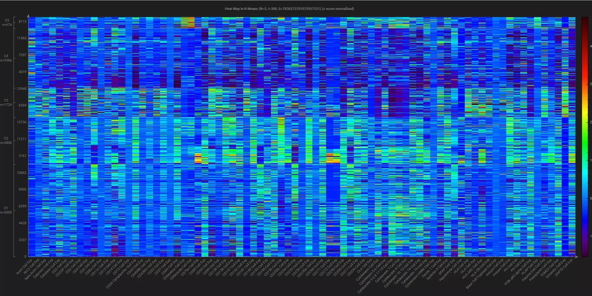
An exemplary approach to spatial data analysis is shown in figure 10.
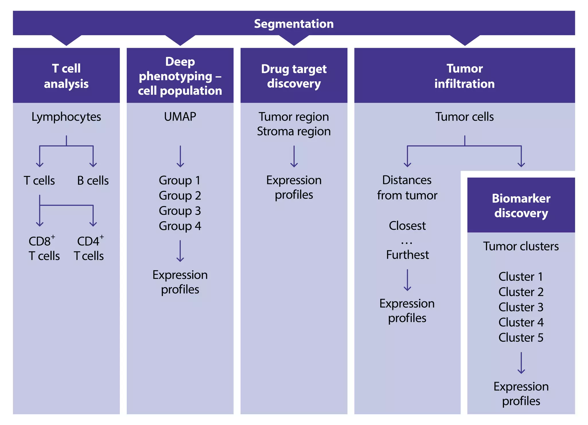
A key advantage of spatial analysis over traditional methods like flow cytometry is the ability to consider spatial relationships between cells and molecules. By analyzing factors such as cell distance, density, and proximity to specific structures, researchers can extract deeper insights from biological processes.
4.4.2 Analyzing spatial data with MACS® iQ View
The MACSima Platform is complemented by the user-friendly MACS iQ View Analysis Software, designed to simplify the analysis of complex spatial biology data. The software empowers researchers of all backgrounds to efficiently extract meaningful insights and generate publication-ready results.
The intuitive interface allows for the visualization of hundreds of markers simultaneously, enabling the creation of diverse image and video formats. The software offers a comprehensive suite of analytical tools: single-cell quantification, spatial analysis, and visualization tools.
Table 11: Analyzing spatial data
| Analytical tools included in MACS® iQ View – Spatial Biology | |
| Image Viewer | Effective data visualization and navigation can be achieved through the utilization of histogram tools, pixel-based correlations, and line intensity profiles. |
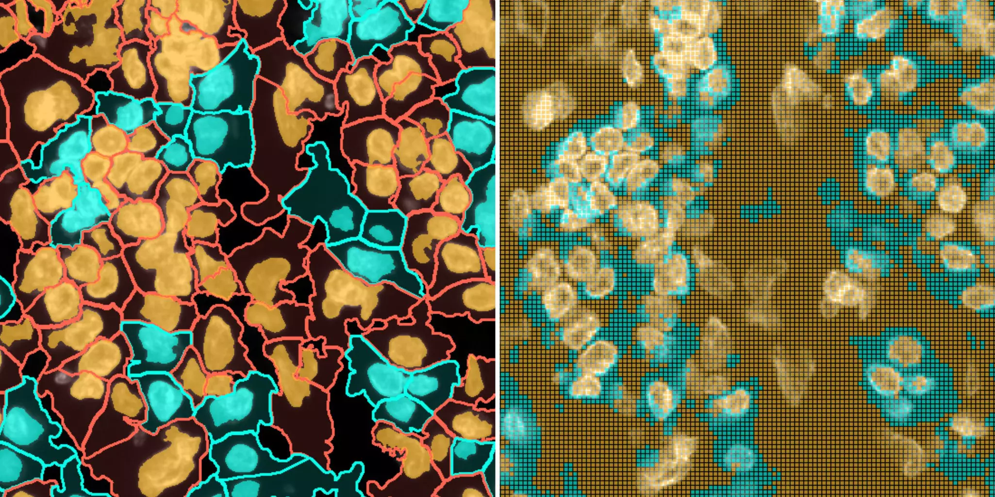
| Segmentation | MACS iQ View enables both classic cell morphology (based on nuclei detection) and superpixel (image-based) segmentation methods. Additionally, it offers the capability to import segmentation masks from external sources. |
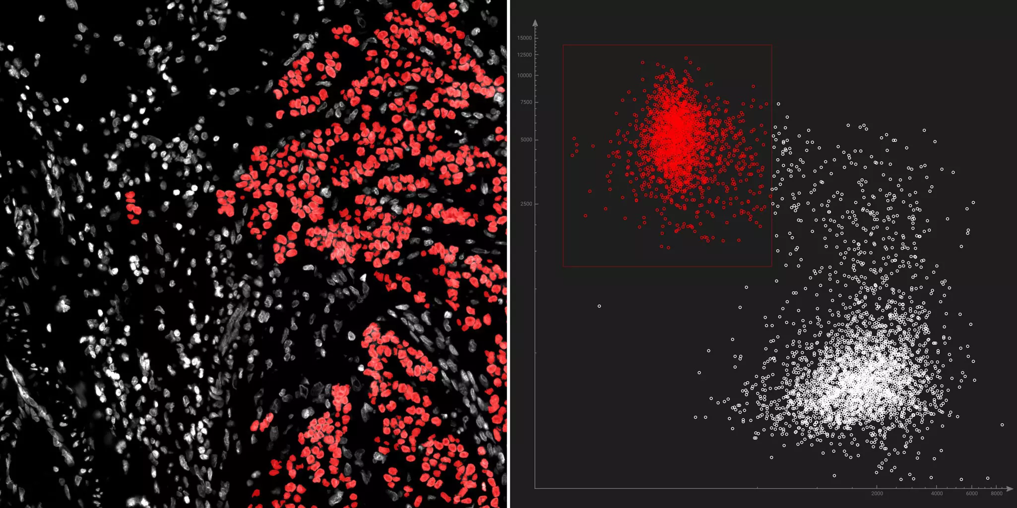
| Gating | The term "gating" is commonly associated with flow cytometry, where it is used to analyze and sort cells based on specific markers. MACS iQ View applies gating to select cells expressing a specific protein marker or exhibiting particular characteristics of interest. It helps researchers focus on regions of interest within a tissue or specific subsets of data that are relevant to their biological questions or hypotheses. The software’s dynamic data display enables you to gate cell populations or even select single cells on your plot and immediately see where they are located in the image or data table and vice versa. |
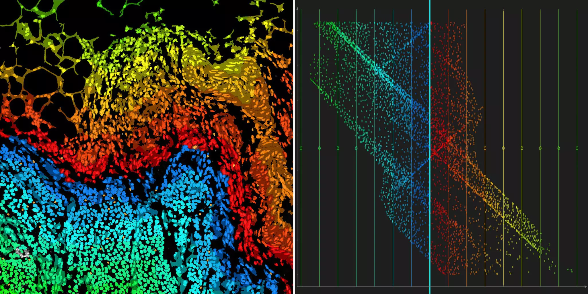
| Spatial analysis and data visualization | MACS iQ View offers standard image analysis tools, including histograms and correlation matrices, along with data extraction and various visualization options. What sets it apart are its advanced spatial analysis capabilities, particularly its precision in relative distance measurements, crucial for accurate tumor analysis, and relative density measurements, essential for biomarker discovery. MACS iQ View provides a comprehensive solution for in-depth tumor characterization and spatial biology analysis. |
| Clustering and dimension reduction | Incorporating a wide range of bioinformatic tools, MACS iQ View goes beyond traditional flow cytometry and image data analysis. Users can employ tools such as UMAPs and t-SNE plots for unbiased and qualitative data analysis. For those with advanced skills, the software features a Python console, providing access to intricate and tailored data analysis. MACS iQ View delivers a comprehensive and flexible solution for data analysis, accommodating users of different expertise levels. |
| Workflow editor | The workflow editor empowers users to design, visualize, and save their workflows. For enhanced automation, this tool enables the duplication of entire workflows for use with different datasets. Furthermore, it supports the cross-analysis of datasets. |
| Data visualization and export | Data can be exported for further analysis in either CSV or standard TIFF formats, allowing for the extraction of all the visual data required for publication or other scientific communication. |
Related PDFs:
Related products:
5 Current trends and developments in spatial biology
5.1 Integration of multiomics technologies in spatial biology
Spatial biology is evolving rapidly through the integration of diverse omics technologies. By combining transcriptomics, proteomics, genomics, and other emerging omics technologies, researchers can now explore biological complexity at an unprecedented scale. Spatial multiomics approaches enable the simultaneous mapping of gene expression, protein abundance, and molecular modifications directly within intact tissues, providing deeper insights into how cellular programs are orchestrated across space and time. This integrated view reveals critical spatial context within tissues and supports the discovery of molecular signatures associated with tissue microenvironments, disease progression, and treatment responses.
By integrating in situ sequencing with spatial proteomics and transcriptomics, researchers can unlock an unprecedented level of detail. This combined approach not only reveals the spatial organization of cells within tissues and identifies detected RNAs, but also enables genomic-level analysis of the precise sequence of individual cells. Ultimately, this delivers a more comprehensive picture of tissue function and architecture.
5.2 3D spatial imaging of organ tissue architecture
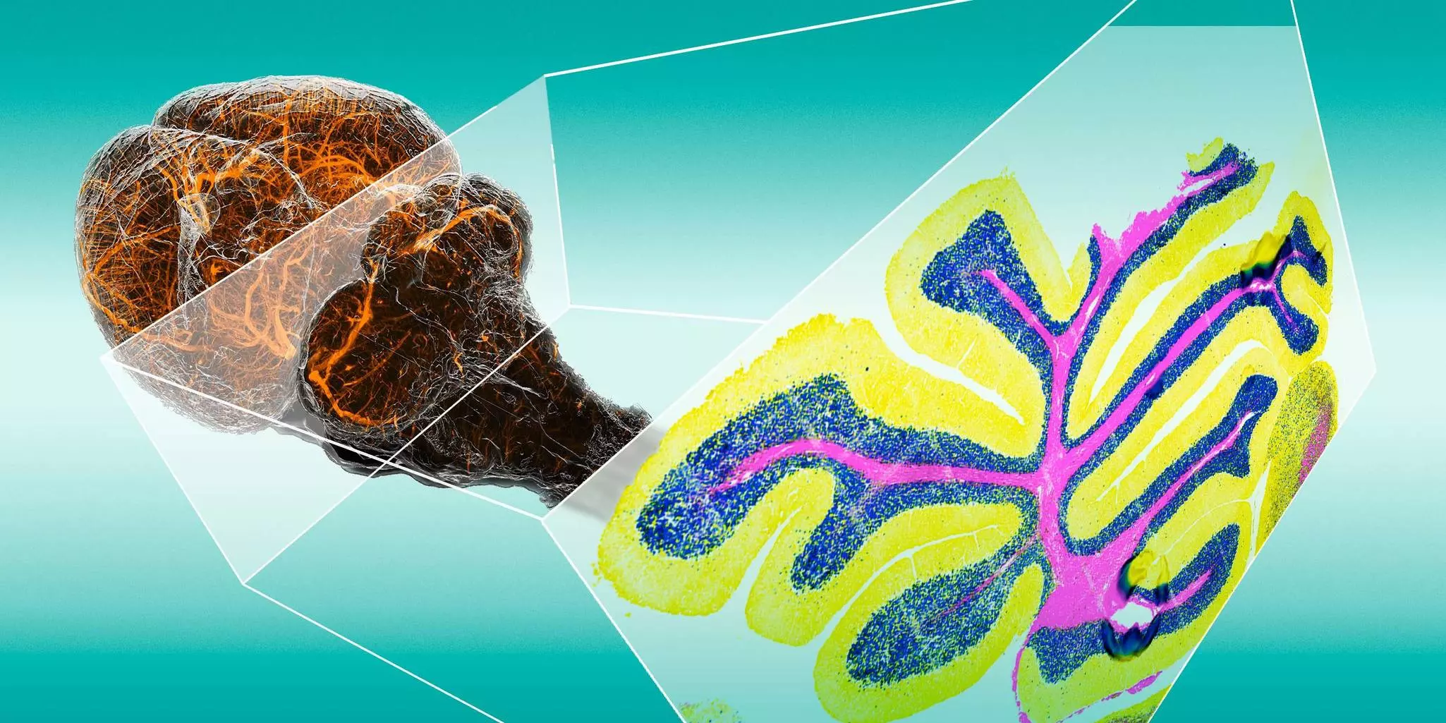
Traditional 2D tissue imaging captures only a partial view of biological complexity. Today, the field is moving boldly toward 3D spatial biology, offering a far more comprehensive and accurate representation of tissue architecture. By bridging whole-tissue 3D imaging with cellular-resolution multiplex analysis, this integrated approach enables powerful multiscale investigation of tissue microenvironments. Tailored for translational and preclinical research in oncology, neurobiology, immunopathology and beyond, 3D spatial imaging empowers scientists to explore complex biological systems with exceptional spatial accuracy and deeper molecular insight.
A major advancement comes from using thicker tissue sections, which improve mechanical stability and preserve the native microenvironment, allowing better correlation between morphology and molecular signatures. Advances in tissue clearing, multiplexed labeling, and volumetric imaging are driving this transition. Ultimately, multiplex 3D spatial biology provides the contextual depth needed to understand complex biological systems and disease mechanisms with unprecedented clarity.
5.3 Computational challenges in spatial omics data analysis
The explosion of spatial omics data has created a critical analysis bottleneck – but also an incredible opportunity. Integrating large, multidimensional datasets requires advanced computational pipelines capable of handling diverse data types and scales. Efficient data storage, sharing, and interoperability are becoming essential to accelerate collaborative research and maximize insights.
Artificial intelligence (AI), machine learning (ML), and deep learning are transforming how spatial data is analyzed, automating segmentation, pattern recognition, and feature extraction from complex spatial data. These powerful tools help uncover cellular phenotypes, tissue structures, and molecular gradients that might otherwise remain hidden – unlocking new discoveries faster than ever before.
At the same time, ensuring algorithm correctness, transparency, and reproducibility remains a key challenge and priority. While commercialization is making these advanced tools more accessible, standardized, and widely adopted, it remains essential to cultivate a diverse ecosystem of technologies and vendors and actively support the open exchange of protocols and software – ensuring that innovation in spatial biology can thrive[6].
5.4 Efficient workflows and time-to-data in spatial biology
As experimental complexity grows, one factor has never been more critical: time to data. Researchers face tight timelines to validate hypotheses, identify therapeutic targets, and unravel disease mechanisms.
In drug development, every preclinical study is truly a race against time. Multiplex imaging delivers rich, multidimensional datasets that reveal safety, efficacy, and mechanisms of action. Reducing time to data ensures these insights are available early, minimizing bottlenecks and enabling timely go/no-go decisions, thereby accelerating the path to new therapies.
That’s why streamlined workflows are essential. By seamlessly integrating every step – from sample preparation and panel design to imaging and analysis – researchers can move faster from raw data to actionable results, turning complex experiments into clear, impactful discoveries.
5.5 Clinical and translational adoption and implementation of spatial biology
Bringing spatial biology into clinical and pathology settings requires solutions that are standardized, reproducible, and fully compliant with regulatory requirements. The focus is shifting from experimental flexibility to workflow robustness and seamless implementation. Automation and user-friendly software are essential for integrating spatial biology smoothly into existing diagnostic pipelines.
Standardized protocols ensure consistent, comparable data across instruments and laboratories – critical for regulatory approval. Reproducibility and traceability further reinforce quality assurance in clinical applications.
As spatial biology technology matures, it is emerging as a cornerstone of precision medicine, enabling the identification of spatial biomarkers, therapeutic targets, and disease-specific microenvironments. By integrating seamlessly into routine workflows, spatial biology is positioned to accelerate breakthroughs in diagnostics, drug discovery, and translational research.
6 Overview / Conclusion
Spatial biology is revolutionizing our understanding of biological systems by providing unprecedented insights into the spatial organization and interactions of molecules within tissues. By combining high-throughput imaging with advanced data analysis, this field is unlocking new avenues for research and discovery.
The MACSima Platform is a powerful tool for advancing spatial biology research. It enables researchers to delve deeply into the complexities of biological processes by providing a comprehensive workflow for sample preparation, data acquisition, and analysis. The platform’s ability to combine spatial proteomics and transcriptomics data offers a unique opportunity to unravel the intricate interplay between genes and proteins within their native tissue context.
Through the application of spatial biology, researchers can gain invaluable insights into a wide range of biological processes, including cancer development, immune responses, and neural function. The ability to identify novel biomarkers and therapeutic targets holds immense potential for the development of innovative treatments and diagnostics.
Ultimately, the integration of spatial biology into research will drive significant advancements in our understanding of health and disease, paving the way for improved patient outcomes.
You have previously chosen International as your location.
What do you want to do?