Walter E. Washington Convention Center
SfN Neuroscience 2026
November 14–18 | Washington, D.C., USA


SfN Neuroscience 2026
Advance your neuroscience discoveries
Join us at the Society for Neuroscience (SfN) annual meeting (Neuroscience 2026) and discover innovative technologies designed to advance neuroscience research with rodent and pluripotent stem cell (PSC)-derived neural models.
We look forward to connecting with you at booth #1809 and showcasing our powerful solutions for gentle cellular analysis, 3D imaging, and spatial biology. Find out more about our full program below.
Exhibit opening hours (EST)
What are you interested in?
Content
- Product Theater
- Scientific posters
- Live demos
- Our solutions
Product Theater
Combining 3D imaging and light sheet–guided 2D spatial multiplexing for deep characterization of a glioblastoma mouse model
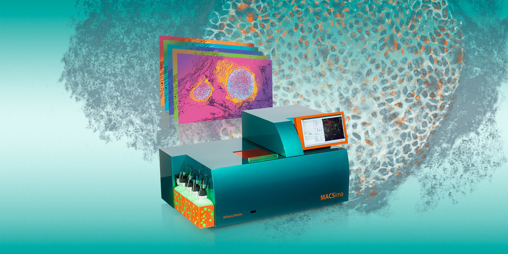
Discover how integrated 3D and 2D imaging workflows can reveal new insights into tissue organization and cellular interactions. Combining the UltraMicroscope Platform for whole-sample visualization with the MACSima® Platform for highly multiplexed analysis, our approach enables researchers to move seamlessly from tissue architecture to deep cellular phenotyping within the same specimen.
Join our presentation to learn how this workflow is being applied in glioblastoma research and how it can help uncover complex biological mechanisms with greater context and precision.
Sunday, November 15
10:00–10:30 a.m. (EST)
Focused Product Theater – Aisle 1100
Session PROT16

Presenter: Kyle Kern, PhD
Product Manager 3D Imaging and Microscopy, Miltenyi Biotec, USA
Product Manager for the 3D imaging and microscopy portfolio at Miltenyi Biotec, Kyle Kern has eight years of experience in sales and marketing specializing in microscopy, tissue clearing, and imaging analysis software. He earned his PhD in Biological Sciences at the Louisiana State University, USA.

Speaker: Kevin Bigott, MSc
R&D Team Coordinator Neurobiology, Miltenyi Biotec, Germany
Team Coordinator in the R&D department in the field of neurobiology, Kevin Bigott joined Miltenyi Biotec in 2018, working on the implementation of new tools and applications for ultramicroscopy, including the development of a refined clearing procedure and the setup of antibody conjugates with improved features for 3D microscopy. Currently, his major interests include the development of advanced applications in 3D imaging, and new tools for deep 3D data analysis in an unobstructed environment. He graduated from the University of Konstanz (Bachelor) and University of Cologne (Master), Germany, with a focus on neurobiology, imaging technology and image analysis.
Scientific posters
Explore the scientific posters in Halls A–C.
#PSTR125
Monday, November 16
8:00 a.m.–12:00 p.m.
Combining 3D imaging and light sheet–guided 2D spatial multiplexing for deep characterization of a glioblastoma mouse model
Kevin Bigott
#PSTR211
Tuesday, November 17
8:00 a.m.–12:00 p.m.
A stage-resolved transcriptomic and cellular atlas of human cerebral organoid development reveals developmental windows of ethanol vulnerability
Muhammed Bishir (Customer poster)
#PSTR352
Wednesday, November 18
3:00–4:00 p.m.
1D, 2D, 3D: cross-scale cell typing and spatial analysis of the APP/PS1 Alzheimer's disease mouse brain using single-nucleus RNA sequencing, cyclic multiplexed immunofluorescence, and light sheet microscopy
Kyle Kern
Live demos
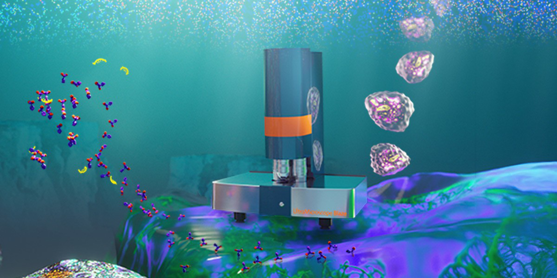
UltraMicroscope Blaze™ live demos on demand
Visit our booth to see the UltraMicroscope Blaze in action and find out how to advance your spatial biology with integrated 3D to 2D tissue profiling.
Miltenyi Biotec
Booth #1809
November 15–18
9:30 a.m.–5:00 p.m. (EST)
3D visualization of mouse glioblastoma architecture
This video shows a whole mouse brain with glioblastoma that was optically cleared and stained using Anti GFP-Alexa Fluor® 647 Nanobody from Chromotek and the Antibody Staining Solution of the MACS® Deep Clearing Kit. The brain was then imaged using the UltraMicroscope Blaze. The 3D rendering and reconstruction were performed with Imaris v10.2.0 (Oxford Instruments).
Our solutions

Contact us
Do you want to get in contact with us? Send your inquiry to:
Karen Cherrez, Product Manager Regenerative Medicine
You have previously chosen International as your location.
What do you want to do?