The CD140a (PDGFRα) MicroBead Kit was developed for the direct isolation or depletion of CD140a (PDGFRα) positive oligodendrocyte precursors from dissociated mouse brain tissue.


Related products for CD140a (PDGFRα) MicroBead Kit, mouse
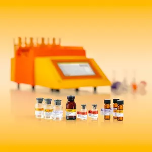
The CD140a (PDGFRα) MicroBead Kit was developed for the direct isolation or depletion of CD140a (PDGFRα) positive oligodendrocyte precursors from dissociated mouse brain tissue.
CD140a (PDGFRα) MicroBeads (PDGFRα: platelet-derived growth factor receptor alpha) have been developed for the separation of mouse cells based on the expression of the CD140a (PDGFRα) antigen. CD140a (PDGFRα) has been reported to be broadly expressed in embryonic tissue, various malignancies and embryonic stem cell-derived cardiomyogenic cells.1,2 Furthermore, CD140a (PDGFRα) is specifically expressed by oligodendrocyte precursor cells, which differentiate into myelinating oligodendrocytes.3–5 CD140a (PDGFRα) MicroBeads were especially optimized for the separation of oligodendrocyte progenitor cells (OPCs) based on the expression of CD140a (PDGFRα). The isolation was tested particularly on dissociated postnatal CD1 mouse brain tissue derived from animals younger than postnatal day eight (P8). In principle, cell isolation is also possible from older mice, but purity of the positive fraction might be lower due to lower frequency of CD140a (PDGFRα) positive cells after tissue dissociation.
For optimal results, the Neural Tissue Dissociation Kit (P) is recommended prior to cell isolation.
CD140a (PDGFRα)+ cells were isolated from P2 CD-1 mouse brain tissue using the CD140a (PDGFRα) MicroBead Kit, the Neural Tissue Dissociation Kit (P), the gentleMACS Dissociator, a MiniMACS Separator, and an MS Column. Cells were fluorecently stained with Labeling Check Reagent-APC and analyzed by flow cytometry using the MACSQuant® Analyzer. Cell debris and dead cells were excluded from the analysis based on scatter signals and propidium iodide fluorescence.
- Nishiyama, A. et al. (1996) Co-localization of NG2 proteoglycan and PDGF alpha-receptor on O2A progenitor cells in the developing rat brain. J. Neurosci. Res. 43: 299-314
- Nishiyama, A. et al. (2009) Polydendrocytes (NG2 cells): multifunctional cells with lineage plasticity. Nat. Rev. Neurosci. 10: 9-22
- Sim, F. J. et al. (2011) CD140a identifies a population of highly myelinogenic migration-competent and efficiently engrafting human oligodendrocyte progenitor cells. Nat. Biotechnol. 29: 934-941
- Takakura, N. et al. (1997) PDGFRα expression during mouse embryogenesis: immunolocalization analyzed by whole-mount immunohistostaining using the monoclonal antibody anti-mouse PDGFR alpha antibody APA5. J. Histochem. Cytochem. 45 (6): 883-893
- Heldin, C. H. and Westermark, B. (1999) Mechanism of action and in vivo role of platelet-derived growth factor. Physiol. Rev. 79: 1283-1316
You have previously chosen International as your location.
What do you want to do?