Isolation, expansion, and phenotyping of bone marrow-derived mesenchymal stem cells (MSCs)
Mesenchymal stem cells or mesenchymal stromal cells (MSCs), are somatic or adult stem cells that can be isolated from various sources, including bone marrow, adipose tissue or umbilical cord. Isolated MSCs are fibroblast-like cells that demonstrate site-specific differentiation. MSCs may regenerate damaged or diseased tissues in vivo and potentially play a role in immunomodulation, which provides the basis for a variety of clinical applications. This application protocol describes the enumeration, isolation, expansion, and quality assessment of bone marrow-derived MSCs.
Protocol
Isolation, expansion, and phenotyping of bone marrow-derived mesenchymal stem cells (MSCs)
1.75 h
Isolation of mononuclear cells
40 min
Enumeration of MSCs
45 min
Isolation of MSCs (research scale)
Expansion of MSCs
varies
Expansion of MSCs
1st passage: 9 d
Additional passages: 3–5 d
20 min
Phenotypic analysis of expanded MSCs
varies
Functional analysis of expanded MSCs
AdipoDiff: 21 d
OsteoDiff: 10 d
ChondroDiff: 28 d
This protocol includes detailed instructions for preparing necessary reagents and materials and then carrying out each step.
PE buffer for isolation of mononuclear cells
- Prepare a solution with phosphate buffered saline (PBS), pH 7.2, and 2 mM EDTA. Keep buffer cold (2–8 °C).
▲ Note: EDTA can be replaced by other supplements such as anticoagulant citrate dextrose formula-A (ACD-A) or citrate phosphate dextrose (CPD).
- Prepare a solution containing phosphate-buffered saline (PBS), pH 7.2, 0.5 % bovine serum albumin (BSA), and 2 mM EDTA by diluting MACS® BSA Stock Solution 1:20 with autoMACS® Rinsing Solution. Keep buffer cold (2−8 °C).
▲ Note: EDTA can be replaced by other supplements such as anticoagulant citrate dextrose formula-A (ACD-A) or citrate phosphate dextrose (CPD). BSA can be replaced by other proteins such as respective serum albumin, respective serum, or fetal bovine serum (FBS). Buffers or media containing Ca2+ or Mg2+ are not recommended for use.
Isolate mononuclear cells by density gradient centrifugation using Ficoll-Paque™. Use the protocol of the following special protocol.
Download the special protocol
The BM-MSC Analysis Cocktail Kit, anti-human is especially designed to quantify human MSCs from fresh bone marrow aspirate (BMA) with a standardized flow cytometry protocol and gating strategy based on the expression of CD271 (LNGFR) and Anti-MSCA-1 (W8B2). The kit allows identification of CD45+ leukocytes and CD271 (LNGFR)+/Anti-MSCA-1 (W8B2)+ MSCs, which are also CD45dim. Follow the protocol of the kit data sheet.
Download the data sheet
Bone marrow-derived human MSCs can be isolated by plastic adherence or by specific surface markers such as CD271. Marker-based MSC isolation provides more uniform cell populations that show higher clonality, compared to plastic adherence. Use the CD271 MicroBead Kit, human to isolate MSCs. Follow the protocol of the kit data sheet.
It is recommended to filter the magnetically labeled cell suspension to guarantee a single-cell suspension before separating cells on the column.
- Place a Pre-Separation filter (30 µm) on the LS or MS Column.
- Rinse the column 3 times with PBE buffer, ensuring that the filter is pre-wetted.
- Apply cell suspension and PBE buffer to the filter on the column.
Download the data sheet
Expand isolated human MSCs using the StemMACS™ MSC Expansion Media Kit XF, an optimized and standardized complete medium for the reproducible and reliable generation and expansion of MSCs from bone marrow and other tissue sources. Follow the protocol of the kit data sheet.
Download the data sheet
Growing interest in using MSCs for clinical research necessitates a fundamental understanding of mechanisms and processes underlying their differentiation into specific cell types, as well MSC phenotype validation of cultivated cells. The MSC Phenotyping Cocktail Kit, anti-human, REAfinity™ is based on ISCT (International Society for Cellular Therapy) markers for human MSCs, and helps easily perform standardized phenotyping of culture-expanded MSCs by flow cytometry. Follow the protocol of the kit data sheet.
Download the data sheets and protocols
MSC Phenotyping Cocktail Kit, anti-human, REAfinity
Mesenchymal stem cell culturing protocols
To plan a dedicated antibody panel for a MSC research project, use the antibody panel builder.
Analysis of differentiation potential
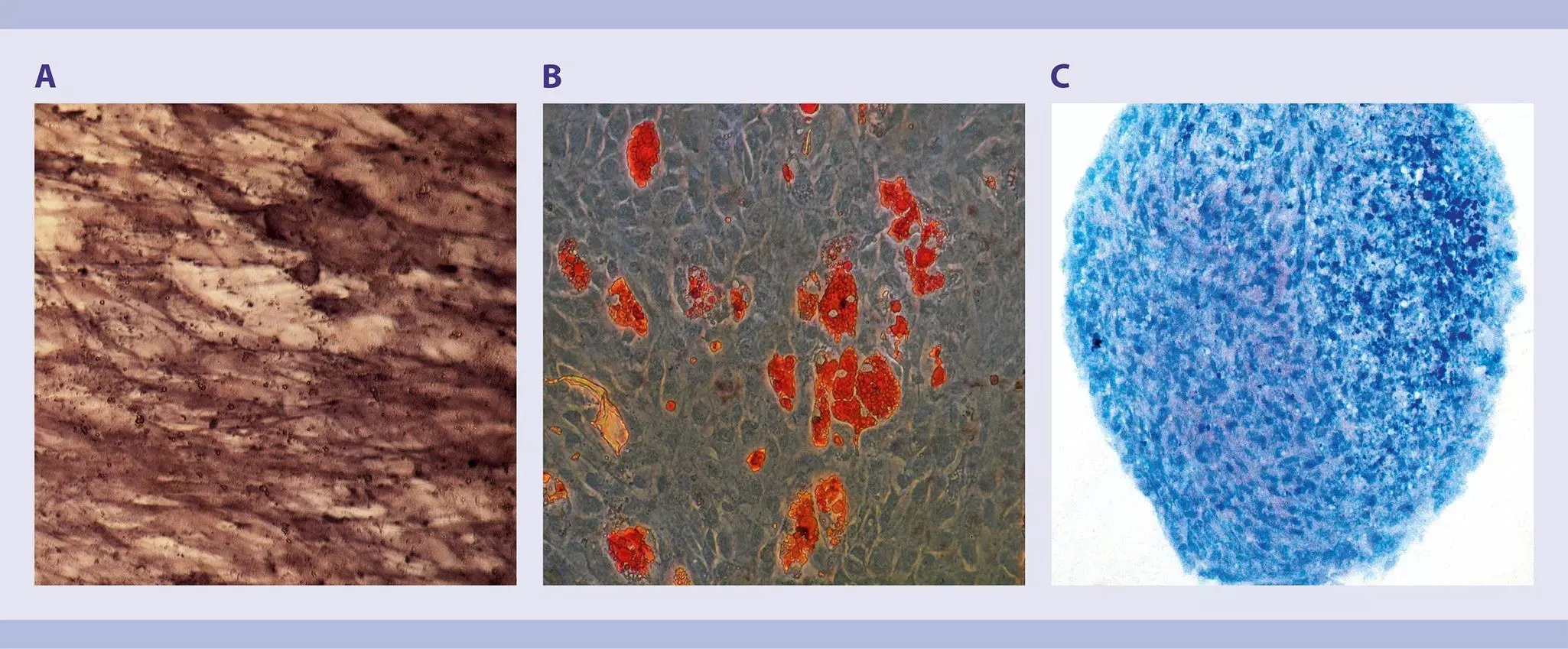
Differentiation potential is a meaningful tool to qualify and define a MSC population. Grade of differentiation into adipocytes, chondrocytes, and osteoblasts is influenced by MSC origin, so for example, MSCs from bone marrow show greater potential to differentiate into osteoblasts than MSCs from umbilical cord. Increased age of donor and passaging of cultured MSCs also decrease differentiation potential.
StemMACS™ AdipoDiff, OsteoDiff, and ChondroDiff Media, human are optimized differentiation media for human MSCs isolated from various tissue sources. Each medium supports differentiation capacity analysis or quality control of expanded MSCs, as well as in vitro studies on MSC differentiation processes, including gene expression and protein profiling. Follow the protocol of the relevant kit data sheet.
Download the respective data sheet
StemMACS AdipoDiff Media, human
Analysis of immunomodulatory potential
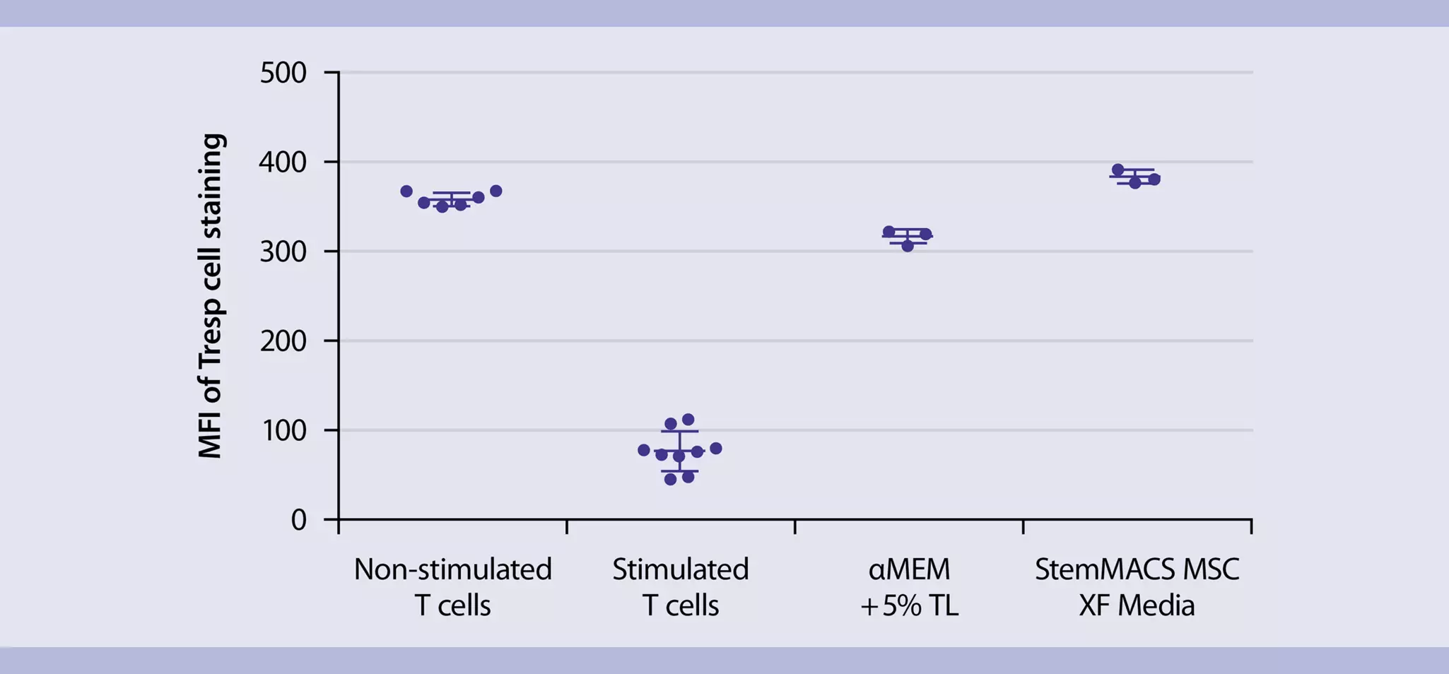
In the last few years, scientists have directed their attention to the immunomodulatory potential of MSCs. MSCs derived from bone marrow have been observed to suppress proliferation of T cells.1,2 This function of MSCs can be analyzed using the MSC Suppression Inspector, human, which contains T cell stimulation reagents optimized for an MSC suppression assay. Follow the protocol of the kit data sheet.
Download the data sheet
Additional resources
- Effective licensing of human mesenchymal stem cells
- Flyer: MSC Phenotyping Kit, human – Confirm compliance with ISCT criteria
- Flyer: MSC Enumeration Kit, human – Standardized, fast quantification of human mesenchymal stem cells
- Flyer: StemMACS™ MSC Expansion Media Kit XF
- Pütsch, K. and Schmitz, J. Clinical-scale isolation of mesenchymal stromal cells from bone marrow with the CliniMACS® System and CD271 antibody–conjugated MicroBeads
- Godthardt, K. et al. (2016) Improved clinical-scale MSC isolation from primary tissue and expansion with a GMP matrix- and xeno-free medium. ISCT, Singapore
- REAfinity™ Recombinant Antibodies – Flow cytometry is in their genes
You have previously chosen International as your location.
What do you want to do?