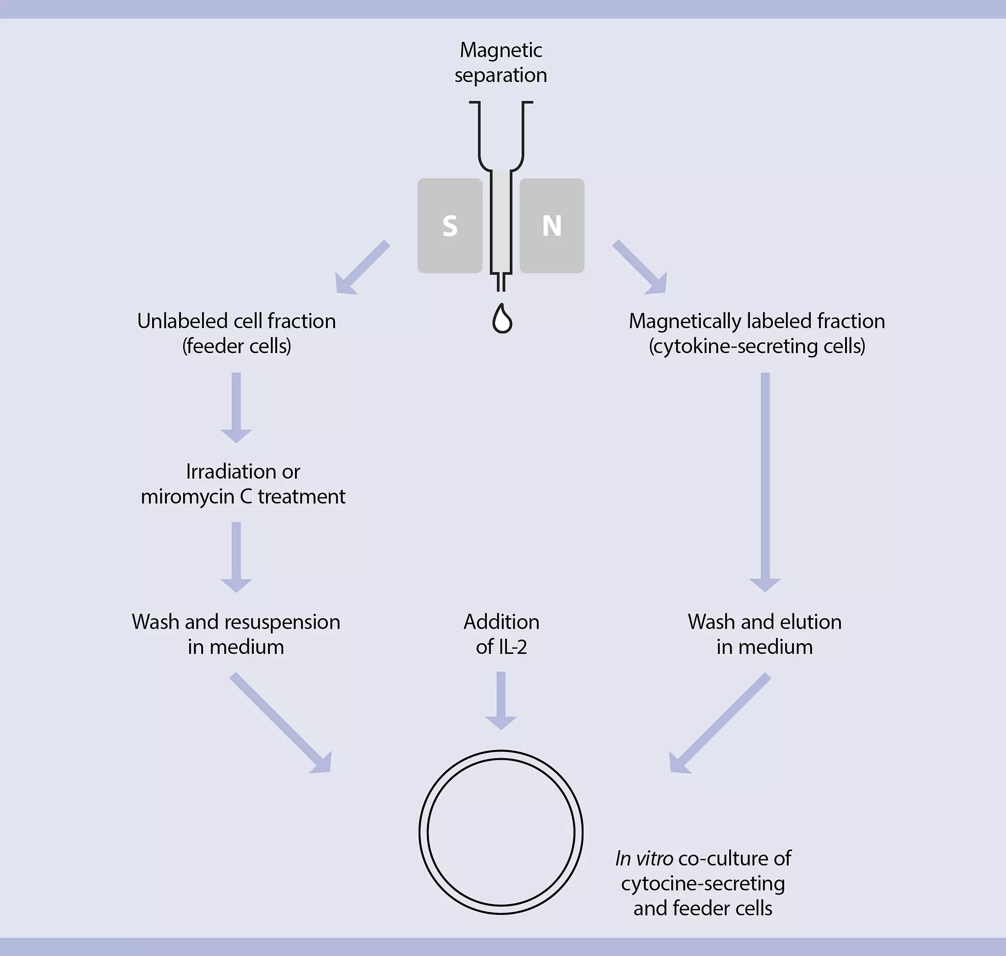
This expansion system is designed for in vitro expansion of cytokine-secreting, antigen-specific T cells. Antigen-specific T cells in peripheral blood mononuclear cells (PBMCs) or leukapheresis are restimulated with antigen. The responding cells are magnetically isolated, according to their secretion of cytokines, using the MACS Cytokine Secretion Assay. The cell fraction not secreting the respective cytokine, i.e., the unlabeled flow through, is used as a source of antigen-presenting cells (referred to as “feeder cells” throughout the protocol) after irradiation or treatment with mitomycin C. The cytokine-secreting cells and the feeder cells are co-cultured in vitro in the presence of interleukin 2 (IL-2).
Expansion of antigen-specific cytokine-secreting T cells
This application protocol describes how to expand antigen-specific T cells which have been isolated using the MACS® Cytokine Secretion Assay after restimulation with antigen. These T cells are therefore of a memory phenotype. The memory phenotype, particularly for CD8+ cells, is heterogeneous. It contains cells, which divide rapidly in response to antigen-“central memory cells” and cells, which exhibit recall effector function, such as cytotoxicity-“effector memory cells”. These subsets are not mutually exclusive, e.g., cells can divide rapidly but still lyse antigen-loaded targets. The mixture of memory cells isolated with the MACS Cytokine Secretion Assay is entirely dependent on the antigen the T cells responded to, e.g., immune responses against chronic virus infections are often characterized by a pre-dominance of effector memory T cells. As a general rule, central memory cells have a higher proliferation rate in vitro, compared to effector memory cells. Therefore, the rate of proliferation of T cells expanded with this method will depend on the memory phenotype of the cells that have been isolated. This protocol has been optimized to expand both central and effector memory T cells very efficiently. However, the individual results obtained may vary depending on the antigen and the Cytokine Secretion Assay used to isolate the cells. This protocol has been established for the expansion of CMV-specific, IFN-γ–secreting T cells. It may require optimization when working with other antigens or expanding cells which were isolated according to the secretion of cytokines other than IFN-γ.
Protocol
This protocol includes detailed instructions for preparing necessary reagents and materials and then carrying out each step.
 View details
- All steps in the following protocol have to be performed under sterile conditions.
- Prepare and restimulate cells with antigen and perform the Cytokine Secretion Assay including magnetic labeling referring to the respective data sheet.
- Choose an appropriate MACS Column and MACS Separator according to the number of total cells and the number of IFN-γ–secreting cells. For details refer to the table in the Materials tab.
- Always wait until the column reservoir is empty before proceeding to the next step.
- When enriching antigen-specific T cells, always perform two consecutive column runs to achieve best results.
Magnetic separation with MS or LS Columns
- Place column in the magnetic field of a suitable MACS Separator. For details refer to the respective MACS Column data sheet.
- Prepare two columns by rinsing each with the appropriate amount of cold buffer:
- MS: 500 μL
- LS: 3 mL
- Apply cell suspension onto the column.
- Collect unlabeled cells that pass through and wash column with the appropriate amount of buffer. Collect total effluent; this is the unlabeled cell fraction. Perform washing steps by adding buffer three times. Only add new buffer when the column reservoir is empty. Keep sterile for further processing as feeder cells.
- MS: 3×500 μL
- LS: 3×3 mL
- Remove first column from the separator, place the second column into the separator and put the first column on top of the second one.
- Pipette the appropriate amount of cold buffer onto the first column. Immediately flush out the magnetically labeled cells by firmly pushing the plunger into the first column directly onto the second column.
- MS: 1 mL
- LS: 5 mL
- Collect unlabeled cells that pass through and wash column with the appropriate amount of cold buffer and medium. Perform the first washing step by adding buffer. Perform two further washing steps by adding cold medium successively once the column reservoir is empty.
- With cold buffer:
- MS: 1×500 μL
- LS: 1×3 mL
- With cold medium:
- MS: 2×500 μL
- LS: 2×3 mL
- With cold buffer:
- Remove the second column from separator and place column on a suitable sterile tube.
- Pipette appropriate amount of cold medium onto the column. Immediately flush out the magnetically labeled T cells by firmly pushing the plunger into the column. Collect T cells in a sterile tube and keep it under sterile conditions.
- MS: 1 mL
- LS: 5 mL
- Take an aliquot for cell counting and flow cytometric analysis and proceed to cell culture (refer to section Expansion protocol, Initiation of T cell expansion).
▲ Note: For flow cytometry cells should be washed with buffer to remove medium before analysis.
▲ Note: The remaining cells may be stored at room temperature until cultured further. - Take an aliquot for cell counting from the unlabeled fraction and proceed to preparation of feeder cells (refer to section Expansion protocol, Preparation of feeder cells).
Magnetic separation with the autoMACS® Pro Separator
- Refer to the respective user manual for instructions on how to use the autoMACS Pro Separator.
- Buffers used for operating the autoMACS Pro Separator should have a temperature of ≥10 °C.
- Prepare and prime the instrument.
- Apply tube containing the sample and provide tubes for collecting the labeled and unlabeled cell fractions. Place sample tube in row A of the tube rack and the fraction collection tubes in rows B and C.
- For a standard separation choose following program:
Positive selection: Posseld
Collect magnetically labeled cells (positive fraction) in a sterile tube in row C of the tube rack. Collect feeder cells (negative fraction) in a sterile tube in row B of the tube rack. - Take aliquots for cell counting and flow cytometric analysis from the labeled and the unlabeled fraction.
- Wash the magnetically labeled cells by adding an appropriate amount of medium and centrifuge at 300×g for 10 minutes. Aspirate supernatant completely. Proceed to cell culture (refer to section Expansion protocol, Initiation of T cell expansion).
▲ Note: The remaining cells may be stored at room temperature until cultured further. - Use the unlabeled cells from the negative fraction for preparation of feeder cells (refer to section Expansion protocol, Preparation of feeder cells).
Magnetic separation with the CliniMACS® Plus Instrument
- For sample preparation refer to the protocol in the data sheet of the Large Scale IFN-γ Secretion Assay – Enrichment Kit, human (# 130-091-329).
- For set-up of the CliniMACS Plus Instrument, selection of the separation program, and installation of the CliniMACS Tubing Set follow the detailed instructions given in the CliniMACS Plus User Manual.
- Connect sample bag and CliniMACS Buffer bag to CliniMACS Tubing Set.
- Separate cells using program version ENRICHMENT 3.1 or higher. The magnetically labeled cell fraction, i.e., the antigen-specific T cells will appear in the target T cell bag and the feeder cells in the non-target T cell bag.
- Take aliquots for cell counting and flow cytometric analysis from the labeled and unlabeled fraction.
- Wash the magnetically labeled cell fraction by adding an appropriate amount of medium and centrifuge at 300×g for 10 minutes. Aspirate supernatant completely. Proceed to cell culture (refer to section Expansion protocol, Initiation of T cell expansion).
▲ Note: The remaining cells may be stored at room temperature until cultured further. - Use the unlabeled cells from the non-target T cell bag for preparation of feeder cells (refer to section Expansion protocol, Preparation of feeder cells)
The protocol is based on standard T cell expansion procedures where specific T cells are cultured with autologous feeder cells, antigen, and IL-2. Using the cells collected from the unlabeled fraction provides a convenient source of antigen pre-loaded feeder cells. As the target T cells are already highly activated and enriched, they have high co-stimulation requirements to grow properly and require more feeder cells than in standard expansion procedure, where the initial frequency of antigen-specific T cells is low. Therefore, in the first phase of expansion, 100 feeder cells are required per antigen-specific T cell in the isolated positive fraction. After two weeks of expansion, the T cells require to be restimulated with feeder cells (which still contain the antigen) at a ratio of 10 feeder cells per T cell. This cycle can be repeated every two weeks.
As there will be only a limited amount of cells from the unlabeled fraction, long term culture or quickly growing cells will require a new source of feeder cells. For this purpose, fresh PBMCs from the same donor may be used. The cells should be used in exactly the same way as for the unlabeled fraction, with the addition of antigen at the same concentration as used to originally stimulate the T cells. It is also possible that restimulation of T cells with frozen cells from the unlabeled fraction can be enhanced by addition of fresh antigen.
Preparation of feeder cells
In order to have feeder cells for later restimulation of T cells, cells of the unlabeled fraction or PBMCs from the same donor can be frozen and later resuscitated.
To prevent the feeder cells from growing out during culture, these cells need to be immobilised using irradiation or mitomycin C treatment.
▲ Note: Cells should not be frozen after irradiation or mitomycin C treatment.
Irradiation
For example, irradiate cells with 35–50 Gy (3500–5000 rads) using a caesium 137 source. Individual irradiators may vary in performance and the optimum degree of irradiation has to be determined.
Mitomycin C
- Mitomycin C is extremely toxic. Observe manufacturer’s safety warnings.
- Determine cell number of the unlabeled fraction.
- Wash cells of the unlabeled fraction once in culture medium and centrifuge at 300×g for 10 minutes. Aspirate supernatant completely.
- Prepare a fresh working solution of 25 μg/mL mitomycin C in culture medium.
- Add 500 μL of mytomycin C working solution per 107 cells.
- Incubate cells for 30 minutes at 37 °C.
- Wash the cells four times in culture medium. Each time centrifuge at 300×g for 10 minutes. Aspirate supernatant completely.
- Determine cell number and adjust cells to a final density of 107 cells/mL.
▲ Note: It is normal to see a reduction in cell numbers by up to 50% after mitomycin C treatment. This is due to the death of many B cells and some general cell loss during the washing steps.
Initiation of T cell expansion
- Determine total number of antigen-specific T cells in the positive fraction.
▲ Note: Determine cell count of antigen-specific T cells, not the total cell count, as monocytes or dead cells may be co-enriched with antigen-specific T cells. Use flow cytometric analysis to get an accurate count. - Adjust the antigen-specific T cell fraction to 105 cells/mL in medium.
- Combine the antigen-specific T cells with an equal volume of prepared feeder cells (at 107 cells/mL). Mix well and plate cells in an appropriate cell culture dish at 2.5×10⁶ cells/cm2, e.g., 1 mL of cell suspension in one well of a 24-well plate (refer to section Appendix: Flask and dish sizes for T cell expansion).
- Add 20 IU of IL-2 per mL of medium.
- Incubate at 37 °C, 5–7.5% CO2.
- The final concentrations are:
- 100 feeder cells per antigen-specific T cell
- 5×10⁶ cells per mL
- 2.5×10⁶ cells per cm2
- 20 IU IL-2/mL
▲ Note: 20 IU IL-2 is a low dose, some cells may need 20–100 IU IL-2.
Maintenance of culture
The following are guidelines on how cells should grow; the rates of growth will depend on the phenotype of the cells generated. After one or two days it is normal that large clumps of cells appear in the culture. Cells should be observed daily to ensure that the medium is not depleted of nutrition. A basic schedule is to feed the T cells every 3–4 days with fresh medium supplemented with 20 IU IL-2 at day 3, 7, and 11 (start is day 0). By day 14, cells need to be restimulated with feeder cells and antigen, followed by a repeated cycle of feeding with medium and IL-2.
In detail
- If at any point the indicator of the medium turns yellow, remove about 70% of the medium with a pipette without resuspending the cells. Replace volume with medium supplemented with 20 IU/mL of IL-2 and transfer half of the culture to a new culture dish.
Day 0:
Start (refer to section Expansion protocol, Initiation of T cell expansion)
Day 3:
Fast-growing cells
Gently resuspend the cells using a pipette and transfer half of the culture to a new culture dish. Add an equivalent volume of medium supplemented with 20 IU/mL of IL-2 to each well.
Slow-growing cells
Gently resuspend the cells using a pipette and add 20 IU/mL IL-2 (no fresh medium).
Day 7:
- Count cells and determine culture viability.
▲ Note: At this point the culture is a mixture of growing antigen-specific T cells and dying feeder cells, so total viabilities of 50% are common. - Adjust the cell concentration to 5×10⁵ viable T cells per mL with medium supplemented with 20 IU/mL of IL-2.
Day 11:
Repeat procedure from Day 3.
Day 14:
- Harvest T cells and determine cell count and viability.
- If desired or if there are large numbers of dead cells in the culture, the cells may be purified by density gradient centrifugation.
- Wash cells by adding medium and centrifuge at 300×g for 10 minutes. Aspirate supernatant.
- Remove supernatant and resuspend cells in 1 mL of medium per 106 cells.
- Prepare feeder cells as before and resuspend the cells in 1 mL of medium per 107 cells (refer to section Expansion protocol, Preparation of feeder cells).
- Combine equal volumes of feeder cells and T cells in a suitable culture vessel and incubate as before.
- Add 20 IU of IL-2 per mL of medium.
This 2-week cycle may be repeated to continue proliferation of T cells.
Good to know
- Long term (> 4 weeks) culture of T cells selects the cells that grow best in culture – the longer the cells are cultured, the less they will reflect the original effector phenotype of the isolated cells (antigen-specificity is unaltered).
- Some T cells may require to be restimulated with feeder cells on a weekly basis after the initial 14-day period.
For T cell expansion the cells should be resuspended in culture medium containing 5% of human serum and 20 IU of IL-2 at 5×10⁶ cells/mL. The cells should be plated at a density of 2.5×10⁶ cells/cm2. Both the dilution and the cell density are important to assure optimal stimulation and cell growth.
The following table lists culture plate, dish, and flask sizes suitable for different cell numbers. It also indicates the appropriate amount of medium to add. For culture in other formats (e.g. culture bags), scale up accordingly.
Table 1: Culture plate, dish, and flask sizes suitable for different cell numbers
| Total cell number | Medium volume to add | Culture plate | Well diameter |
| 0.75×10⁶ | 0.15 mL | 96 well | 0.64 cm |
| 2. 5×10⁶ | 0.5 mL | 48 well | 1.13 cm |
| 5×10⁶ | 1 mL | 24 well | 1.60 cm |
| 1×10⁷ | 2 mL | 12 well | 2.26 cm |
| 2.5×10⁷ | 5 mL | 6 well | 3.50 cm |
| Total cell number | Medium volume to add | Culture dish | Dish diameter |
| 2.5×10⁷ | 5 mL | small | 3.5 cm |
| 7.5×10⁷ | 15 mL | medium | 6 cm |
| 20×107 | 40 mL | large | 10 cm |
| 45×107 | 90 mL | extra large | 15 cm |
| Total cell number | Medium volume to add | Culture flask | Growth area |
| 6×10⁷ | 12 mL | 50 mL | 25 cm2 |
| 20×107 | 40 mL | 250 mL | 75 cm2 |
| 40×107 | 80 mL | 720 mL | 162 cm2 |
| 55×107 | 110 mL | 900 mL | 225 cm2 |
You have previously chosen International as your location.
What do you want to do?