Bead-based immunoassay for detection of patient-derived SARS-CoV-2 antibodies
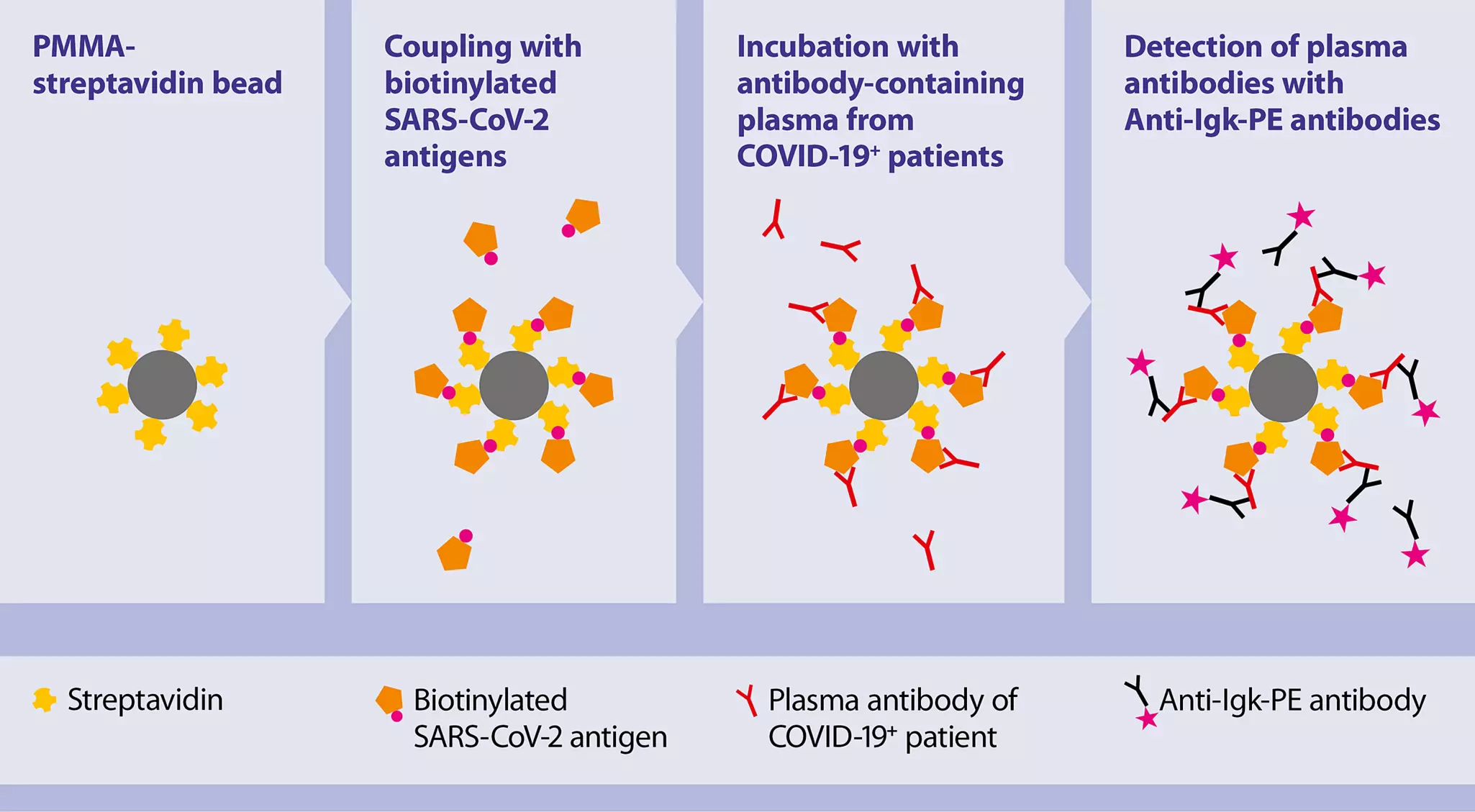
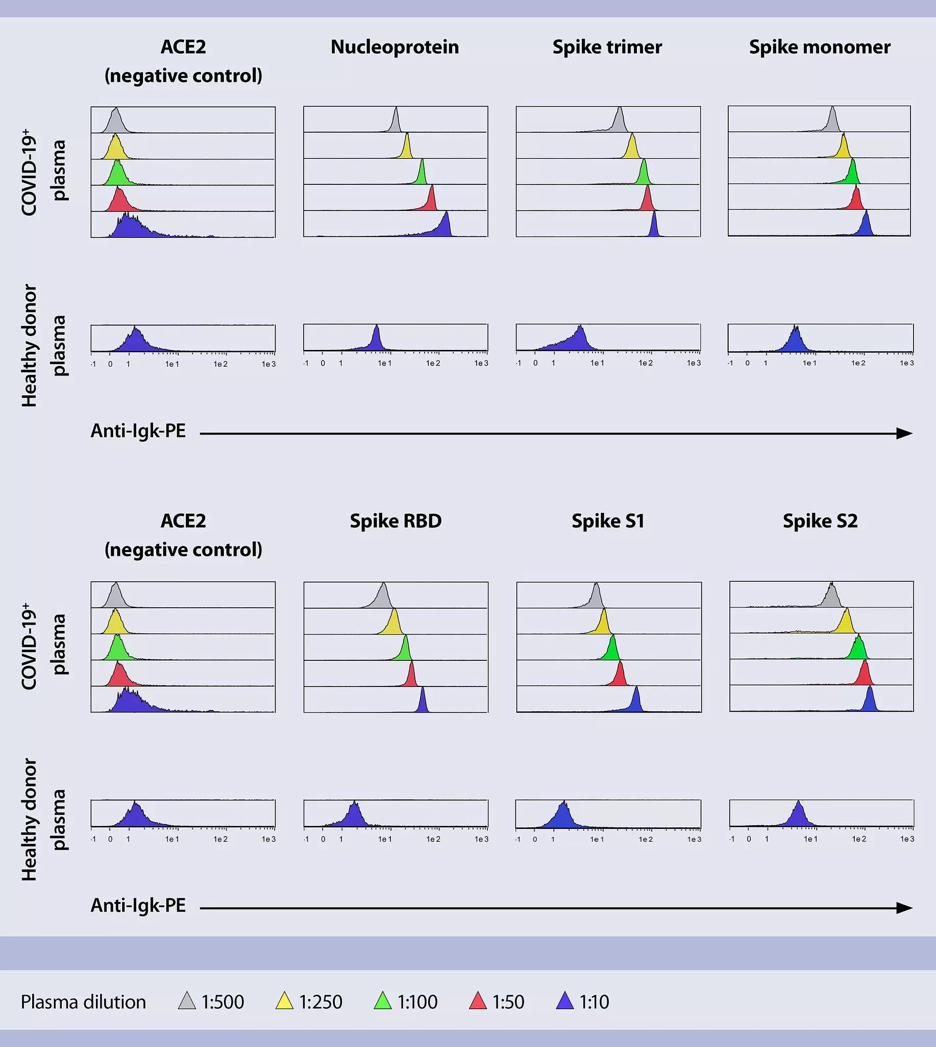
This application protocol describes how SARS-CoV-2–specific antibodies can be detected in plasma or serum samples of COVID-19 Convalescents using our biotinylated recombinant SARS-CoV-2 proteins in a bead-based flow cytometric assay. Knowing the humoral response of SARS-CoV-2 convalescent patients is an essential background information to analyze the cellular responses, such as antigen-specific B cell analysis, more in detail.
The materials and methods described in this protocol are for detection of patient-derived SARS-CoV-2 antibodies.
- Assay buffer
Prepare a solution containing phosphate-buffered saline (PBS), pH 7.2, 0.5% bovine serum albumin (BSA), and 2 mM EDTA by diluting MACS® BSA Stock Solution (# 130-091-376) 1:20 with autoMACS® Rinsing Solution (# 130-091-222). - Dilute selected Detection Reagent 1:50 with assay buffer.
- Recombinant proteins
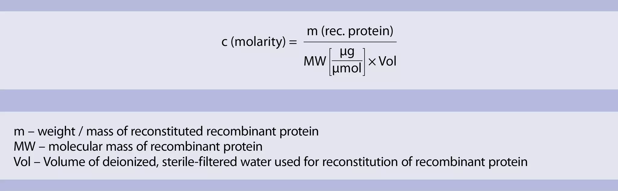
- Reconstitute lyophilized recombinant proteins to a final concentration of 0.1 mg/mL with deionized, sterile-filtered water.
- Calculate the molar concentration (molarity) of the protein solution (see formula below).
- Prepare 10 nM solutions for each protein by diluting stock solution of recombinant protein with assay buffer.
Table 1: Exemplary concentration calculations of reconstituted recombinant proteins
| Recombinant protein variant | Concentration after reconstitution (mg/mL) | MW (kDa) | Corresponding molar concentration (µM) |
| Recombinant SARS-CoV-2 RBD (HEK)-Biotin | 0.1 | 33.8 | 2.96 |
| Recombinant SARS-CoV-2 RBD (Insect cells)-Biotin | 0.1 | 34.2 | 2.92 |
| Recombinant SARS-CoV-2 Nucleoprotein-Biotin | 0.1 | 49.1 | 2.04 |
| Recombinant SARS-CoV-2 Spike-Trimer (HEK)-Biotin | 0.1 | 418.0 | 0.24 |
| Recombinant SARS-CoV-2 Spike-S2 (insect cells) | 0.1 | 61.2 | 1.63 |
| Recombinant SARS-CoV-2 Spike-Prot (HEK)- Biotin | 0.1 | 136.0 | 0.74 |
| Recombinant Human ACE2 (HEK)-Biotin | 0.1 | 87.2 | 1.15 |
| Recombinant Human ACE2 (insect cells) | 0.1 | 72.8 | 1.37 |
Sample preparation
Dilute serum or plasma samples with assay buffer (recommended is 1:10 to 1:500 dilution).
Protocol overview
- Transfer 1x10⁶ Streptavidin-coated beads to suitable polypropylene or polystyrene reagent tubes. Prepare one tube for each recombinant protein to be used in the assay.
- Wash Streptavidin-coated beads 2x with 1 mL assay buffer.
▲ Note: For washing, centrifuge at room temperature (RT) at 1000xg for 1 minute, aspirate supernatant, and resuspend beads in 1 mL assay buffer. - Resuspend Streptavidin-coated beads in 500 µL of 10 nM solution of recombinant proteins.
- Incubate for at least 15 minutes at RT using the MACSmix™ Tube Rotator. Set on maximum permanent run speed.
- Centrifuge beads coupled to recombinant proteins at 1000xg for 1 minute at RT.
- Wash beads coupled to recombinant proteins 1x with assay buffer as described above.
- Resuspend beads coupled to recombinant proteins in 1 mL of assay buffer.
- Per each sample transfer 50 µL of beads coupled to recombinant protein to suitable polypropylene or polystyrene reagent tube or appropriate well of MACSPlex Filter Plate.
- To remove the assay buffer from the sample: centrifuge at 1000xg for 1minute at RT and aspirate afterwards the assay buffer.
- Resuspend beads in 100 µL of diluted plasma or serum.
- Incubate for 1 hour at RT while shaking.
▲ Note: When working with tubes: use MACSmix™ Tube Rotator which is set on maximum permanent run speed. When working with MACSPlex Filter Plate: use orbital shaker for microtiter plates with a shaking speed set at 500 rpm. - To remove the plasma / serum after incubation: centrifuge at 1000xg for 1minute at RT and aspirate the supernatant.
- Wash beads 4x with assay buffer.
▲ Note: For example, when using standard reagent tubes wash as described above: centrifuge at 1000xg for 1 minute at RT, aspirate supernatant, and resuspend beads in 1 mL assay buffer. - Resuspend beads in 100 µL of diluted Detection Reagent.
- Incubate in the dark for 10 minutes at RT while shaking.
▲ Note: When working with tubes: use MACSmix™ Tube Rotator which is set on maximum permanent run speed. When working with MACSPlex Filter Plate: use orbital shaker for microtiter plates with a shaking speed set at 500 rpm. - Remove unbound Detection Reagent.
▲ Note: For example, when using standard reagent tubes centrifuge at 1000xg for 1 minute at RT and aspirate supernatant. - Wash beads 1x with assay buffer as described above.
- Resuspend beads in 100 µL of assay buffer and proceed to flow cytometry analysis.
Prior acquisition
- Make sure lasers in the flow cytometer, e.g., the MACSQuant® Analyzer 10, are properly calibrated (by using, e.g., the MACSQuant Calibration Beads), and in case more than one fluorochrome is used, compensation of the spillover of fluorochrome emissions has been established in advance using cells or the MACS® Comp Bead Kit, anti-REA.
- Adjust forward scatter (FSC) and side scatter (SSC) parameters.
You have previously chosen International as your location.
What do you want to do?