

Synonyms: TGFB1, CED, DPD1
- Clone:
- CH6-17E5.1
- Type of antibody:
- Primary antibodies
- Isotype:
- mouse IgG1κ
- Applications:
- FC Glossary
- Select a conjugate:
- Select a size:


Other sizes
Related products for LAP (TGF-β1) Antibody, anti-human


Flow cytometry applications
APC
Biotin
PE
Vio Bright FITC
Recommended dilution:
1:50
Remark:
Cells should be stained prior to fixation, if formaldehyde is used as a fixative.
Data source:
Miltenyi Biotec
Conjugate:
APC
Used products:
130-125-197, 130-125-235
Description:
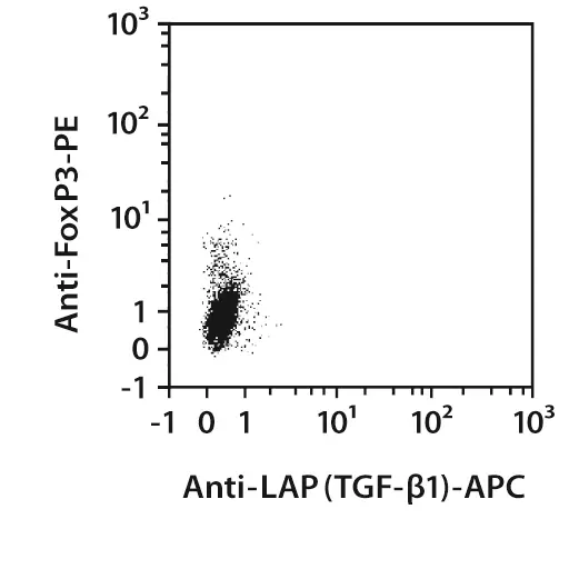
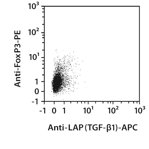
Human PBMCs were either left unstimulated (left image) or stimulated with Treg Expansion CD3/CD28 MACSiBead Particles for 48 hours. The cells were stained with Anti-LAP (TGF-β1) antibodies. LAP-TGF-β1–staining was controlled by incubating cells with unconjugated Anti-LAP (TGF-β1) antibodies prior to labeling with fluorochrome-conjugated antibodies. After that the cells were fixed, permeabilized, and intracellularly stained with Anti-FoxP3‑APC or -PE and analyzed by flow cytometry using the MACSQuant® Analyzer. Gating was performed according to the CD4 expression and side scatter properties of the cells. Cell debris was excluded from the analysis in an fluorescence 3 versus fluorescence 4 dot plot.
 view details
 view details
Specifications for LAP (TGF-β1) Antibody, anti-human
Overview
Clone CH6-17E5.1 reacts with the N-terminal latency-associated peptide (LAP) of the transforming growth factor β1 (TGF-β1) dimer. TGF-β1 belongs to a family of homologous, disulfide-linked, homodimeric proteins. These highly pleiotropic cytokines inhibit proliferation of most cells, but can promote the growth of mesenchymal cells and enhance extracellular matrix formation. The pivotal function of TGF-β1 in the immune system is to mediate immunosuppression and maintain tolerance by regulating lymphocyte proliferation, differentiation, and survival. TGF-β1 is produced by many cell types, but is reported to be most abundant in mammalian platelets and bone. It is secreted predominantly as an inactive latent complex. After proteolytical processing of the TGF-β1 precursor, the resulting N-terminal latency-associated peptide (LAP) remains non-covalently associated with the TGF-β1 dimer. Mature and biologically active TGF-β1 can be released from the complex by action of proteases and/or conformational changes. LAP-TGF-β1 was recently described to be expressed on the cell surface of activated human regulatory T cells.
TGFB1, CED, DPD1
Detailed product information
Technical specifications
Clone
CH6-17E5.1
Clonality
monoclonal
Isotype
mouse IgG1κ
Isotype control
Isotype Control Antibody, mouse IgG1
Host
mouse
Type of antibody
Primary antibodies
Species reactivity
human
Antigen
LAP (TGF-β1)
Molecular mass of antigen [kDa]
28
Distribution of antigen
platelets, bone
Entrez Gene ID
RRID
AB_2659987, AB_2857747, AB_2857760, AB_2659985, AB_2659984, AB_2659986
Resources for LAP (TGF-β1) Antibody, anti-human
Certificates
To access a Certificate of Analysis (CoA) follow the link below and enter the specific lot of your product.
Should you wish to request additional documentation, such as a Safety Data Sheet (SDS) in other languages than English or Swedish, an SDS-replacement document, or a Certificate of Origin (CoO), please direct your inquiry to your local technical support team.
 Powered by Bioz
You have previously chosen International as your location.
What do you want to do?