The updated CD4+CD62L+ T Cell Isolation Kit is the most recent development for the isolation of mouse CD4+CD62L+ T helper cells from spleen and lymph nodes. The isolation kit has been updated for faster cell isolation and increased purity, compared to previous kit (# 130-093-227).


Related products for CD4+CD62L+ T Cell Isolation Kit, mouse

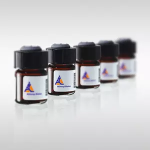
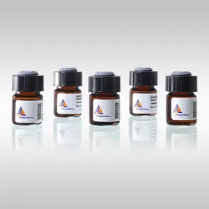

The updated CD4+CD62L+ T Cell Isolation Kit is the most recent development for the isolation of mouse CD4+CD62L+ T helper cells from spleen and lymph nodes. The isolation kit has been updated for faster cell isolation and increased purity, compared to previous kit (# 130-093-227).
This updated kit version comes with a fast two-step procedure and increased purity, compared to the previous kit (# 130-093-227). Benefit from our shortened protocol with reduced incubation times, less centrifugation and handling steps (total manual isolation time 55 minutes).
For a simple, fast and convenient way to isolate untouched and pure naive CD4+ T cells in as little as 18 minutes it is recommend to use the Naive CD4+ T Cell Isolation Kit, mouse (# 130-104-453).
CD62L (L-selectin) is highly expressed on naive T cells and down-regulated upon activation. It is also expressed on a small subset of memory T helper cells, the central memory T cells, which can be distinguished from naive T helper cells by their high expression of CD44. Furthermore, CD62L is expressed on most thymocytes, naive CD8+ T cells, B cells, dendritic cells, macrophages, NK cells, neutrophils, eosinophils, regulatory T cells, and TCRγ/δ+ T cells.
CD4+CD62L+ T helper cells isolated with the CD4+CD62L+ T Cell Isolation Kit can be used for the analysis of T cell activation by antigen-presenting cells, for studies on cytokine expression and receptor signaling, or for adoptive transfer experiments. Naive T cells, isolated with the CD4+CD62L+ T Cell Isolation Kit, have been used for in vitro polarization into Th1 and Th2 cells and were further analyzed for cytokine production after treatment with IL-33.1 In addition, isolated naive T cells were used in a study about the influence of the golli protein (an alternatively spliced product of the myelin basic protein gene) on the regulation of calcium influx in T cells. Isolated naive T cells were e.g. analyzed by calcium imaging as well as in patch clamp experiments.2
For the first magnetic separation (depletion): LS or autoMACS® Columns. For the second magnetic separation (positive selection): MS or autoMACS Columns.
CD4+CD62L+ T cells were isolated from a mouse spleen cell suspension using the CD4+CD62L+ T Cell Isolation Kit. The cells were fluorescently stained with CD45-VioGreen, CD4-VioBlue and CD62L-PE for detection of naive T cells (A) and with CD62L-PE and CD44-FITC for detection of central memory T cells (B). Additionally, cells were stained with CD25-APC to illustrate the excellent purity by removal of CD25+CD62L+ regulatory T cells (C). Analysis is performed for CD45+ cells. Cell debris and dead cells were excluded from the analysis based on scatter signals and PI fluorescence.
- Schmitz, J. et al. (2005) IL-33, an interleukin-1-like cytokine that signals via the IL-1 receptor-related protein ST2 and induces T helper type 2-associated cytokines. Immunity 23: 479-490
- Feng, J. M. et al. (2006) Golli protein negatively regulates store depletion-induced calcium influx in T cells. Immunity 24: 717-727
You have previously chosen International as your location.
What do you want to do?