Choose the right FFPE Tissue Dissociation Kit for your RNA and DNA workflows

Formalin-fixed paraffin-embedded (FFPE) tissues are an easily accessible but challenging resource for genomic profiling or transcriptomics. On this page you can explore our solutions for processing FFPE samples for RNA and DNA sequencing.

  • FFPE Tissue Dissociation Kit for RNA Profiling: Designed for RNA-based applications, such as single-nucleus RNA sequencing (snRNA-seq). Provides gentle and efficient nuclei isolation from FFPE material, preserving transcriptomic integrity of extracted nuclei and improving data quality across diverse tissue types.

  • FFPE Tissue Dissociation Kit (DNA workflow): Optimized for the preparation of FFPE samples for DNA-based analyses, including mutational profiling and targeted sequencing. During the process, epitopes are recovered for specific tumor cells, allowing higher sequencing accuracy and reduced false-positive variant cells.

Deep-dive into our two FFPE workflows and find the ideal kit for your application.

FFPE Tissue Dissociation Kit for RNA Profiling

FFPE tissue archives contain decades of clinical and research material, representing a valuable resource for molecular studies. These specimens preserve the cellular and molecular features of numerous diseases but are challenging to use due to the crosslinking and degradation caused by fixation. Extracting intact nuclei from FFPE material has therefore been a major limitation for transcriptomic analyses.

The development of snRNA-seq enables gene expression profiling at the nuclear level, providing access to transcriptomic information even from complex or partially degraded tissues. The FFPE Tissue Dissociation Kit for RNA Profiling, in combination with the gentleMACS® Octo Dissociator with Heaters, allows efficient, automated, and gentle extraction of nuclei from FFPE samples, supporting reliable snRNA-seq workflows, including the GEM-X Flex Gene Expression assay from 10× Genomics®. This approach makes archived specimens accessible for transcriptomics, facilitating studies, biomarker identification, and translational research.

Sample preparation

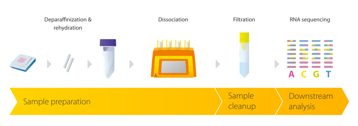
Prepare your 25–50 µm thick FFPE slices using a gentleMACS C Tube and the gentleMACS Octo Dissociator with Heaters through automated enzymatic and mechanical dissociation.

Infographic showing sample preparation of FFPE samples for RNA sequencing

Sample preparation

Prepare your 25–50 µm thick FFPE slices using a gentleMACS C Tube and the gentleMACS Octo Dissociator with Heaters through automated enzymatic and mechanical dissociation.

Sample cleanup

After dissociation, larger debris particles are removed using a 20 µm Pre-Separation Filter.

Infographic showing a tube with a strainer used for sample filtration

Sample cleanup

After dissociation, larger debris particles are removed using a 20 µm Pre-Separation Filter.

Downstream analysis

The isolated nuclei are then ready for downstream RNA sequencing workflows.

Infographic showing RNA sequencing results

Downstream analysis

The isolated nuclei are then ready for downstream RNA sequencing workflows.

Infographic showing a workflow for RNA analysis done after using the FFPE Tissue Dissociation Kit

Sample preparation

Prepare your 25–50 µm thick FFPE slices using a gentleMACS C Tube and the gentleMACS Octo Dissociator with Heaters through automated enzymatic and mechanical dissociation.

Infographic showing sample preparation of FFPE samples for RNA sequencing

Sample preparation

Prepare your 25–50 µm thick FFPE slices using a gentleMACS C Tube and the gentleMACS Octo Dissociator with Heaters through automated enzymatic and mechanical dissociation.

Sample cleanup

After dissociation, larger debris particles are removed using a 20 µm Pre-Separation Filter.

Infographic showing a tube with a strainer used for sample filtration

Sample cleanup

After dissociation, larger debris particles are removed using a 20 µm Pre-Separation Filter.

Downstream analysis

The isolated nuclei are then ready for downstream RNA sequencing workflows.

Infographic showing RNA sequencing results

Downstream analysis

The isolated nuclei are then ready for downstream RNA sequencing workflows.

Infographic showing improved snRNA-seq results using the FFPE tissue dissociation kit
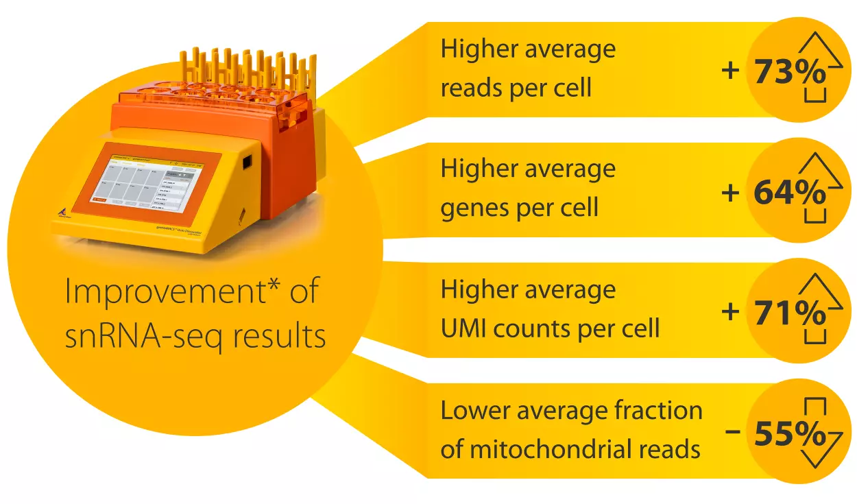
*Results from five independent experiments using 10× Genomics GEM-X Flex Gene Expression assay.**

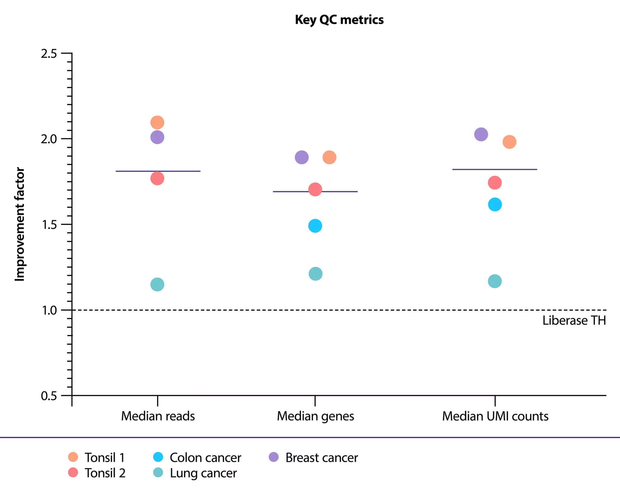
Accurate snRNA-seq from FFPE tissue requires stringent quality control. Internal and external validations across different tissue types revealed that the FFPE Tissue Dissociation Kit for RNA Profiling improves key QC parameters – including median reads, median genes, and UMI counts per cell – by an average factor of more than 1.5 over the use of Liberase™ TH Research Grade (Roche®).**

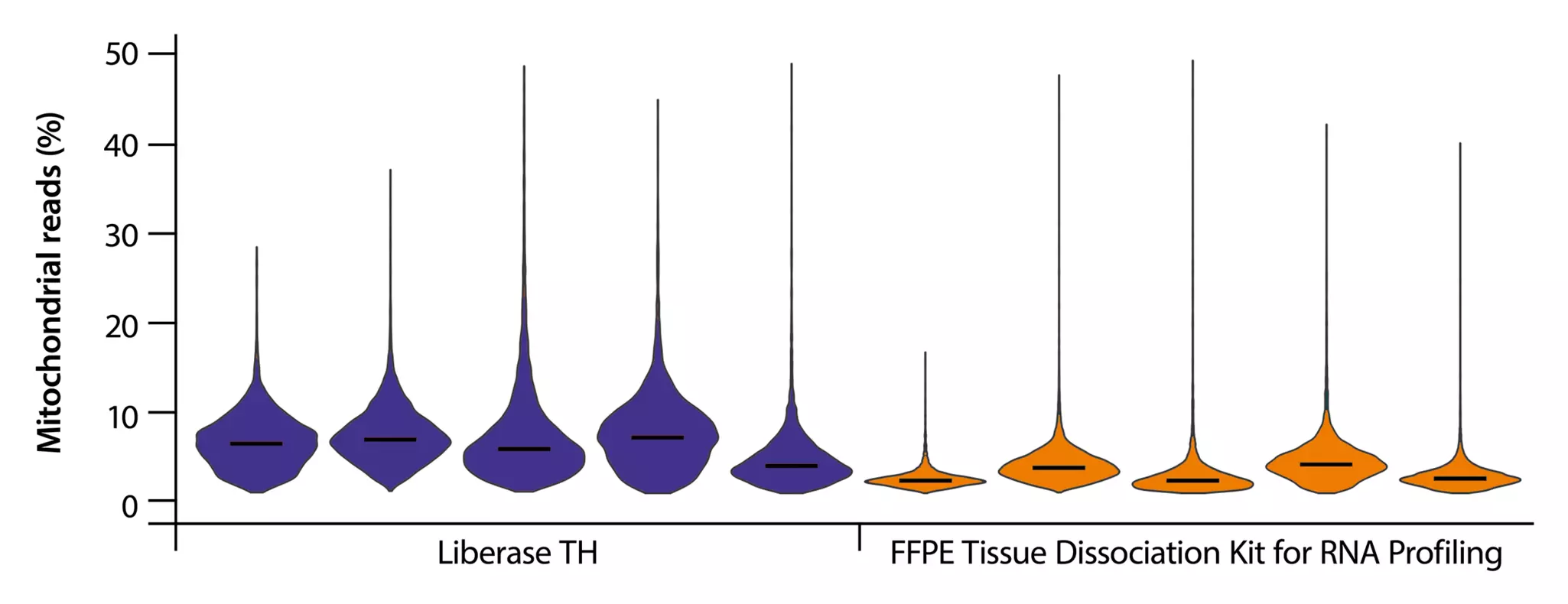
In addition, the kit significantly reduces the amount of mitochondrial mRNA contamination. This reduces background noise and ensures a higher proportion of informative, confidently mapped nuclear transcripts, providing robust, reproducible sequencing results.

A plot is shown on the factor of improvment in QC metrics for snRNA-seq using the FFPE Tissue Dissociation Kit for RNA Profiling normalized to Liberase TH.
Sequencing QC metrics comparison.

Factor of improvement in QC metrics for snRNA-seq using the FFPE Tissue Dissociation Kit for RNA Profiling (circles) normalized to Liberase TH (dotted line) on median reads, median genes, and median UMI counts per cell across three FFPE tumor samples (breast, colon, and lung cancer) and two healthy FFPE tonsil tissues.

Comparison of mitochondrial read percentage actross five different tissue sampels of different donors are shown, that were either processed with the FFPE Tissue Dissociaton Kit for RNA Profiling for Liberase TH.
Comparison of mitochondrial read percentages across five different tissue samples of different donors processed with either FFPE Tissue Dissociation Kit for RNA Profiling or Liberase TH.

Comparison of mitochondrial read percentages across five different tissue samples of different donors processed with either FFPE Tissue Dissociation Kit for RNA Profiling or Liberase TH. Reduction in mitochondrial read content when using the FFPE Tissue Dissociation Kit for RNA Profiling highlights the impact of dissociation methods on data quality.

** Comparison was performed against Liberase TH Research Grade which is used in an established tissue dissociation workflow for FFPE samples, as described in the demonstrated protocol CG000784 | Rev B, 10× Genomics: https://cdn.10xgenomics.com/image/upload/v1744998411/support-documents/CG000784_DemonstratedProtocol_SamplePrep_from__FFPETissueSections_GEM-X_Flex_GeneExpression_Rev_B.pdf (accessed May 2026)

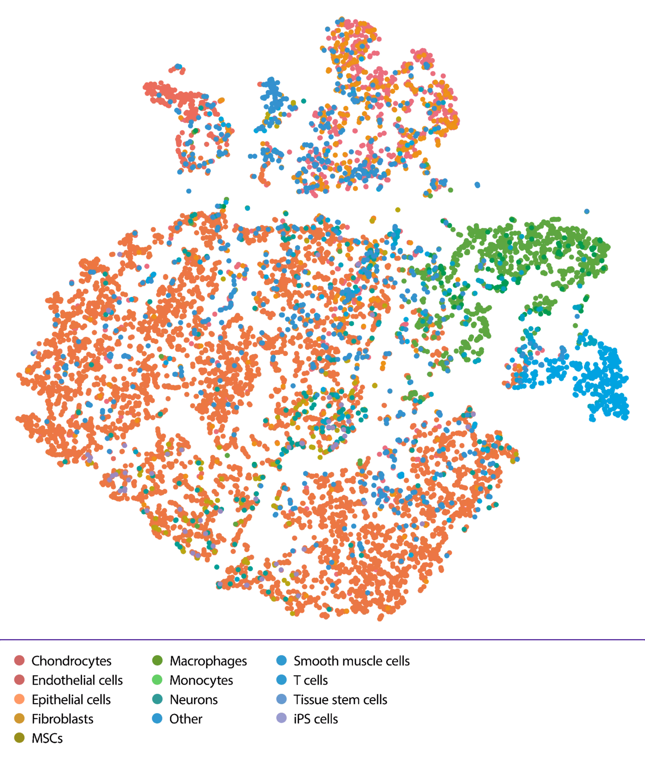
Exemplary t-SNE plots displaying cell type annotations of a breast cancer tumor sample, such as Epithelial cells or T cells.
Exemplary t-SNE plot displaying cell type annotations of a breast cancer sample.

Improved data quality is evident in downstream analyses. UMAP and t-SNE projections show clearer spatial separation of annotated cell populations, enabling the identification and characterization of even rare cell types with higher confidence.

FFPE Tissue Dissociation Kit for DNA analysis

Mutational profiling of FFPE samples with low tumor cell content or diffuse tumor morphology remains a major limitation in modern tumor diagnostics. Laboratories without microscope-assisted laser microdissection lack the means to selectively isolate tumor cells. As a result, analyses either require deep sequencing, which consumes significant resources, or outsourcing to specialized facilities, leading to extended turnaround times.

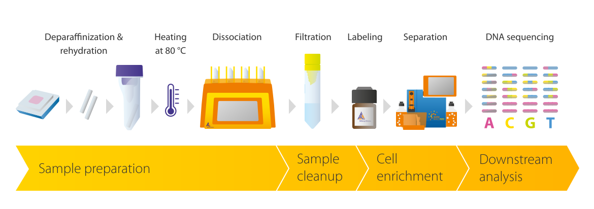
The presented workflow for the FFPE Tissue Dissociation Kit provides an optimized solution for FFPE tissue processing in standard laboratory settings. Following deparaffinization and rehydration, the gentleMACS Octo Dissociator with Heaters, in combination with the FFPE Tissue Dissociation Kit, gently generates single-cell suspensions with intact epitopes. These suspensions can then be used for subsequent cell isolation with Anti-Cytokeratin MicroBeads, human, utilizing MACS® Cell Separation Technology or flow sorting. This workflow enables efficient tumor cell enrichment without the need for specialized microdissection equipment. Even diffuse tumors, which are difficult to enrich by laser microdissection, can be readily processed for reliable DNA sequencing.

Sample preparation

FFPE tissue sections (25–50 µm thick) are cut from an FFPE tissue block and transferred to a gentleMACS C Tube. They are then dissociated using a gentleMACS Octo Dissociator with Heaters.

Infographic showing sample preparation of FFPE samples for DNA sequencing

Sample preparation

FFPE tissue sections (25–50 µm thick) are cut from an FFPE tissue block and transferred to a gentleMACS C Tube. They are then dissociated using a gentleMACS Octo Dissociator with Heaters.

Sample cleanup

Once the automated dissociation process is complete, larger debris particles are removed using a 20 µm Pre-Separation Filter.

Infographic showing a tube with a strainer used for sample filtration

Sample cleanup

Once the automated dissociation process is complete, larger debris particles are removed using a 20 µm Pre-Separation Filter.

Tumor cell enrichment

Tumor cells can subsequently be enriched using MACS Cell Separation Technology.

Infographic showing cell enrichment with MACS technology

Tumor cell enrichment

Tumor cells can subsequently be enriched using MACS Cell Separation Technology.

Downstream analysis

Samples can then be processed downstream, e.g., for DNA sequencing.

Infographic showing sequencing results

Downstream analysis

Samples can then be processed downstream, e.g., for DNA sequencing.

Infographic showing a workflow for DNA analysis done after using the FFPE Tissue Dissociation Kit

Sample preparation

FFPE tissue sections (25–50 µm thick) are cut from an FFPE tissue block and transferred to a gentleMACS C Tube. They are then dissociated using a gentleMACS Octo Dissociator with Heaters.

Infographic showing sample preparation of FFPE samples for DNA sequencing

Sample preparation

FFPE tissue sections (25–50 µm thick) are cut from an FFPE tissue block and transferred to a gentleMACS C Tube. They are then dissociated using a gentleMACS Octo Dissociator with Heaters.

Sample cleanup

Once the automated dissociation process is complete, larger debris particles are removed using a 20 µm Pre-Separation Filter.

Infographic showing a tube with a strainer used for sample filtration

Sample cleanup

Once the automated dissociation process is complete, larger debris particles are removed using a 20 µm Pre-Separation Filter.

Tumor cell enrichment

Tumor cells can subsequently be enriched using MACS Cell Separation Technology.

Infographic showing cell enrichment with MACS technology

Tumor cell enrichment

Tumor cells can subsequently be enriched using MACS Cell Separation Technology.

Downstream analysis

Samples can then be processed downstream, e.g., for DNA sequencing.

Infographic showing sequencing results

Downstream analysis

Samples can then be processed downstream, e.g., for DNA sequencing.

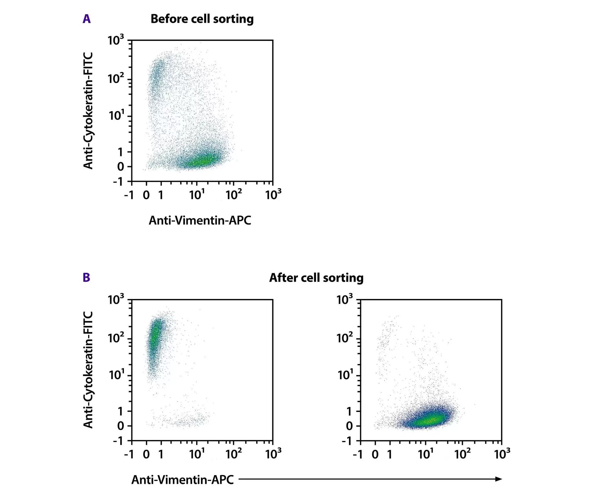
Flow cytometry analysis of cytokeratin-positive and vimentin-positive cells.

Increase sensitivity for your DNA analysis

After enzymatic dissociation of FFPE tissue sections, cell-surface epitopes such as cytokeratins and vimentins are recovered. These epitopes can then be used for flow cytometric sorting or MACS Technology. This allows distinction between carcinoma cells, stromal cells, and tumor-infiltrating lymphocytes (TILs) based on cytoskeletal marker expression. Cytokeratin is expressed in epithelial-derived carcinoma cells, while vimentin is expressed in stromal cells and TILs. This marker-based approach enables precise cell enrichment and improves the accuracy of downstream molecular analyses.

After isolation of tumor cells using cytokeratin-based selection, the enriched tumor cell fraction can be directly used for DNA sequencing.

Sequencing of tumor cells previously enriched using cytokeratin-based selection reveals several advantages over a macrodissection approach. Higher tumor cell purity increases sequencing accuracy, allowing the identification of relevant somatic mutations even for samples with a low tumor cell count.

This approach enhances sensitivity without requiring greater sequencing depth, thereby optimizing resource use by focusing analysis on the relevant target cells.

Sequencing results from DNA extracted after macrodissection or from enriched tumor cells:

Specimen IDPre-processing prior DNA extractionConcentration of extracted DNA (ng/μL)DNA input for NGSDriver mutation identifiedCoverage
Allele frequency
Specimen 1 (70% tumor cells)
Macrodissection
48.8040EGFR: c.2236_2250del; p.E746_A750del147152.3%
Enriched tumor cells38.0040EGFR: c.2236_2250del; p.E746_A750del252259.9%
Specimen 2 (10% tumor cells)Macrodissection
4.7820KRAS: c.34G>T; p.G12C38811%
Enriched tumor cells0.4620KRAS: c.34G>T; p.G12C62328%
Specimen 3 (< 5% tumor cells)
Macrodissection
5.6440KRAS: c.34G>T; p.G12C4051%
Enriched tumor cells2.1240KRAS: c.34G>T; p.G12C9517%

Application note

Sensitive genetic analysis of enriched cell populations from FFPE carcinoma samples by NGS

In this application note, we describe a complete workflow for the efficient isolation of tumor cells from FFPE samples using reagents and instruments from Miltenyi Biotec.
Application note

Enrichment of tumor cells from FFPE lung adenocarcinoma resection specimens increases sensitivity of mutation analysis

A complete workflow to enrich tumor cells from bulk FFPE NSCLC sections to improve NGS-based cancer diagnostics is described within this application note.

Reserve your demo spot

A person working in the lab putting a gentleMACS Heater on the gentleMACS Octo Dissociator with Heaters, a MACS Multistand, a MACSmix Tube Rotator and a tube rack with tubes are next on the lab bench

Experience the gentleMACS Octo Dissociator with Heaters in action with a demo tailored to your laboratory’s FFPE tissue preparation workflow.

Discover all products for FFPE tissue dissociation

5 products found

|

You have previously chosen International as your location.
What do you want to do?