Mouse spleen


1 Introduction
The spleen is the largest organ in the lymphatic system and supports both immune defense and blood filtration. It triggers immune responses to blood-borne antigens by producing white blood cells and by filtering the blood for old or damaged red blood cells, pathogens, and foreign material. These functions take place in two distinct regions: the red pulp and the white pulp, which differ in structure and cellular composition based on their roles.
The red pulp acts primarily as a blood filter and reservoir. It regulates the number of red blood cells in circulation and stores erythrocytes and platelets. Healthy erythrocytes are able to move through the spleen’s narrow internal passages, while older or damaged erythrocytes are trapped, broken down, and digested. This allows for the recycling of iron and the protein components of hemoglobin. In rodents, the red pulp is also a key site of hematopoiesis, meaning it supports the production of new blood cells.
The white pulp functions as an immune control center. It contains lymphocytes, macrophages, dendritic cells, and plasma cells, all of which work together to eliminate filtered pathogens and produce antibodies. During infections, the spleen may increase in size due to the rapid multiplication of white blood cells and heightened immune activity.
2 Cell types, frequencies, and marker expression
The spleen plays a major role in the lymphatic system, and as a result, it contains a wide range of blood cells, including erythrocytes, leukocytes, and platelets, as well as dead cells and cellular debris. This cellular diversity makes the spleen an important tissue for analyzing immune cell subpopulations and the functional mechanisms that shape immune reactivity.
At a glance: Cell types in spleen tissue
| Cell type or subset | Frequency (%) of total cells | Markers | Function |
| T cells | 21–25 | CD90, CD3, CD4, CD8 | Adaptive immune response |
| B cells | 44–58 | CD19, CD45R/B220 | Antibody and cytokine secretion Antigen presentation |
| Monocytes | 3.5–5 | CD11b, CD115, CCR2, Ly6C | Migrate to the site of infection Differentiate into macrophages or dendritic cells |
| Granulocytes | 1–2 | CD11b, Ly6C, SiglecF, CD49b, CD193 | Among first responders at the site of inflammation Pathogen destruction Recruitment of immune cells |
| Dendritic cells | 1–3 | CD11c, MHCII, XCR1, | Tissue-based sentinels that activate the immune response |
| Natural killer cells | 1–2 | NKp46, NK1.1 | Recognize and destroy infected cells without antigen presentation on MHC |
| Marcophages | 1–2 | F4/80 | Phagocytosis of cellular debris, microbes, cancer cells, etc. |
3 Sample preparation
At a glance: Kits, reagents, and hardware for the preparation of spleen samples
| Use | Comments | Product |
| Kits and reagents | ||
| Dissociation of spleen tissue | The gentlest way to generate single-cell suspensions while preserving cell surface epitopes yield and function using the gentleMACS® Dissociators and C Tubes. | Spleen Dissociation Kit, mouse |
| Hardware and consumables | ||
| Tissue dissociation | Preset programs to dissociate different tissues with Miltenyi Biotec enzyme kits for high cell yield and viability. | gentleMACS Octo Dissociator with Heaters |
| Tissue dissociation | Precision tool coupled with gentleMACS Dissociators that generates optimal shearing to efficiently disrupt tissue while keeping cells intact. | gentleMACS C Tube |
| Filtration | Smart design prevents clogging and enables one-step filtration with decreasing filter sizes while accommodating both 50 mL and 15 mL tubes | MACS SmartStrainer, 70 µm |
Lymphocytes can be efficiently released from spleen tissue using the gentleMACS Tissue Dissociator, followed by filtration through a MACS® SmartStrainer to remove remaining tissue fragments before cell isolation. Tissue-resident immune cells, including dendritic cells and macrophages, require a more thorough dissociation process that combines both mechanical disruption and enzymatic digestion to fully free embedded and infiltrating cells.
Crude digestion enzymes differ widely in their activity profiles. Certain enzymes can cleave cell surface epitopes, which can negatively affect downstream applications, including cell isolation, flow sorting, and flow cytometry analysis. Using validated and optimized enzymes, helps ensure reproducible and high-quality tissue dissociation, supporting reliable results in subsequent experiments.
A fast and gentle approach for generating high-quality single-cell suspensions from mouse spleen combines automated mechanical dissociation using the gentleMACS Dissociator and the gentleMACS C Tubes with the Spleen Dissociation Kit, mouse. This method consistently yields high numbers of viable leukocytes, including dendritic cells, and supports reproducible results for downstream applications, including flow cytometry and flow sorting analysis.
 MACS Handbook:
 Related videos:
4 StraightFrom® Spleen Kits: Combine tissue dissociation and cell separation into one simple step
At a glance: Kits, reagents, and hardware for the tissue dissociation and cell separation with StraightFrom Spleen Kits
| Use | Comments | Products |
| Kits and reagents | ||
| Cell separation | Negative isolation of lymphocyte subsets directly from spleen | StraightFrom Spleen Isolation Kits |
| Positive isolation of lymphocyte subsets directly from spleen | StraightFrom Spleen MicroBead Kits | |
| Instruments and consumables | ||
| Tissue dissociation | Preset programs to dissociate and label cells from spleen tissue with our StraightFrom Spleen MicroBead and Isolation Kits | gentleMACS Octo Dissociator with Heaters |
| Cell separation | Fully automated cell separation of target cells after dissociation | autoMACS® NEO Separator |
| Semi-automated cell separation of target cells after dissociation | MultiMACS™ Cell 24 Separator Plus | |
| Manual cell separation of target cells after dissociation | MACS Columns | |
| Filtration | Smart design prevents clogging, enables one-step filtering with decreasing filter sizes, while accommodating 50 mL or 15 mL tubes. | MACS SmartStrainer, 70µm |
Our StraightFrom Spleen Kits combine mechanical tissue dissociation, enzymatic digestion, and magnetic labeling in a single format, reducing sample processing time by up to 30 minutes per isolation compared with conventional protocols.
The kits are designed to preserve primary cells in a naive and unstimulated state, maintaining responsiveness to activation, proliferative capacity, and surface marker integrity. They support both positive and negative isolation strategies. StraightFrom Spleen MicroBeads enable positive selection by directly labeling target cells, while StraightFrom Spleen Isolation Kits use negative isolation to remove unwanted cells and leave the target population untouched and label free for downstream applications.
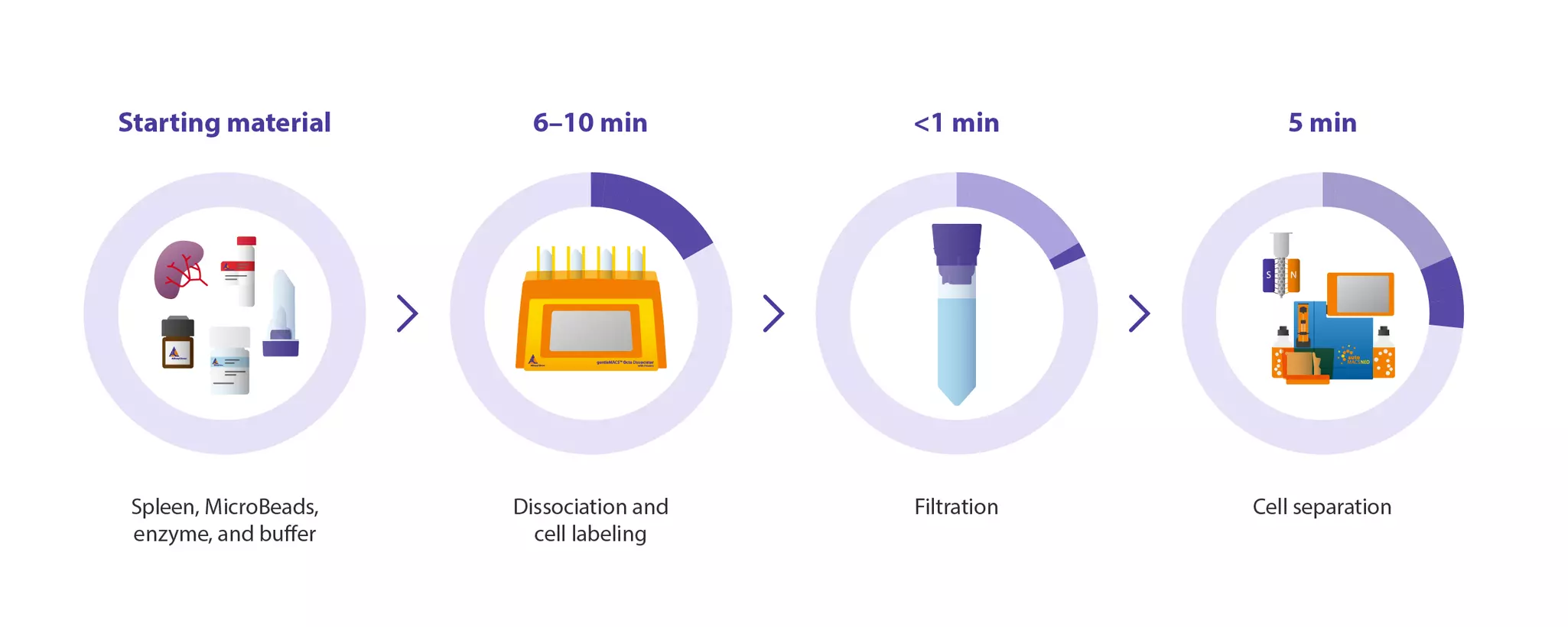
All dissociation and labeling steps take place in a single gentleMACS C Tube using the gentleMACS Octo Dissociator with Heaters, producing a single-cell suspension from mouse spleen in as little as 15 minutes. The workflow eliminates the need for cell counting, erythrocyte lysis, and centrifugation, requiring only one filtration step with a Pre-Separation Filter before manual or automated cell separation. This makes it easy to integrate into existing workflows.
The overview below shows the general workflow, where the starting materials depends on the selected isolation strategy. StraightFrom Spleen MicroBeads for positive isolation require up to 10 minutes for dissociation and labeling, while StraightFrom Spleen Isolation Kits for negative isolation take about 6 minutes. The final separation step can be performed manually or automated to fit your workflow needs.


You have previously chosen International as your location.
What do you want to do?