

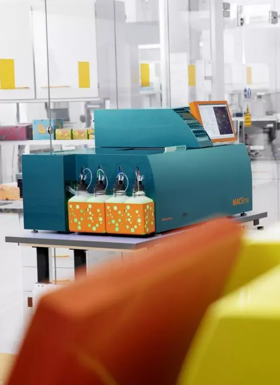


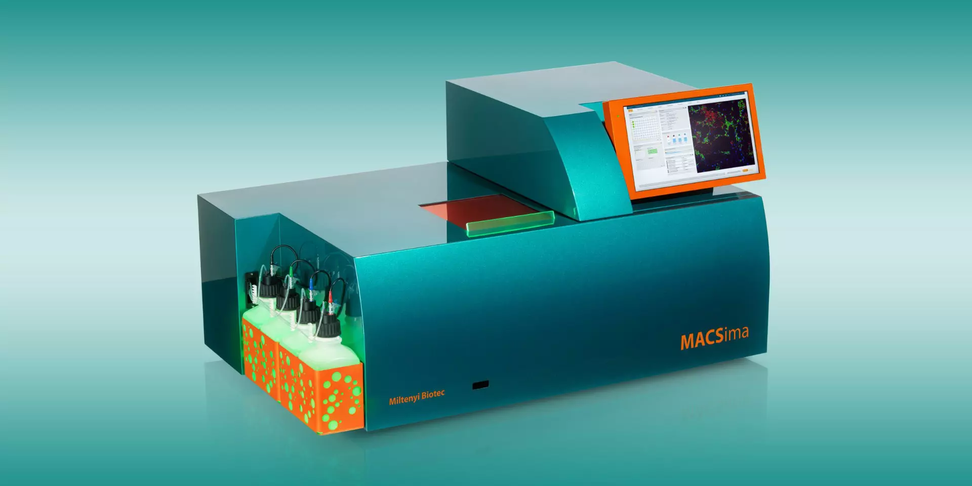
| Microscope | |
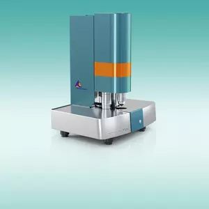
| Instrument type | Widefield epifluorescence microscope |
| Camera | Equipped with a latest-generation sCMOS camera with high resolution and thermoelectrically cooled sensor(15 megapixel, large 25 mm diagonal field of view) |
| Excitation | Six high-power excitation LEDs (filters: 386/23 nm, 420/10 nm, 470/40 nm, 531/46 nm, 628/32 nm, 725/40 nm) |
| Emission | Five emission filters (470/40 nm, 530/43 nm, 580/25 nm, 698/70 nm, 809/81 nm) |
| Autofocus | Dual approach of hardware- and image-based autofocus mechanisms |
| Objectives | • 2× objective to generate overview images; NA 0.1 • 20× long working distance objective (designed for 1 mm thick slides); NA 0.45 • 20× objective with high numerical aperture (designed for 170 μm thick cover glass); NA 0.75 |
| Bleaching unit | • Separate bleaching unit for optimized signal erasure • Illuminated area: 3 mm × 3 mm • Light intensity: 2 W |
| Liquid handling system | |
| Needle arm | Robotic needle arm allowing fully automated liquid transfer |
| Steel needle | Washable stainless steel needle with minimized spillover |
| Syringe pump | Syringe pump drive for accurate volumetric staining of samples |
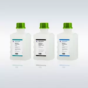
| Fluid containers | 1.5 L fluid containers for automatic operation over several days without the need to exchange containers |
| Sample and reagent stage | |
| Automated stage | Stage with sub-micron positioning accuracy of 100 nm (automated image registration for different cycles) |
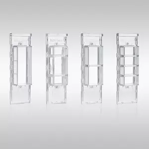
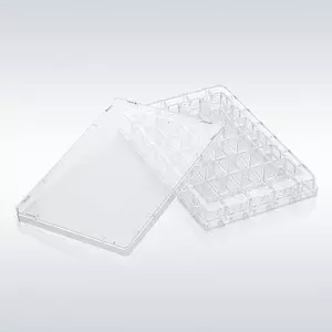
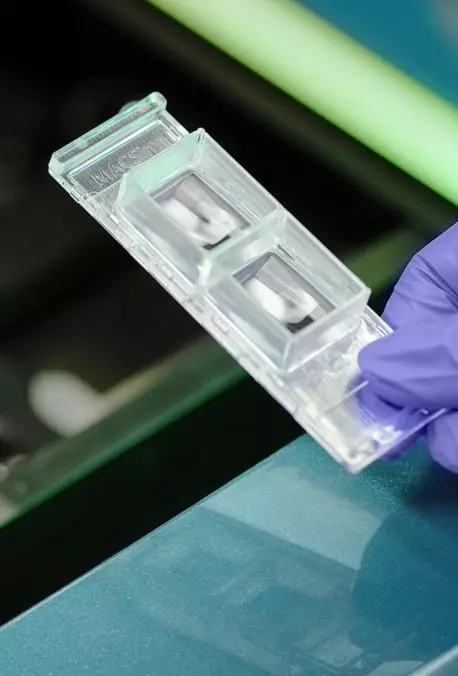
| Automated sample and reagent carrier positioning | Convenient loading and clamping of all supported sample carriers and reagents |
| Technical data | |
| Computer | Two integrated computers for experiment planning/execution and image analysis |
| Monitors | • Tiltable integrated touch display (Full HD) for PC1 • External 4K monitor for PC2 |
| Storage/ports | • 25 TB integrated storage to store data of several experiments • 2× USB 3.0 • HDMI port |
| Network | 1× 10 GbE 1× RJ45 GbE/10GbE |
| Power requirement and consumption | 100–240 V, 50/60 Hz, max. 7/13A, max. 1,300 W |
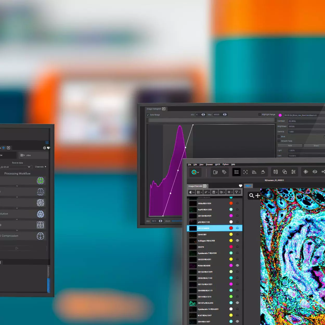
| MACS iQ View Software | |
| Control software | • Comprehensive sample and reagent management system • Easy planning and design of both simple and complex experiments • User-friendly execution and monitoring of experiments |
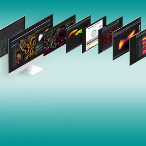
| Analysis software | • Easy display of hundreds of markers from high-dimensional datasets • Fast and flexible segmentation • Interactive gating • Unbiased data analysis (k-means clustering, UMAP and t-SNE calculation) • Multiple plotting options (histogram, scatter plot, strip and violin plots, heatmaps) • Workflow editor • Distance analyses |
| Size and weight | |
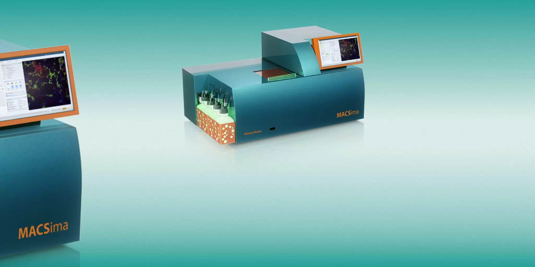
| Dimensions | • Instrument dimension: 1,220 mm × 780 mm × 650 mm (w × d × h); footprint: 1,210 mm × 750 mm (w × d) • MACSima Table1: 1,250 mm × 765 mm × 813 mm (w × d × h) |
| Weight | • Instrument: 170 kg • MACSima Table1: 120 kg |
1 The MACSima System is delivered with a trolley table specifically designed to support the system.