
Understanding TILs and TIL therapy

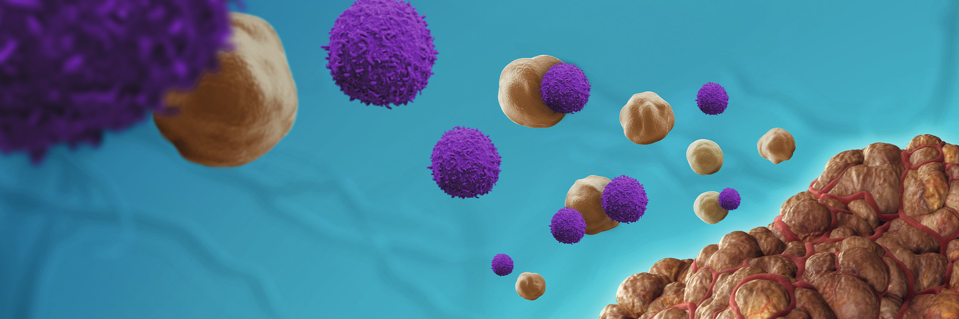
3D graphic showing tumor-infiltrating leukocytes (TILs) and a tumor
Logo

Understanding TILs and TIL therapy

Cancer treatment has undergone a significant transformation over the past decade, with immunotherapy emerging as one of the most promising avenues. Among the cutting-edge therapies, tumor-infiltrating lymphocytes (TILs) have gained attention for their ability to harness the body's immune system to fight cancer. This approach offers hope, especially for patients with advanced or resistant tumors. But what exactly are TILs, and how are they changing the landscape of cancer treatment?

In this post, we'll explore the science behind TILs, their role in cancer immunotherapy, and why they are considered a game-changer in the battle against cancer.

Read time: 5–10 minutes

This information is intended for biological researchers and should not be viewed as advice on the use of TILs in the treatment of cancer.

Check out our blog and get up to speed on the science and clinical promise of tumor-infiltrating lymphocytes (TILs) and TIL therapy

Content overview

Content

  • What are tumor-infiltrating lymphocytes (TILs)?
  • What are “hot” and “cold” tumors?
  • What are the different TIL subtypes and their markers?
  • How does TIL cancer treatment work?
  • What are some success stories and clinical trials of TIL therapy?
  • What challenges and limitations does TIL therapy currently face?
  • What new developments are there in TIL cancer treatment research?
What are tumor-infiltrating lymphocytes (TILs)?

TILs are a specific type of immune cell, primarily T cells, that are found naturally within tumors. These cells are part of the body's immune response and play a critical role in recognizing and attacking cancer cells. However, in many cancers, the tumor microenvironment suppresses the activity of TILs, allowing the cancer to evade the immune system. Despite this suppression, the presence of TILs in a tumor indicates that the immune system is trying to mount a defense against the cancer.

Pathologists often assess the percentage of the tumor area that is infiltrated by lymphocytes. There are scoring systems like the TIL Working Group or the International TILs Working Group recommendations, which suggest scoring TILs as a percentage of stromal area in the tumor. The scoring system for TILs is important because it helps assess the immune system's response to cancer by measuring how many immune cells are present within and around the tumor.

The TIL scoring system (often categorized as low, medium, or high infiltration) provides insight into:

  • Tumor immunogenicity – how visible or "foreign" the tumor appears to the immune system;
  • Immune activity – the extent to which the immune system is attempting to fight the tumor; and
  • Prognosis and treatment decisions – especially for immunotherapies like checkpoint inhibitors.

What are “hot” and “cold” tumors?

The terms "hot" and "cold" tumors describe the level of immune activity and level of immune cell infiltration within the tumor microenvironment (TME).1 This classification helps guide immunotherapy strategies, especially with immune checkpoint inhibitors.

  • Hot tumors are those that are highly infiltrated by immune cells, particularly T cells. These tumors already show signs that the immune system has recognized them as a threat and is attempting to fight them. They are characterized by:
    • High levels of immune cell infiltration.
    • High levels of immune signaling molecules, such as pro-inflammatory cytokines (e.g., IFN-γ), as well as the presence of PD-L1.
    • Often have a high mutational burden (many genetic mutations), which may make them more recognizable by the immune system.2
  • Cold tumors, on the other hand, are those with little to no immune cell infiltration. These tumors evade the immune system and do not provoke much of an immune response, making them more challenging to treat with immunotherapy. They are characterized by:3
    • Low levels of T cell infiltration.
    • Lack of immune system recognition.
    • Often have fewer mutations (lower mutational burden), making them harder for the immune system to detect.

What are the different TIL subtypes and their markers?

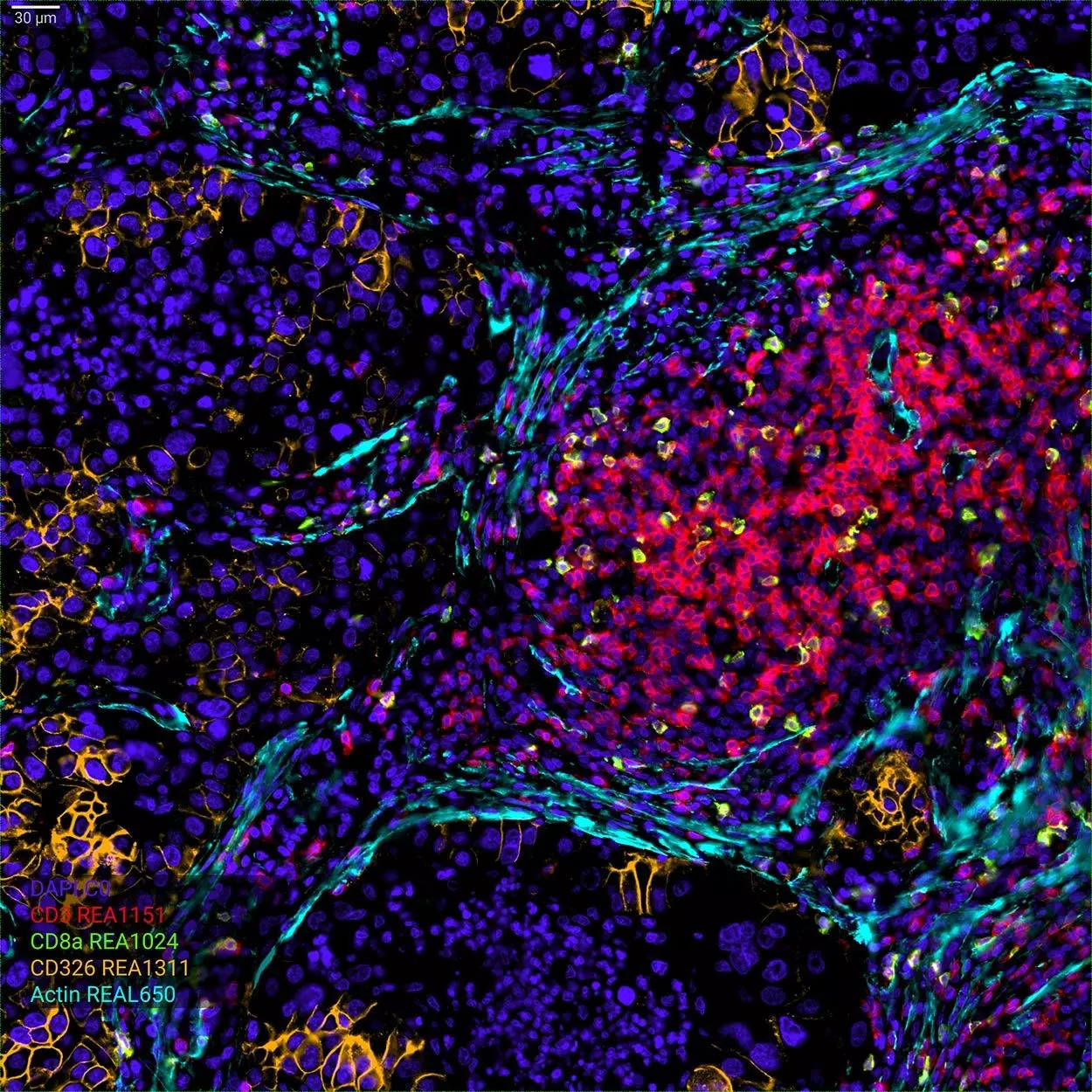
FFPE tissue section of a human donor with NSCLC stained for infiltrating CD3-positive and CD8a-positive T cells
FFPE tissue section a human donor with non-small cell lung cancer (NSCLC) stained for infiltrating CD3+ and CD8a+ T cells

Identifying and characterizing TILs is crucial for understanding their role in the immune response to tumors and for developing therapies like TIL-based immunotherapy. TILs are primarily composed of various subtypes of T cells, but other immune cells like B cells and NK (natural killer) cells can also be present in the tumor microenvironment.4

TIL subtype: T cells

The relative numbers of different types of TILs can vary significantly depending on the tumor type, its stage, and the immune microenvironment. Generally, the number of specific TILs is dynamic and reflects the ongoing immune response to the tumor. Typically, 20–40% of the TILs can be CD8+ T cells in tumors with high immune activity, but in tumors with a strong immune response (e.g., melanoma, ovarian cancer), the proportion may be even higher.5–7

CD4+ helper T cells play a key role in activating other immune cells, including CD8+ T cells, macrophages, and B cells. Their numbers may be lower compared to CD8+ T cells, but still significant in many immune-competent tumors.7,8

The proportion of Tregs among TILs can vary widely. In some cancers, Tregs can make up 20–50% of the TILs, particularly in advanced or metastatic tumors, where immune suppression is a key feature.9–11

Overview of the key markers for identifying different types of T cells TILs

 General markers for T cell TILsCD8⁺ cytotoxic T lymphocytes (CTLs)CD4⁺ helper T cellsRegulatory T cells (Tregs)Exhausted and dysfunctional TILs
MarkersCD45: A pan-leukocyte marker, used to identify all immune cells in the TME.

CD3: A common marker for T cells, expressed by both.

CD8⁺ and CD4⁺ T cells infiltrating tumors.

CD69: Activation marker, often found on early activated T cells in tissues.
CD8: Identifies cytotoxic T cells, key to tumor cell killing.

Granzyme B, Perforin:
Effector molecules involved in direct cytotoxicity.

NKG2D, T-bet:
Activating receptors and transcription factors for effector function.

CD103 (ITGAE):
enables cells to adhere to and remain within epithelial tumor tissues, enhancing their cancer-killing ability.
CD4: Helper T cells that assist other immune cells.

T-bet (for Th1), GATA3 (for Th2), RORγt (for Th17): Transcription factors indicating polarization.

IL-2, IFN-γ: Key cytokines involved in immune responses.
CD25, FoxP3: Master regulator of Tregs, critical for immune suppression.

CD4: Tregs are usually CD4.
PD-1, CTLA-4, TIM-3, LAG-3: Immune checkpoint molecules involved in suppressing anti-tumor immune responses, markers of T cell exhaustion.

CD39, CD73
: Adenosine-producing enzymes that contribute to immune suppression.
LocalizationTumor borders, stroma, and sometimes within tumor nests. TILs predominantly reside at the tumor periphery but can also localize within tumor nodules.Tumor core and periphery, often at sites of active cytotoxic killing.Tumor stroma and periphery, supporting CD8⁺ T cell activation and macrophage function.Predominantly in tumor stroma, where they suppress immune activation and facilitate tumor progression.Exhausted TILs tend to accumulate in tumor areas with low oxygen and nutrient supply, often in the tumor core or regions of active immune evasion.

TIL subtype: B cells, natural killer (NK) cells, macrophages, and dendritic cells (DCs)

Besides T cells, B cells, natural killer (NK) cells, macrophages, and dendritic cells (DCs) can also infiltrate tumors. In the table below, you will find an overview of markers for tumor-infiltrating B cells, NK cells, macrophages, and dendritic cells.

B cells infiltrate tumors and can contribute to the immune response through antibody production and antigen presentation. However, their numbers tend to be lower compared to T cells and usually account for 5–10% of the total TIL population. In tumors like Hodgkin lymphoma, melanoma, and some breast cancers, B cells can sometimes make up a higher proportion of TILs, particularly in the context of tertiary lymphoid structures (TLS).12–14

NK cells usually represent 5–10% of the TILs in many tumors. However, in tumors like melanoma and hepatocellular carcinoma, NK cell infiltration can be higher and contribute significantly to tumor surveillance.15–18

Tumor-associated macrophages (TAMs) are one of the most abundant immune cell types within the tumor microenvironment, especially in cancers with an immunosuppressive phenotype. They typically represent 30–50% of TILS, though this can vary depending on the tumor type and its immune microenvironment. TAMs tend to be more numerous in breast cancer, colon cancer, and non-small cell lung cancer.19

DCs usually represent 5–10% of the TILs, but their presence can increase in tumors with a high level of immune activation.20

Overview of markers for tumor-infiltrating B cells, NK cells, macrophages, and dendritic cells

 B cellsNatural Killer (NK)Tumor-associated macrophages (TAMs)Dendritic Cells (DCs)
MarkersCD19, CD20: general markers for B cells.

IgM, IgG: antibodies produced by B cells.

CD27:
a marker for memory B cells.
CD56: classic marker for NK cells.

NKp46, CD16: NK cell surface markers.

NKG2D, KIRs
: Receptors involved in tumor recognition.

Perforin, Granzyme B
: Cytotoxic molecules involved in tumor cell killing.
M1 Macrophages (anti-tumor): CD68, iNOS (inducible nitric oxide synthase), TNF-α (pro-inflammatory cytokine).

M2 Macrophages (pro-tumor): CD163, CD206, IL-10 (anti-inflammatory cytokine).
CD11c, HLA-DR: markers for mature dendritic cells.

CD80, CD86: co-stimulatory molecules for T cell activation.
LocalizationOften localized in peritumoral areas or form tertiary lymphoid structures (TLS), which resemble lymph nodes.Found in both the tumor core and periphery, playing a role in early tumor defense.Abundant in the tumor stroma and can be located in areas of necrosis or near blood vessels, where they help in tissue remodeling and immune suppression.Found in tumor stroma, peritumoral areas, and sometimes in tertiary lymphoid structures (TLS), where they interact with T cells and help activate them.

Icon showing a shining lightbulb with an exclamaiton mark on it

Did you know?

The “immune contexture” refers to the composition, density, functional state, and spatial organization of immune cells within a tissue or tumor. The presence of more T cells inside a tumor is often correlated with better patient outcomes in many cancers, such as melanoma and ovarian cancer.21

How does TIL cancer treatment work?

TIL cancer treatment involves extracting TILs from a patient’s tumor, expanding them in the lab to increase their numbers, and reactivating them to enhance their cancer-fighting ability. TIL cancer therapy involves several steps22:

  • Tumor biopsy and isolation: A sample of the patient's tumor is taken through a biopsy. This sample contains TILs, which have already been trying to fight the cancer.
  • Expansion in the lab: In the laboratory, the TILs are separated from the tumor and stimulated to grow. By exposing the cells to interleukin-2 (IL-2) and other growth factors, the number of TILs is dramatically expanded – sometimes into the billions.
  • Activation and modification: These TILs can be further engineered or activated to enhance their cancer-fighting capabilities. For instance, they may be optimized to target specific antigens present on the cancer cells.
  • Reinfusion into the patient: Once the TILs are grown and activated, they are reinfused into the patient in large numbers. Before this, patients often undergo a conditioning regimen (usually chemotherapy) to deplete their existing immune cells, which allows the newly infused TILs to function more effectively.
  • Ongoing support with IL-2: After reinfusion, patients may receive IL-2 to help the TILs survive and continue to attack cancer cells.

What are some success stories and clinical trials of TIL therapy?

TIL therapy has been most successful in the treatment of advanced melanoma, with some patients experiencing complete remission even after other treatments, such as chemotherapy or immune checkpoint inhibitors, have failed. Other cancers, including cervical and lung cancers, are also being tested with TIL therapy, with promising early outcomes.

Approved or late-stage clinical TIL therapy trials, April 2025.

 Lifileucel (LN-144)LN-145
StatusThe first FDA-approved TIL therapy, granted approval in February 2024.23Investigational, in clinical trials (phase II).
IndicationAdvanced metastatic melanoma.Cervical cancer and other solid tumors.
ManufacturerIovance Biotherapeutics.Iovance Biotherapeutics.
DescriptionLifileucel is a tumor-derived autologous T cell immunotherapy for adult patients with unresectable or metastatic melanoma previously treated with a PD-1 blocking antibody and, if BRAF V600 positive, a BRAF inhibitor with or without a MEK inhibitor.LN-145 is another TIL therapy under development, primarily for advanced cervical cancer but also being tested in other solid tumors, such as non-small cell lung cancer (NSCLC). Like lifileucel, LN-145 involves the extraction and expansion of a patient's own TILs, which are then reinfused to boost the anti-tumor immune response.
Regulatory progressApproved.LN-145 received Fast Track Designation from the FDA and is currently being investigated in a phase 2 study IOV-LUN-202 (NCT04614103) in patients with metastatic NSCLC and the IOV-COM-202 (NCT03645928) in patients with solid tumors.

Beyond Lifileucel and LN-145, various other TIL therapies are currently under investigation for different types of solid tumors, including breast cancer, colorectal cancer, ovarian cancer, and NSCLC. These therapies are still in early- to mid-stage clinical trials.

What challenges and limitations does TIL therapy currently face?

While TIL therapy holds great promise, it's not without challenges:

  • Complexity and cost: The process of isolating, expanding, and reinfusing TILs is labor-intensive and requires highly specialized facilities. As a result, it is expensive and not yet widely available.
  • Not effective for all patients: Although many patients have responded well, TIL therapy doesn’t work for everyone. Researchers are still trying to understand why some patients benefit more than others and how to enhance its efficacy.
  • Side effects: Like other immunotherapies, TIL therapy can cause side effects, particularly due to the immune system's activation. Common side effects include fever, low blood pressure, and in some cases, serious inflammatory reactions.

Icon publication
Research highlight

Read more about how the REAfinity® Antibodies, live/dead Viobility™ Dyes, and the MACSQuant® 10 Analyzer contribute to developing a non-invasive biomarker assay to determine the apoptotic cell fraction of tumor-infiltrating T cells.

Method for estimation of apoptotic cell fraction of cytotherapy using in vivo fluorine-19 magnetic resonance: pilot study in a patient with head and neck carcinoma receiving tumor-infiltrating lymphocytes labeled with perfluorocarbon nanoemulsion

Ahrens, ET. et al. (2023) J Immunother Cancer. 11(8);11(6):e007015. | Read article

Icon publication
Research highlight

Read more about how Miltenyi Biotec Interleukins, REAfinity Antibodies, and the MACSQuant 10 Analyzer contribute to developing patient-specific tumor-infiltrating T cells for colorectal cancer.

Generation of colon cancer-derived tumor-infiltrating T cells (TILs) for adoptive cell therapy
Albrecht, HC. et al. (2023) Cytotherapy. 25(5):537-547. | Read article

What new developments are there in TIL cancer treatment research?

With a future of more refined and accessible therapies, TILs are shaping up to be one of the most exciting developments in the fight against cancer. Recent advancements in TIL research have focused on enhancing TIL scoring methodologies and understanding the role of PD-1 expression on TILs to improve cancer treatment strategies.

Automated TIL scoring in breast cancer

Advancements in artificial intelligence have led to the development of automated pipelines for scoring TILs in breast cancer. These deep learning models can accurately quantify TILs in whole-slide images, providing a reliable and efficient method for assessing TILs in clinical settings.

Automated detection and scoring of tumor-infiltrating lymphocytes in breast cancer histopathology slides.
Yosofvand, M. et al. (2023) Cancers (Basel). 15(14):3635. | Read article

Prognostic value of TILs

TILs are recognized as crucial components in anti-tumor immunity. Their presence within tumors has been associated with favorable prognostic outcomes, serving as both prognostic and predictive biomarkers in various cancers. A meta-analysis concluded that high levels of TILs in TNBC are associated with better short-term and long-term prognoses. Specifically, elevated levels of CD4+, CD8+, and FOXP3+ TILs were predictive of favorable long-term outcomes.

Prognostic value of tumor-infiltrating lymphocytes in patients with triple-negative breast cancer: a systematic review and meta-analysis.
Gao, G. et.al. (2020) BMC Cancer. 20(1):179. | Read article

Similarly, a study utilizing deep learning on pathology images found that higher TIL densities correlate with better disease progression and overall survival in colorectal cancer patients, underscoring the prognostic value of TILs in this context.

Prognostic significance of tumor-infiltrating lymphocytes determined using LinkNet on colorectal cancer pathology images.
Liu, A. et al. (2023) JCO Precis Oncol. 7:e2200522. | Read article

PD-1 expression on TILs and therapeutic implications

The expression of programmed cell death protein 1 (PD-1) on TILs has been linked to patient outcomes. In a cohort study including various solid tumors, patients with PD-1–positive TILs exhibited longer progression-free survival (7.0 vs. 1.9 months) and overall survival (18.1 vs. 8.0 months) compared to those with PD-1–negative TILs following immune checkpoint blockade therapy.

Tumor Infiltrating Lymphocyte Expression of PD-1 Predicts Response to Anti-PD-1/PD-L1 Immunotherapy
Bevins, NJ. et al. (2022) J Immunother Precis Oncol. 5(4):90-97. | Read article

Similarly, research indicates higher PD-1 expression on TILs has been associated with increased TIL infiltration, higher TIL grading, while PD-L1 expression in tumors showed an inverse correlation with TIL grading in colorectal adenocarcinoma tumors, suggesting a more active immune microenvironment. This relationship underscores the potential of PD-1 as a biomarker for selecting patients likely to benefit from PD-1/PD-L1–targeted therapies.

The Potential of PD-1 and PD-L1 as Prognostic and Predictive Biomarkers in Colorectal Adenocarcinoma Based on TILs Grading
Rasyid, NR. et al. (2024) Curr Oncol. 31(12):7476-7493. | Read article

You have previously chosen International as your location.
What do you want to do?