Picture MACS iQ View Product Page Header

MACS® iQ View – 3D Large Volume

Light sheet microscopy image processing software – unlocking superior quality with just a few clicks

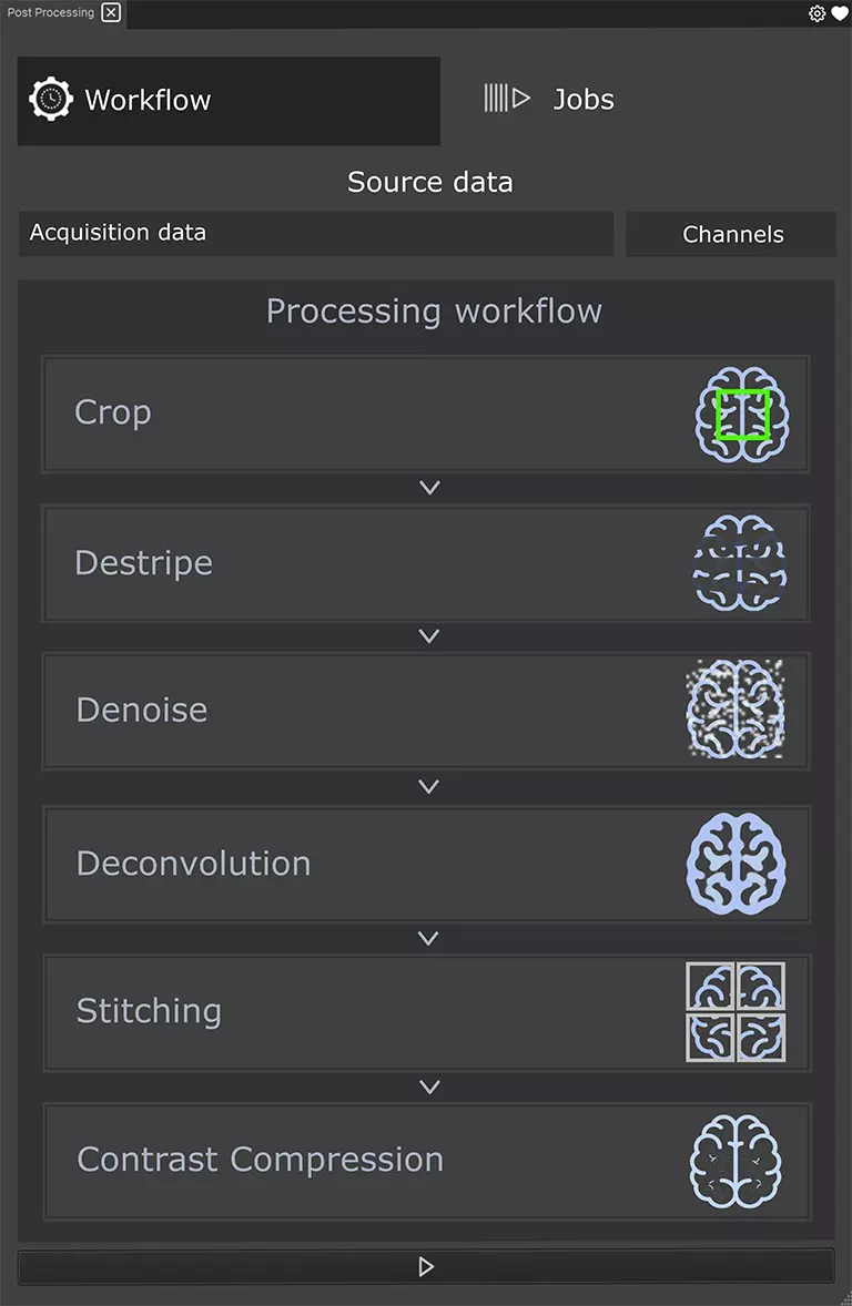
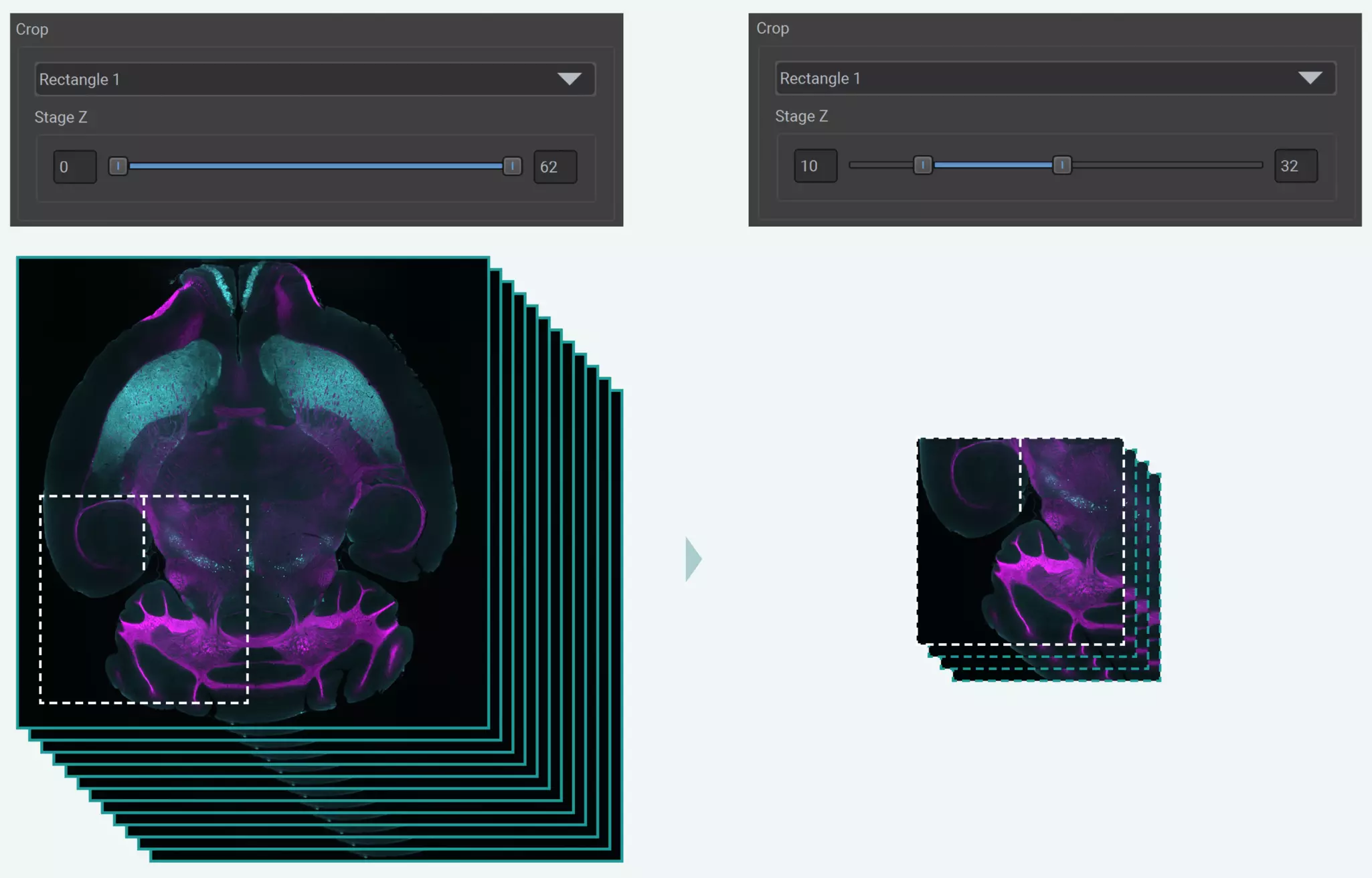
MACS iQ View – 3D Large Volume is a user-friendly, light sheet microscopy image-processing software tailored for managing, stitching, and processing large 3D datasets captured with the UltraMicroscope Platform – featuring UltraMicroscope Blaze™ and UltraMicroscope Choros™. It empowers you to gain meaningful insights and enhance image quality with ease. Its user-friendly interface simplifies navigation and operation, making it accessible to users of all skill levels. Its comprehensive toolkit supports a variety of tasks, including 3D cropping, destriping, denoising, deconvolution, stitching, and contrast compression.

icon

Benefits

Picture MACS iQ View PP Benefit Segmentation

Full automation from acquisition to results

By integrating with ImSpector, UltraMicroscope Platform’s acquisition software, MACS iQ View – 3D Large Volume automatically starts processing right after acquisition, with no manual steps needed.

Picture MACS iQ View PP Benefit Selection

Streamlined image processing workflow

Effortlessly manage and process images by selecting and arranging dedicated modules within the workflow.

Picture MACS iQ View PP Benefit Workflow

Effortless stitching with a single click

Simply click the stitching button and let the software automatically assemble your 3D mosaic stacks, eliminating the need for any manual step.

Picture MACS iQ View PP Benefit Plots

Tailored deconvolution

Enhance image quality and reveal hidden details with deconvolution, tailored to the specific optical characteristics of your UltraMicroscope.

Discover the capabilities of MACS iQ View – 3D Large Volume

MACS iQ View – 3D Large Volume is an essential light sheet microscopy image processing software for researchers seeking to unlock the full potential of their 3D imaging data with ease and efficiency. Unlock the hidden potential of your microscope images and gain deeper insights into biological processes. See here how MACS iQ View – 3D Large Volume elevates microscopy data to an unprecedented level of clarity.

Stitching

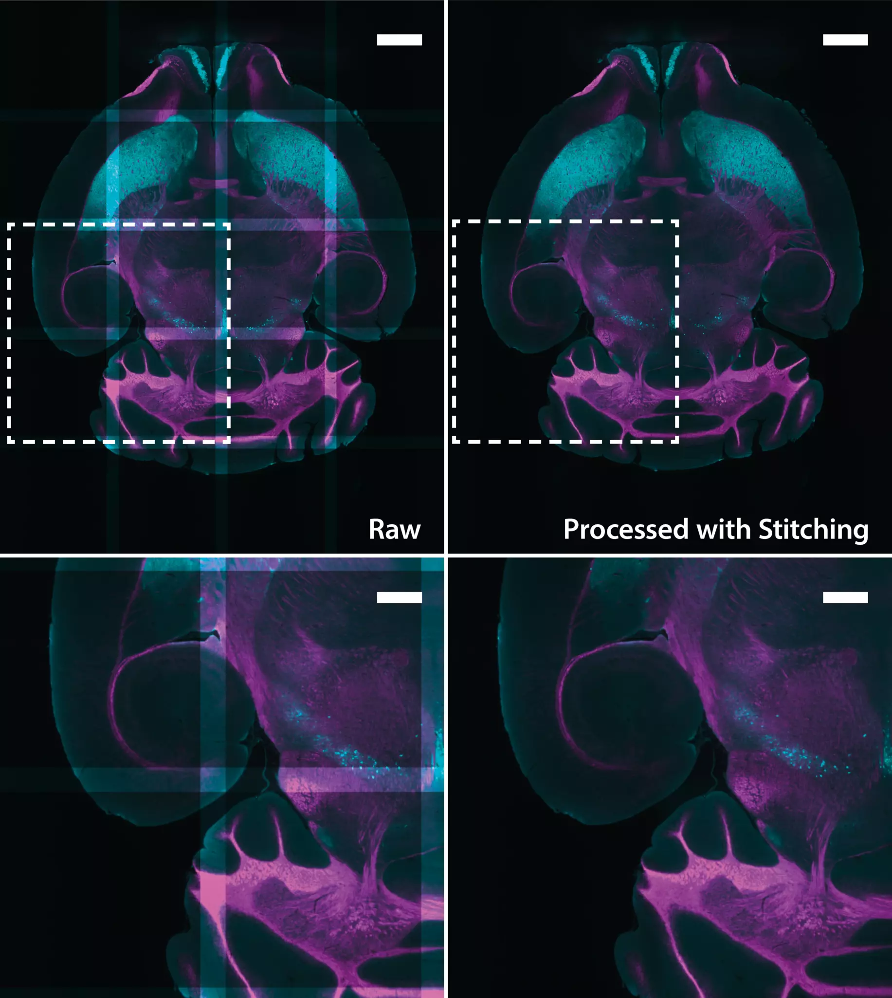
XY-plane of data from a mouse brain before (left) and after (right) processing with the Stitching module of MACS iQ View – 3D Large Volume. The brain was labeled with antibodies against myelin basic protein (magenta, MBP Antibody, anti-human/mouse, Vio® R667, REAfinity™) and Tyrosine hydroxylase (cyan, Tyrosine Hydroxylase Antibody, anti-human/mouse/rat, Vio® G570, REAfinity™), to visualize the myelin basic protein and dopaminergic neurons, respectively.

Destripe, Deconvolution, and Contrast Compression

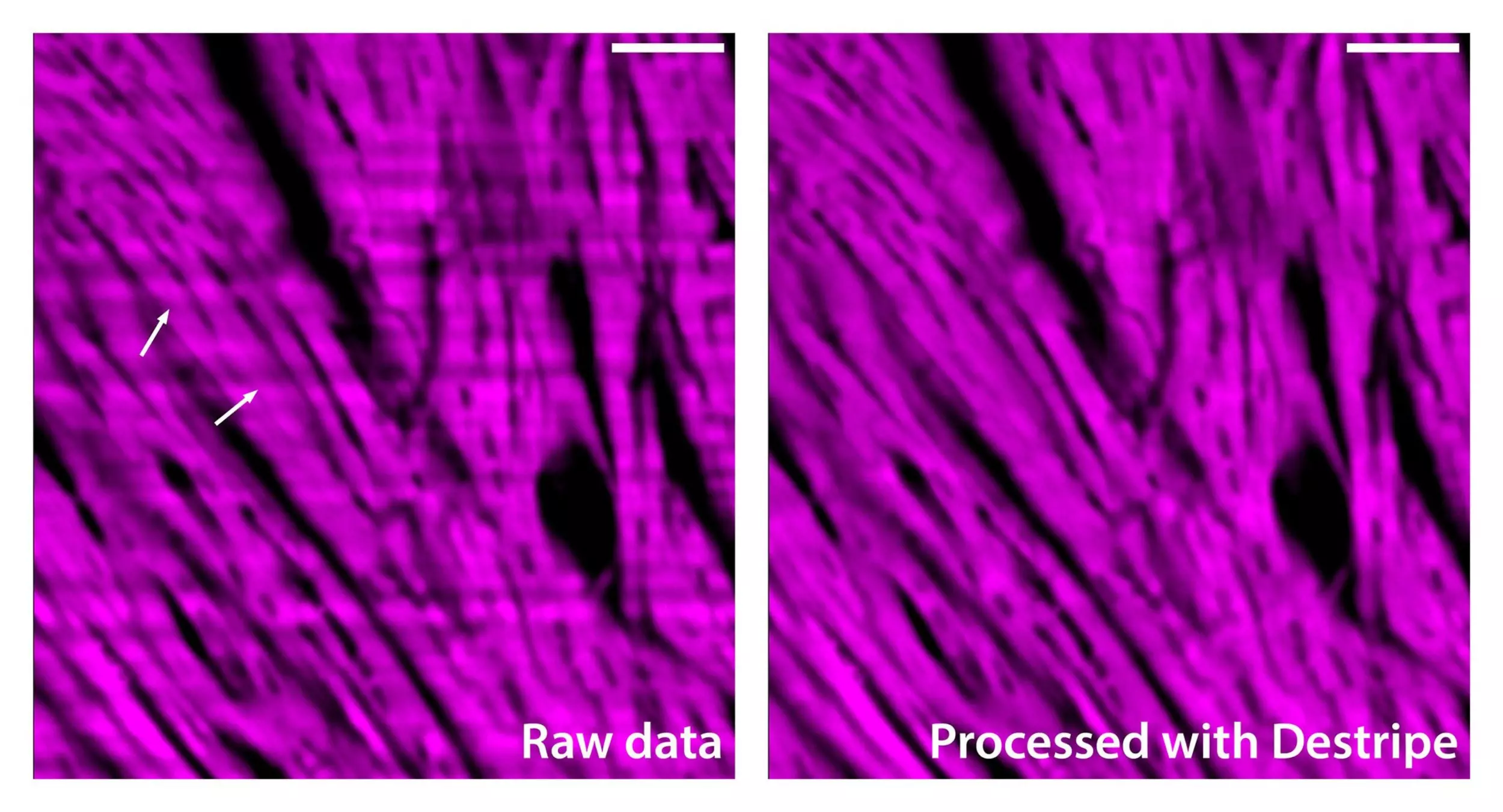
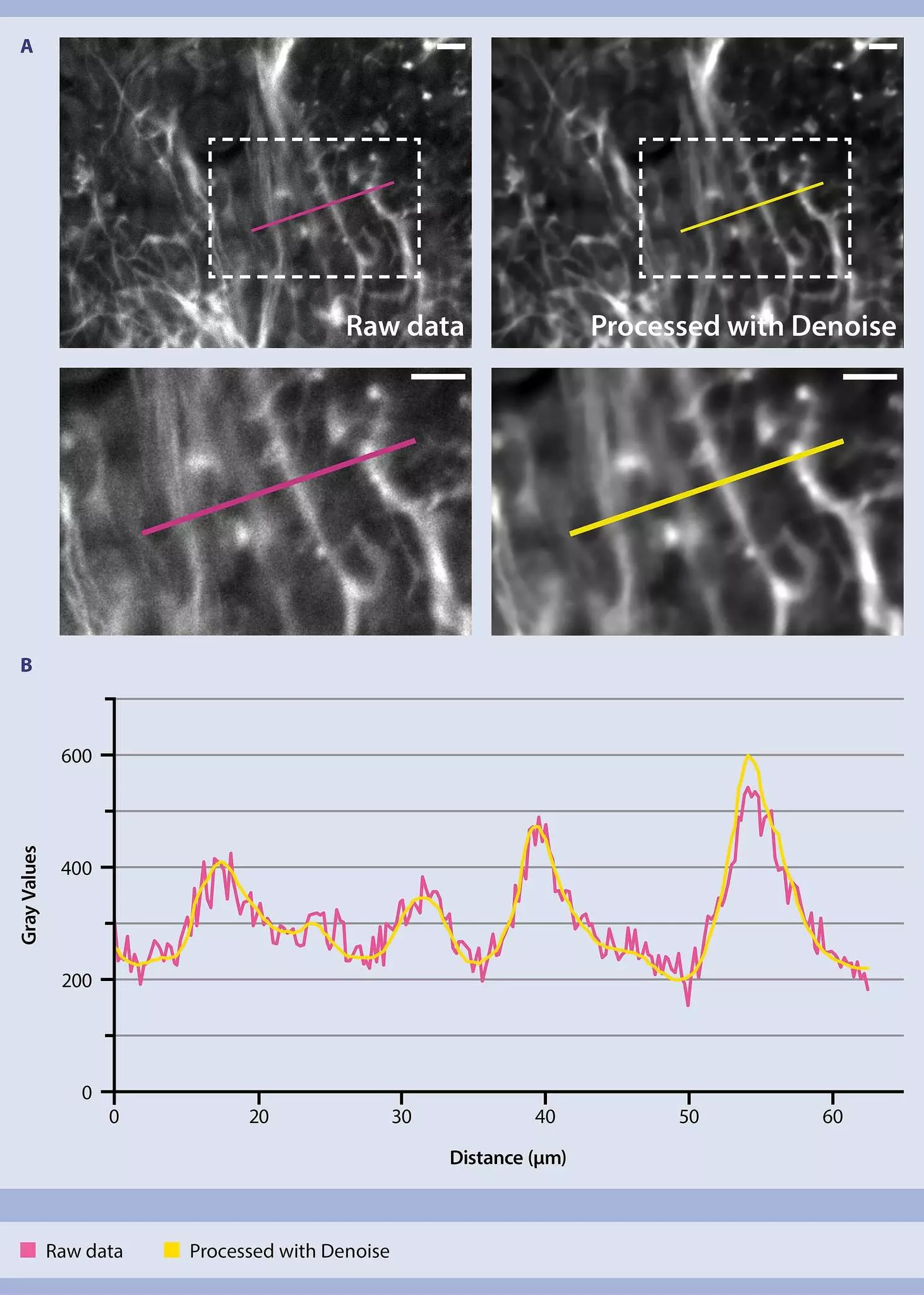
XY-plane of data from dopaminergic neurons in the whole mouse brain (cyan, dopaminergic neurons labeled with Tyrosine Hydroxylase Antibody, anti-human/mouse/rat, Vio® G570, REAfinity™) before (left) and after (right) processing with MACS iQ View – 3D Large Volume. The image was processed with Destripe (standard setting and 20-pixel stripe size) to remove striping artifacts, Deconvolution (subtle setting) to enhance resolution), and Contrast Compression to visualize both dim and bright structures at the same time (Structure mode).

Destripe and Deconvolution

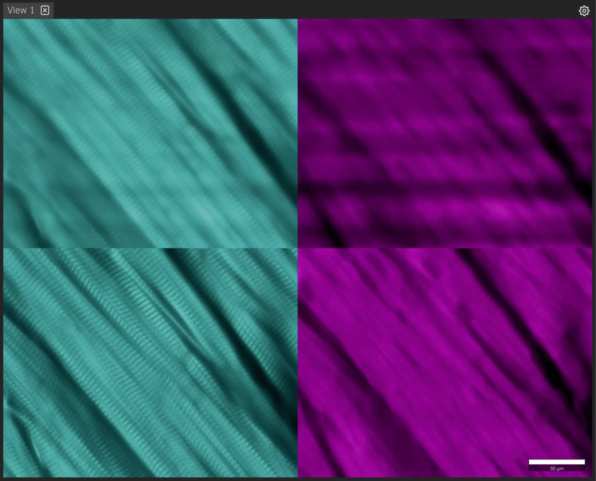
XY-plane of data from a mouse heart labeled with an antibody against murine cardiac troponin T (cyan, Cardiac Troponin T Antibody, anti-human/mouse/rat, Vio®R667 , REAfinity™) before (left) and after (right) processing with MACS iQ View – 3D Large Volume. The data was processed with Destripe (standard setting and 25 pixel stripe size) to remove striping artifacts and Deconvolution (subtle setting) to improve resolution. The image reveals cardiomyocytes and the presence of Troponin T in the cardiac sarcomeres.

3D visualization of Deconvolution

This video showcases a stunning 3D visualization of nucleoli in a murine kidney, acquired using the UltraMicroscope Blaze and processed through deconvolution with the MACS iQ View – 3D Large Volume software to enhance image clarity and enable detailed visualization of subcellular structures such as nuclei and nucleoli. The final visualization was rendered in Imaris.

icon

Features

MACS iQ View – 3D Large Volume is a great new tool that enables us to quickly transition from raw images to 3D reconstructions. Its processing functions, particularly those specific for light-sheet images like stripe artifact removal, simplify the segmentation of images for quantitative analysis. Additionally, stitching works very well and very fast, even when handling large files.

Portrait of a light sheet microscopy researcher Prof. Dr. Bettina Weigelin
icon

License options

License options

MACS iQ View – 3D Large Volume is dedicated to processing images acquired with the UltraMicroscope Platform.We offer permanent licenses with updates and hotfixes included. These licenses are the perfect solution for long-term projects, assuring that you have unrestricted access to your software and all your valuable settings.
Request further information
icon

Related instruments

The UltraMicroscope Blaze for next level light sheet imaging

The UltraMicroscope Platform

  • Features UltraMicroscopes Blaze and Choros that make light sheet microscopy fast and easy.
  • Designed for imaging large or multiple cleared samples at subcellular resolution.
  • Captures 3D images in record time using the LightSpeed Mode: A whole mouse brain takes just a few minutes!

Get to know the UM Platform
icon

Support

Milentyi Biotec’s technical support experts are helping customers via phone

Service where you need it


    • 3-hour training session – introductory training on MACS iQ View – 3D Large Volume. Contact your sales representative for more details.
    • Complimentary video tutorials – available on Miltenyi University with every software release, exclusively for UltraMicroscope Blaze and Choros users.
    • Technical support – our global experts are ready to assist you with any questions or issues.

    Picture MACS iQ View Product Page Footer

    MACS iQ View –
    3D Large Volume

    Light sheet microscopy image processing software – unlocking superior quality with just a few clicks

    You have previously chosen International as your location.
    What do you want to do?