Predefined REAplex antibody kits for spatial mapping and multiplexing of protein markers

  • Predefined 20-plex panels for MICS technology on the MACSima® Platform.
  • Complete REAplex kits including application-focused protocols and data analysis guidelines.
  • Run it straight out of the box, or add more markers to fit your research needs.

Skip panel design and dive straight into discovery
with our predefined REAplex antibody panels

Miltenyi Biotec’s MACSima Imaging Cyclic Staining (MICS) is a cyclic immunofluorescence-based staining technology that enables high-plex spatial analysis by visualizing hundreds of markers in a single tissue section with the fully automated MACSima Platform. These experiments can be very challenging and time consuming, often demanding extensive experimental planning, complex antibody panel design, and workflow optimization.

Our REAplex kits simplify this process by combining predefined REAplex antibody panels with the supporting workflow needed for multiplex spatial mapping using the MACSima Platform. Each kit includes pretested panels and ready-to-use protocols to reduce setup time and streamline experimental planning, while remaining flexible for adding additional markers as needed.

Predefined REAplex antibody panels

Expert-curated, application-tailored liquid antibody panels targeting cell populations and tissue structures most relevant for each application. Easily expandable with additional antibodies to fit your research needs.

Illustration showing the

Predefined REAplex antibody panels

Expert-curated, application-tailored liquid antibody panels targeting cell populations and tissue structures most relevant for each application. Easily expandable with additional antibodies to fit your research needs.

Detailed experimental guidance

Detailed application-focused protocol including recommended additional conjugates and instructions for panel customization, as well as guidelines for sample preparation, staining, and instrument setup.

Detailed experimental guidance

Detailed application-focused protocol including recommended additional conjugates and instructions for panel customization, as well as guidelines for sample preparation, staining, and instrument setup.

Panel-optimized imaging parameters

Pretested imaging parameters for running your panel on the MACSima Platform, including dilution factors and detailed instrument procedures.

Panel-optimized imaging parameters

Pretested imaging parameters for running your panel on the MACSima Platform, including dilution factors and detailed instrument procedures.

Guided workflow for data analysis

Step-by-step guide for efficient data analysis using the MACS® iQ View Software, including preconfigured analysis templates.

Guided workflow for data analysis

Step-by-step guide for efficient data analysis using the MACS® iQ View Software, including preconfigured analysis templates.

Illustration depicting the four components of the REAplex Kits for MACSima Imaging Cyclic Staining (MICS), which are pretested antibody panels, application-focused protocols, optimized imaging settings, and guided data analysis.

Predefined REAplex antibody panels

Expert-curated, application-tailored liquid antibody panels targeting cell populations and tissue structures most relevant for each application. Easily expandable with additional antibodies to fit your research needs.

Illustration showing the

Predefined REAplex antibody panels

Expert-curated, application-tailored liquid antibody panels targeting cell populations and tissue structures most relevant for each application. Easily expandable with additional antibodies to fit your research needs.

Detailed experimental guidance

Detailed application-focused protocol including recommended additional conjugates and instructions for panel customization, as well as guidelines for sample preparation, staining, and instrument setup.

Detailed experimental guidance

Detailed application-focused protocol including recommended additional conjugates and instructions for panel customization, as well as guidelines for sample preparation, staining, and instrument setup.

Panel-optimized imaging parameters

Pretested imaging parameters for running your panel on the MACSima Platform, including dilution factors and detailed instrument procedures.

Panel-optimized imaging parameters

Pretested imaging parameters for running your panel on the MACSima Platform, including dilution factors and detailed instrument procedures.

Guided workflow for data analysis

Step-by-step guide for efficient data analysis using the MACS® iQ View Software, including preconfigured analysis templates.

Guided workflow for data analysis

Step-by-step guide for efficient data analysis using the MACS® iQ View Software, including preconfigured analysis templates.

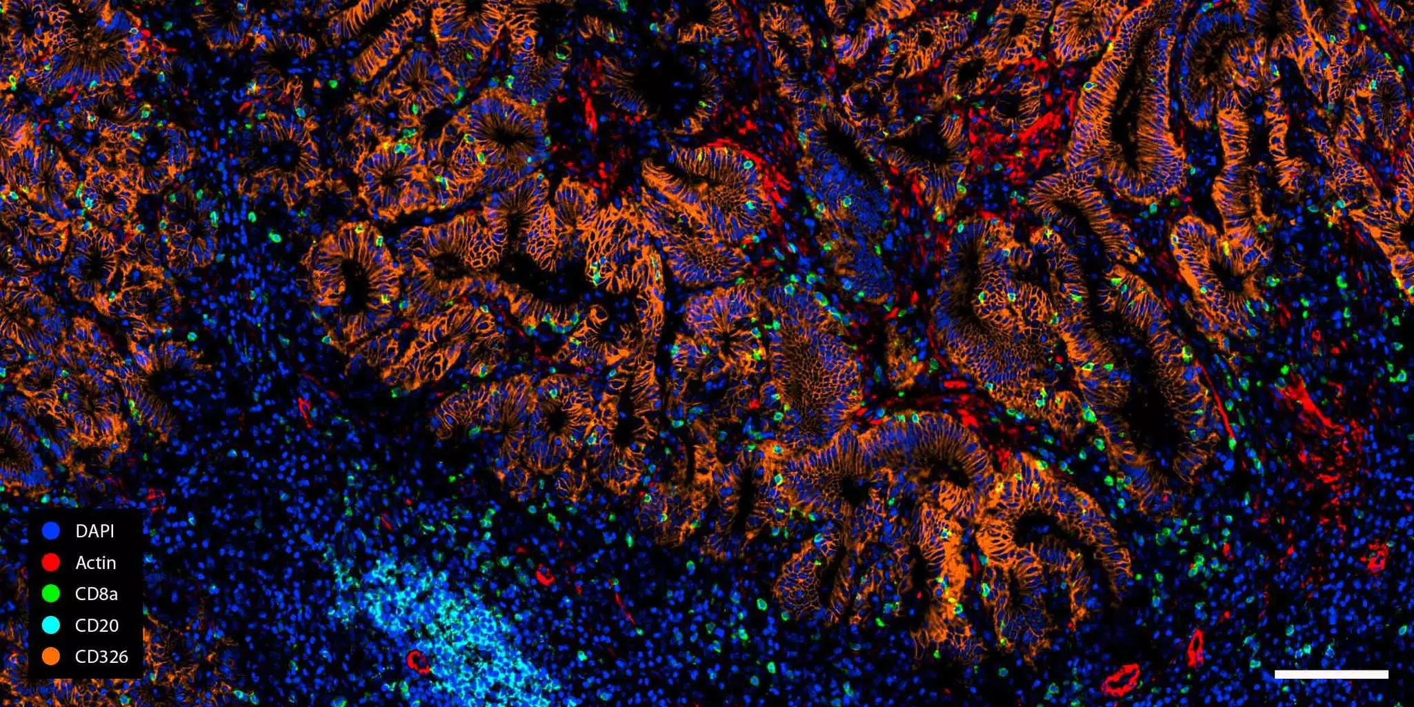
REAplex Immuno-oncology Core kit: diving deep into the tumor microenvironment

The REAplex Immuno-oncology Core Kit provides an expert-curated panel of 20 markers to reveal tumor and immune cells, as well as key structural and functional tissue features in formalin-fixed, paraffin-embedded (FFPE) human tumor samples.

With the REAplex Immuno-oncology Core Kit, you can:

  • Get a comprehensive overview of the tumor microenvironment: Distinguish tumor from healthy tissue and visualize invading immune cells, including T, B, NK cells, and macrophages.
  • Define tumor boundaries and invasiveness: Stain for CD326 (EpCAM) and cytokeratins to visualize epithelial tissue and evaluate the degree of invasiveness.
  • Explore tumor activity and immune regulation: Track proliferation, apoptosis, or neovascularization, and assess immune regulation through the PD-1/PD-L1 pathway.

Want to know more?

Explore the full immuno-oncology workflow.

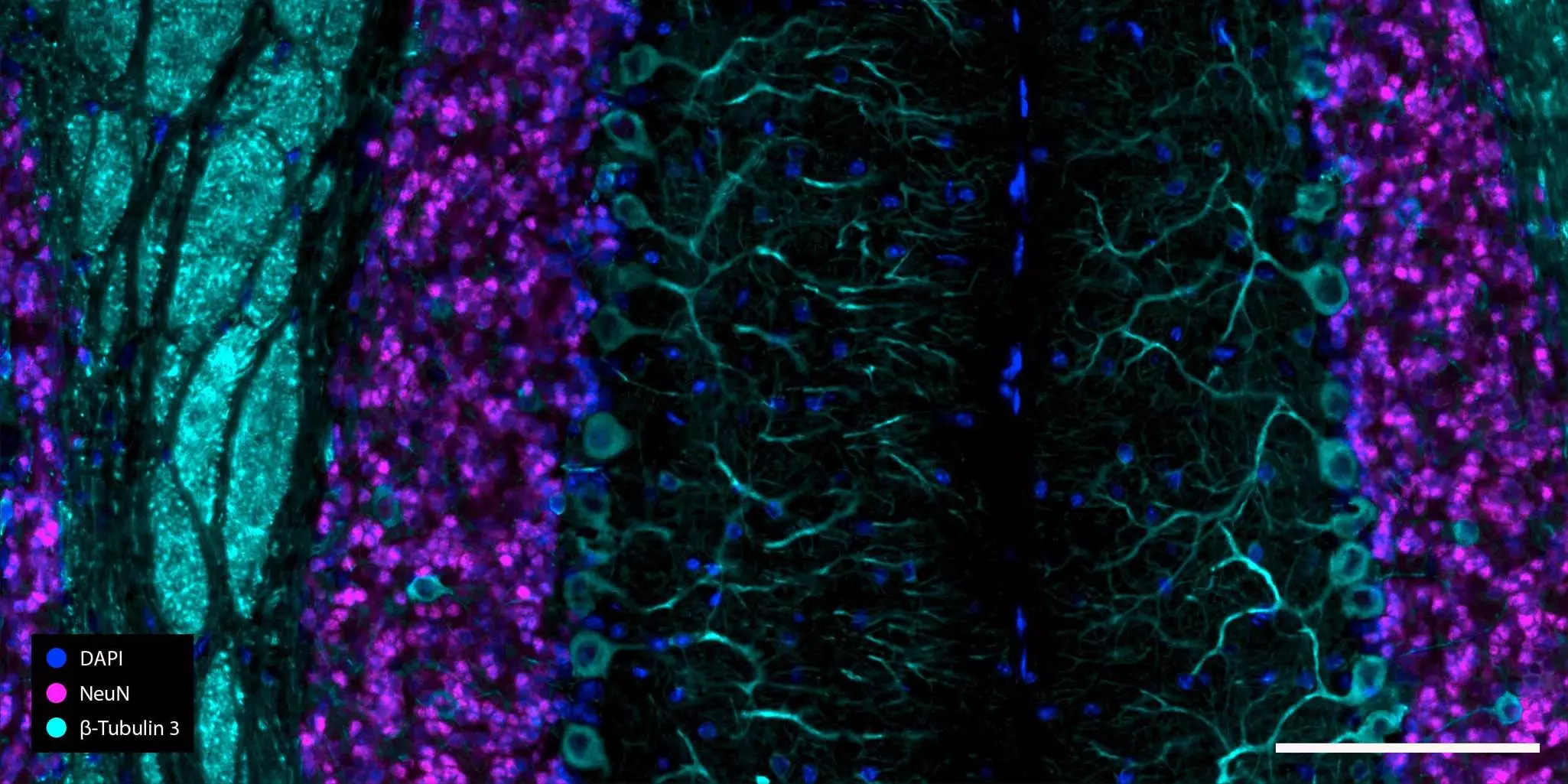
REAplex Neuro Core kit: mapping neuronal complexity with spatial detail

The REAplex Neuro Core Kit provides an expert-curated panel of 20 markers specifically designed to identify key cell types in frozen mouse brain sections.

With the REAplex Neuro Core Kit, you can:

  • Get deeper insights into neuronal architecture: Map brain circuits in detail by visualizing neurons, tracking their maturation state, and examining synaptic structures.
  • Characterize the role of glia: Identify astrocytes, oligodendrocytes, and radial glia and study their roles in shaping network development and plasticity.
  • Assess immune activity in the brain: Detect activated microglia and macrophages to investigate neuroinflammation and its spatial distribution.
  • Study neurodegenerative processes: Evaluate neurofibrillary tangles through Tau staining in Alzheimer's disease, or map the loss of tyrosine hydroxylase expression in Parkinson’s disease

Want to know more?

Explore the full neurobiology workflow.

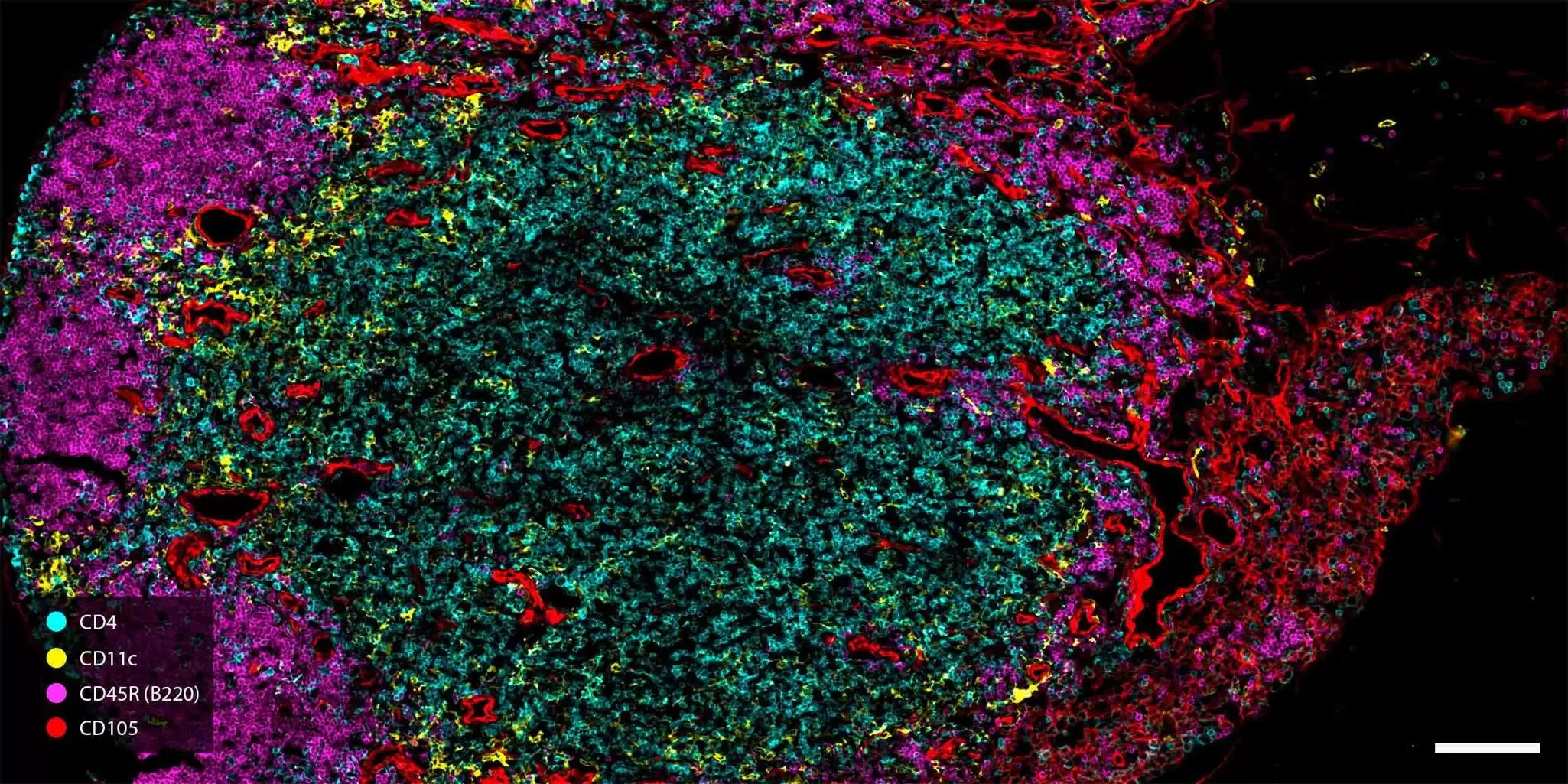
REAplex Immune Phenotyping kit: profiling immune cells in their tissue context

The REAplex Immune Phenotyping kit provides an expert-curated panel of 22 markers designed to identify and visualize key immune cell populations within their tissue context in PFA-fixed, frozen sections of mouse tissues.

With the REAplex Immune Phenotyping kit, you can:

  • Identify key immune cell populations: Visualize and distinguish major immune subsets to characterize immune composition across physiological and pathological contexts, such as tumor microenvironments, inflammatory responses, or infection models.
  • Interrogate tissue architecture and spatial immune organization: Visualize tissue vascularization and neovascularization to assess tissue architecture; map immune infiltration and tissue-resident cells in their spatial context to study disease progression.
  • Examine cellular activity: Track cell proliferation and T cell activation to get insights into dynamic immune responses such as clonal expansion during infection or activation of infiltrating lymphocytes in chronic disease models.

Want to know more?

Explore the full immune phenotyping workflow.

REAplex kits

3 products found

|

You have previously chosen International as your location.
What do you want to do?