Antibodies for tissue clearing and 3D immunofluorescence microscopy
3D-IF-validated antibodies for 3D imaging
Skip the validation step and dive directly into tissue clearing and 3D imaging.
Speed up your experiment with direct immunostaining.
Achieve high signal-to-background ratios for deeper insights.
Ensure efficient and consistent results with the power of recombinant engineering.
Successful 3D-immunofluorescence (3D-IF) imaging of whole large samples relies on proper sample preparation. The first critical step is finding suitable antibodies that penetrate deeply into the sample and provide specific binding with minimal background noise.
Miltenyi Biotec offers 3D-IF antibodies designed and validated specifically for 3D imaging of large samples, compatible with light sheet and confocal microscopy. These primary antibodies, conjugated to bright Vio® Dyes, enable direct immunostaining, saving 50% staining time compared to using secondary antibodies. Easy-to-follow protocols get you up and running quickly. For exceptional results, use our 3D-IF validated antibodies in combination with our compatible tissue clearing reagents.
Why 3D-IF validated antibodies make a difference:
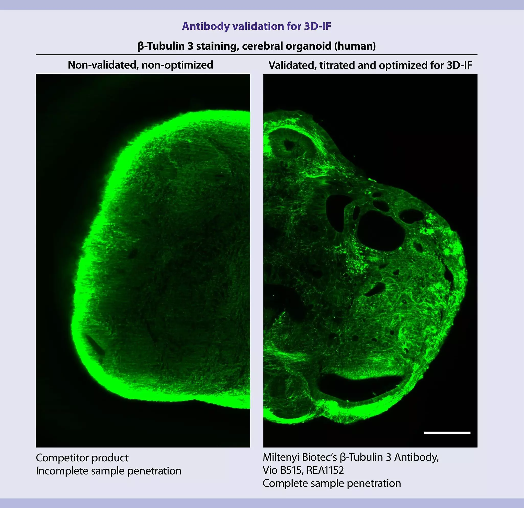
Figure 1: Antibody validation for 3D-Immunofluorescence
1. Validated and optimized for deep tissue penetration of large samples
One of the biggest challenges in staining thick tissue sections or entire organs is achieving complete antibody penetration throughout the sample. Non-validated antibodies often bind non-specifically to the outer surface, leading to unsatisfactory signals (Fig. 1). This necessitates screening most antibodies for compatibility and adapting protocols, resulting in lengthy validation processes and high costs. To ease this burden, our 3D-IF validated antibodies are not only tested for functionality and optimized for tissue penetration, but also validated for use with a wide range of tissue clearing methods.
Figure 2: Antibody diffusion through thick samples.
Our 3D-IF validated antibodies enable deep tissue labeling of large, cleared samples across a wide size range, from spheroids (200–1,000 µm in diameter) to entire mouse brain hemispheres (~4,000 µm wide).
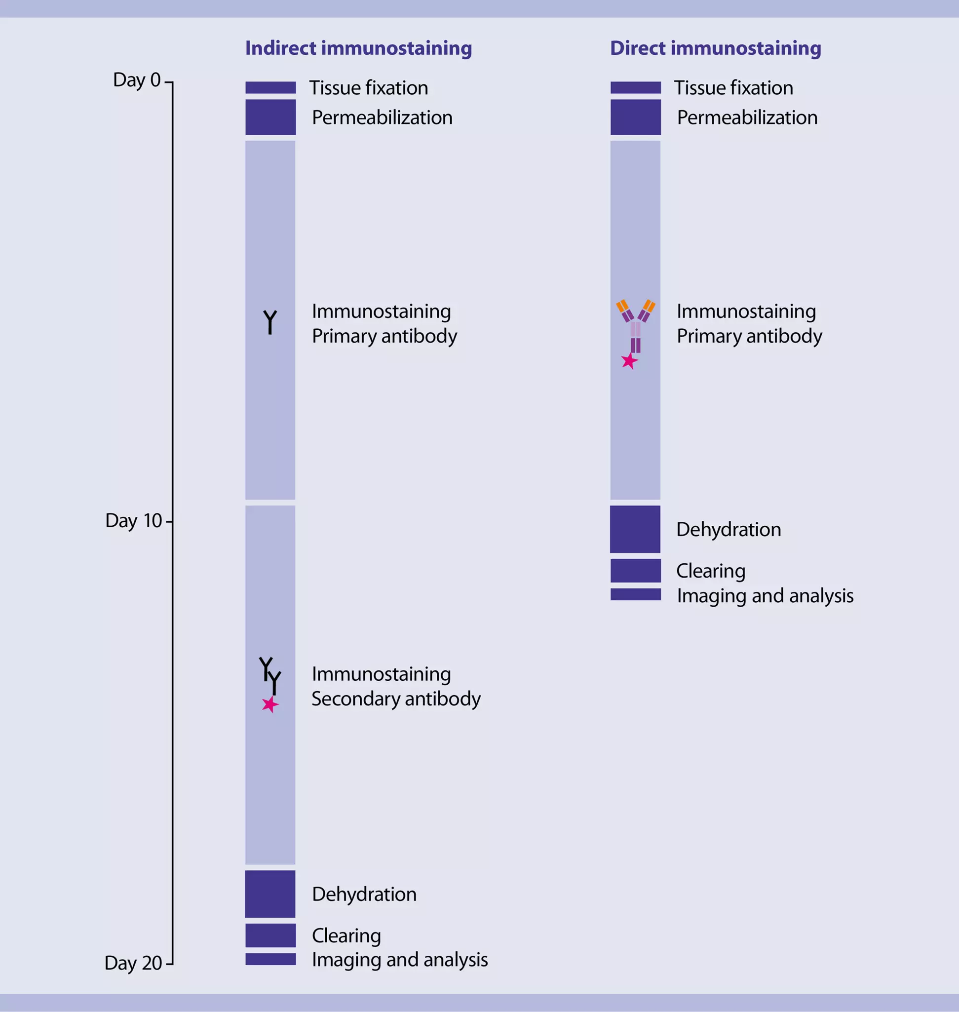
Figure 3: Duration of 3D imaging experiments
2. 50% shorter staining duration due to fluorochrome-conjugated primary antibodies
The immunostaining step, often the most time-consuming part of 3D-IF protocols for whole-mount samples, can be significantly accelerated using 3D-IF validated fluorochrome-conjugated antibodies. For example, indirect immunofluorescence staining of a mouse brain hemisphere with secondary antibodies takes about 14 days (total sample preparation = 20 days). In contrast, direct staining with our fluorochrome-conjugated antibodies eliminates the need for secondary antibodies, cutting staining duration by 50% (7 days) and significantly accelerating your imaging experiments (Fig. 3). For optimized staining and clearing protocols, visit our protocol website.
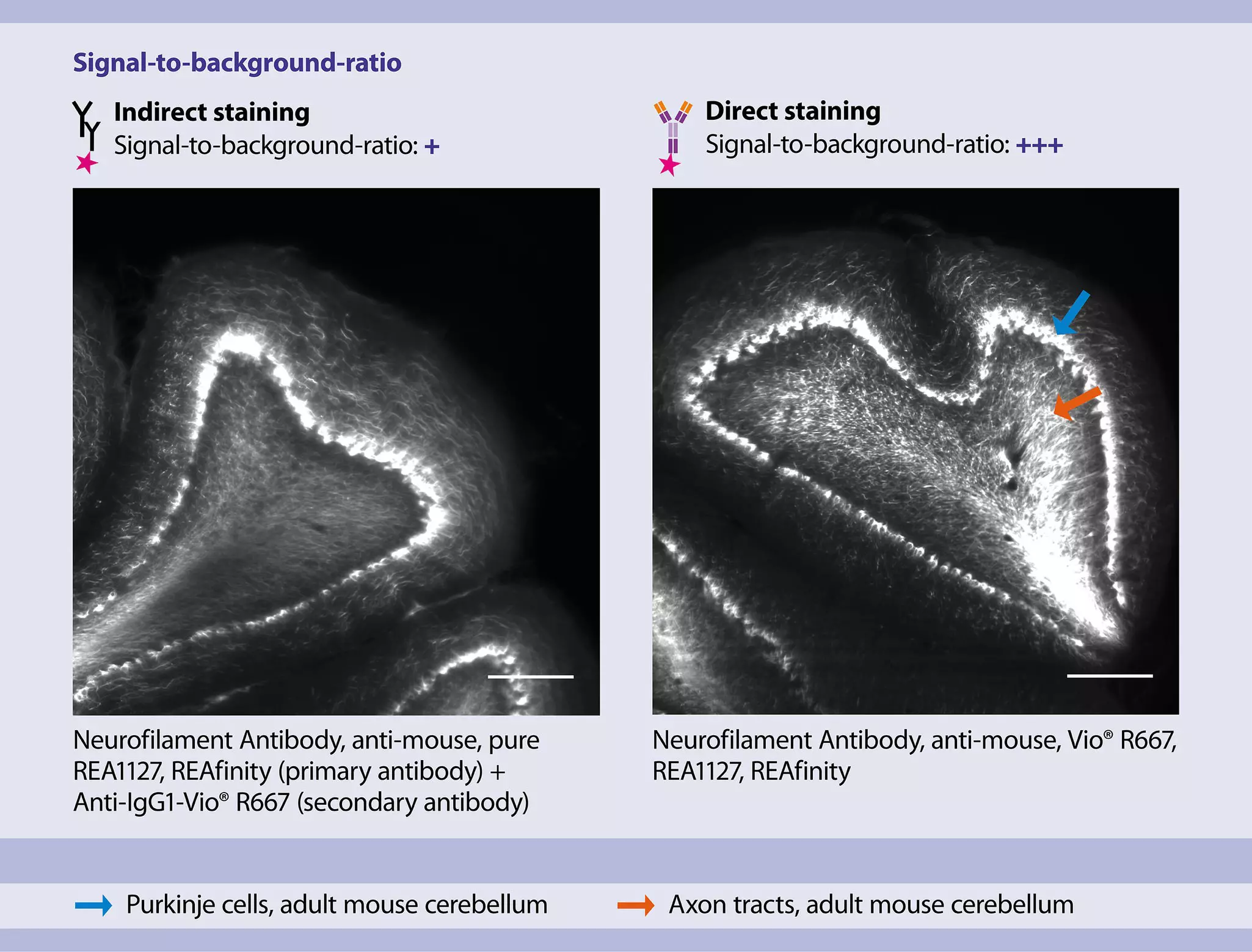
Figure 4: Signal-to-background ratios of large, cleared samples stained using direct vs indirect immunofluorescence staining.
3. Optimal signal-to-background ratios with primary antibodies conjugated to bright and photostable Vio Dyes
Immunostaining with secondary antibodies can suffer from non-specific binding, leading to low signal-to-background ratios. A better option is direct staining with primary antibodies conjugated to bright fluorochromes.
Our 3D-IF validated antibodies use Vio Dyes, known for their brightness and photostability, resulting in excellent signal-to-background ratios and minimal photobleaching. These antibodies allow the acquisition of images with an even better signal-to-noise ratio as compared to the use of secondary antibodies (fig. 4).
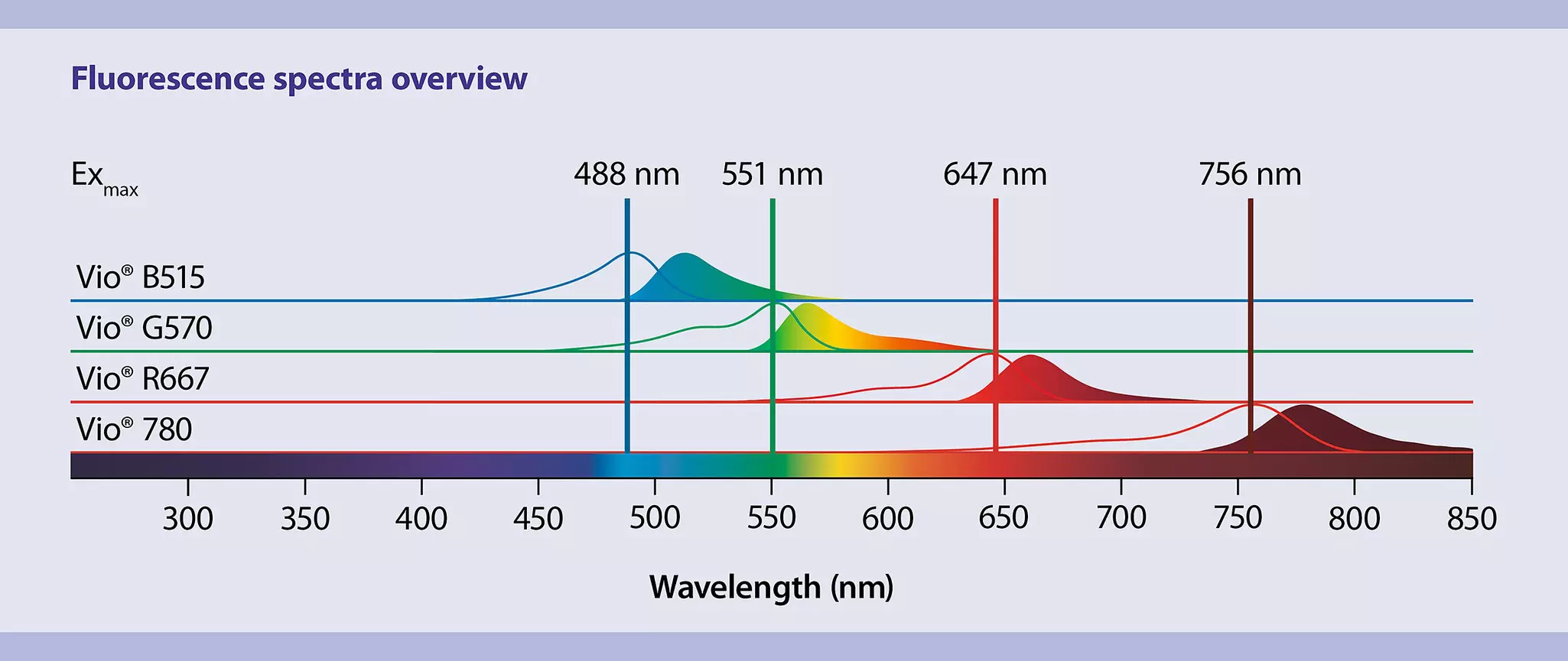
Excitation (empty curve) and emission (filled curve) spectra of Vio® Dyes with respective excitation maxima (Exmax) across the visible and near-infrared spectrum.
Our Vio Dyes used for 3D-IF antibodies show comparable characteristics to commonly used Alexa Fluor dyes but come with increased photostability, making them highly suited for widely used laser/filter combinations.
Name
Exmax
Emmax
Emission color*
Comparable Alexa Fluor
Vio B515
488
515
Green
Alexa Fluor 488
Vio G570
551
570
Yellow
Alexa Fluor 555
Vio R667
645
667
Near-IR**
Alexa Fluor 647
Vio 780
756
777
Near-IR**
Alexa Fluor 750
* Typical emission colors as viewed through conventional fluorescence microscopes with appropriate filters. ** Human vision is insensitive to light beyond ~650 nm; for this reason, it is not possible to view near-IR fluorescent dyes.
Use our fluorescence spectra viewer to compare Vio Dyes with your commonly used dyes and find the perfect combination for your specific needs.
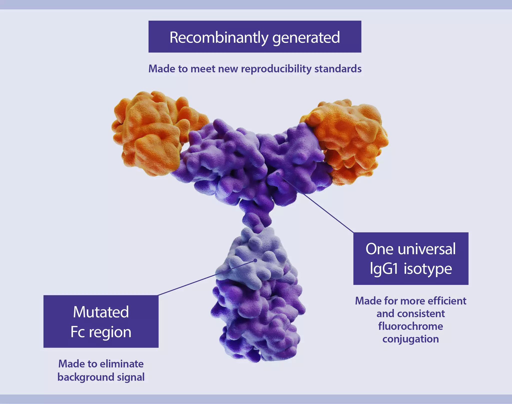
Figure 5: REAfinity recombinant antibody model
4. Recombinantly engineered antibodies for reproducible results and minimal background signals
REAfinity® Antibodies offer superior lot-to-lot consistency and purity compared to monoclonal antibodies derived from mouse or rat hybridomas. These recombinantly engineered antibodies are highly specific and do not bind to Fc receptors on immune cells in your tissue samples. Additionally, their consistent human IgG1 isotype ensures consistent fluorochrome conjugation.
Slide 1 of 2
Choosing the best antibodies is the most important step in 3D-IF. I can confidently say that Miltenyi Biotec's 3D-IF antibodies have exceeded our expectations. With their impressive specificity and sensitivity, my 3D imaging experiments will produce trustworthy results every time.
Dr. Alain Chédotal
Sorbonne Université, INSERM, CNRS, Institut de la Vision, Paris, France
Slide 2 of 2
It is by far the best TH antibody we have ever tested.