Anti-TOM22 Microbeads, mouse have been developed to be used in combination with the Mitochondria Isolation Kit, mouse tissue for the efficient isolation of functional and viable mitochondria. The isolation protocol is based on the renowned MACS Technology, which enables fast isolation of high purity and high yield mitochondria.


Related products for Anti-TOM22 MicroBeads, mouse

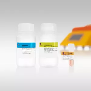
Integrated tissue dissociation followed by mitochondria isolation.
Enrichment of functional mitochondria. Mitochondria were prepared from various mouse tissues using the Mitochondria Isolation Kit, mouse tissue. The mitochondria preparations from muscle, liver, and brain, as well as the flow-through fractions were analyzed by a coupled enzymatic citrate synthase assay. The data indicate that citrate synthase activity, normalized to the activity per mg protein assayed in the flow-through fraction, is highly enriched in the eluate.
Mitochondrial protein import is maintained after magnetic isolation. Mitochondria were prepared using the Mitochondria Isolation Kit, human or by differential centrifugation (DC) and were then incubated for 1 h with 35S-labeled TFAM. Import was studied by SDS-PAGE and fluorography. Lane 1: Control lysate of in vitro translated TFAM. Lane 2-7: Proteins from sedimented mitochondria. P= Precursor protein. M= Imported mature protein. (Courtesy of Dr. H.-T. Hornig-Do, Newcastle, UK)
Anti-TOM22 Microbeads, mouse have been developed to be used in combination with the Mitochondria Isolation Kit, mouse tissue for the efficient isolation of functional and viable mitochondria. The isolation protocol is based on the renowned MACS Technology, which enables fast isolation of high purity and high yield mitochondria.
Tissues can be homogenized with the gentleMACS or gentleMACS Octo Dissociator in combination with the Mitochondria Extraction Kit – Tissue. After one-step homogenization of the tissue and lysis of the cell suspension, mitochondria are magnetically labeled with Anti-TOM22 MicroBeads, mouse, which bind to the translocase of the outer mitochondrial membrane 22 protein (TOM22). The sample is loaded onto an LS Column placed in a MidiMACS or QuadroMACS Separator. After washing, only the magnetically labeled mitochondria are retained on the column. The column is removed from the separator and functional mouse mitochondria are eluted from the column.
After isolation mitochondria can be further subjected to Western blot analysis1,6 or downstream functional assays, including measurement of O2, membrane potential, and ATP4-5,7, or respiratory control4. Growing evidence also suggests that isolated mitochondria are well suited for mitochondrial RNA expression profiling studies2,3.
- Sacchi, S. et al. (2011) Evidence for the interaction of d-amino acid oxidase with pLG72 in a glial cell line. Mol. Cell. Neurosci. 48 (1): 20-28
- Minet, A. D. and Gaster, M. (2010) ATP synthesis is impaired in isolated mitochondria from myotubes established from type 2 diabetic subjects. Biochem. Biophys. Res. Commun. 402 (1): 70-74
- Hornig-Do, H. T. et al. (2009) Isolation of functional pure mitochondria by superparamagnetic microbeads. Anal. Biochem. 389 (1): 1-5
- Papkovskaia, T. D. et al. (2012) G2019S leucine-rich repeat kinase 2 causes uncoupling protein-mediated mitochondrial depolarization. Hum. Mol. Genet. 21 (19): 4201-4213
- Eriksen, M. B. et al. (2011) Intact primary mitochondrial function in myotubes established from women with PCOS. J. Clin. Endocrinol. Metab. 96 (8): E1298-1302
- Hubbard, W. B. et al. (2019) Fractionated mitochondrial magnetic separation for isolation of synaptic mitochondria from brain tissue. Sci Rep 9 (1): 9656
- Minet, A. D. and Gaster, M. (2011) The dynamic equilibrium between ATP synthesis and ATP consumption is lower in isolated mitochondria from myotubes established from type 2 diabetic subjects compared to lean control. Biochem. Biophys. Res. Commun. 409 (4): 591-595
- Bandiera, S. et al. (2011) Nuclear outsourcing of RNA interference components to human mitochondria. PLoS One 6 (6): e20746
- Hornig-Do, H. T. et al. (2012) Nonsense mutations in the COX1 subunit impair the stability of respiratory chain complexes rather than their assembly. EMBO J. 31 (5): 1293-1307
- Barrey, E. et al. (2011) Pre-microRNA and mature microRNA in human mitochondria. PLoS One 6 (5): e20220
- Guo, T. et al. (2010) Quantitative proteomics discloses MET expression in mitochondria as a direct target of MET kinase inhibitor in cancer cells. Mol. Cell. Proteomics 9 (12): 2629-2641
- Schiller, M. et al. (2012) Induction of type I IFN is a physiological immune reaction to apoptotic cell-derived membrane microparticles. J Immunol 189 (4): 1747-1756
- Mukhopadhyay, P. et al. (2012) Mitochondrial reactive oxygen species generation triggers inflammatory response and tissue injury associated with hepatic ischemia-reperfusion: therapeutic potential of mitochondrially-targeted antioxidants. Free Radic. Biol. Med. 53 (5): 1123-1138
You have previously chosen International as your location.
What do you want to do?