The Annexin V MicroBead Kit was developed for positive selection or depletion of cells based on their ability to bind Annexin V. The Annexin V MicroBead Kit includes Annexin V MicroBeads and Annexin V Binding Buffer.


Related products for Annexin V MicroBead Kit

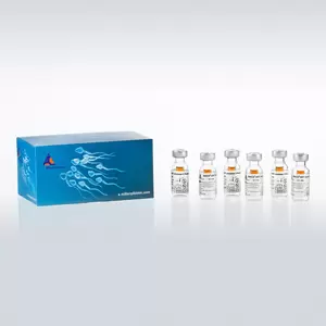
The Annexin V MicroBead Kit was developed for positive selection or depletion of cells based on their ability to bind Annexin V. The Annexin V MicroBead Kit includes Annexin V MicroBeads and Annexin V Binding Buffer.
In most normal, viable eukaryotic cells the negatively charged phospholipid phosphatidylserine (PS) is located in the cytosolic leaflet of the plasma membrane lipid bilayer1. PS redistribution from the inner to the outer leaflet is an early and widespread event during apoptosis1,2. However, in necrosis, PS becomes accessible due to the disruption of membrane integrity2. Apart from necrosis and apoptosis, PS also becomes accessible in activated platelets3, in certain cell anomalies such as sickle cell anaemia4, in erythrocyte senescence5, upon degranulation of mast cells10, and in certain stages of B cell differentiation11. PS exposure also serves as a trigger for the recognition and removal of cells by macrophages6,9.
Annexin V is a 35 kDa phospholipid-binding protein and a major cell membrane component of macrophages and other phagocytic cell types. Annexin V has a high affinity to PS in the presence of physiological concentrations of calcium (CA2+)7 and has already been used to isolate cells with exposed PS using MACS® MicroBeads.4,8
The Annexin V MicroBead Kit is used for the isolation of cells with exposed PS, including sickle cells and apoptotic cells. The Annexin V MicroBead Kit has been further applied to the purification of apoptotic and non-apoptotic neutrophils to study the intracellular signaling pathways that regulate neutrophil survival. In a study on the roles of different cytosolic phospholipase A2s in apoptosis, the Annexin V MicroBead Kit has been used for sensitive assessment of PS externalization.
For positive selection: MS, LS or XS Columns. For depletion: LD, CS or D Columns.
Enrichment of dead (apoptotic/necrotic) cells using the Annexin V MicroBead Kit. Murine thymocytes were treated with CD95 (anti-Fas) monoclonal antibody for 3 hours at 37 °C. PS-exposing cells were enriched by labeling with Annexin V MicroBeads followed by separation on an MS Column in the magnetic field of a MiniMACS™ Separator. Cell fractions were stained with Annexin V-FITC.
Isolation of PS-exposing cells using the Annexin V MicroBead Kit.
- Kuypers, F. A. et al. (1996) Detection of altered membrane phospholipid asymmetry in subpopulations of human red blood cells using fluorescently labeled annexin V. Blood 87: 1179-1187
- Fadok, V. A. et al. (2000) A receptor for phosphatidylserine-specific clearance of apoptotic cells. Nature 405: 85-90
- Khwaja, A. and Tatton, L. (1999) Caspase-mediated proteolysis and activation of protein kinase Cdelta plays a central role in neutrophil apoptosis. Blood 94: 291-301
- Demo, S. D. et al. (1999) Quantitative measurement of mast cell degranulation using a novel flow cytometric annexin-V binding assay. Cytometry 36: 340-348
- Atsumi, G. et al. (2000) Distinct roles of two intracellular phospholipase A2s in fatty acid release in the cell death pathway. Proteolytic fragment of type IVA cytosolic phospholipase A2alpha inhibits stimulus-induced arachidonate release, whereas that of type VI Ca2+-independent phospholipase A2 augments spontaneous fatty acid release. J. Biol. Chem. 275 (24): 18248-18258
- Moss, S. E. et al. (1991) Diversity in the Annexin family. In: Novel Calcium Binding Proteins, Springer Verlag : 535-566
- Fadok, V. A. et al. (1992) Exposure of phosphatidylserine on the surface of apoptotic lymphocytes triggers specific recognition and removal by macrophages. J Immunol 148: 2207-2216
- Martin, S. J. et al. (1995) Early redistribution of plasma membrane phosphatidylserine is a general feature of apoptosis regardless of the initiating stimulus: inhibition by overexpression of Bcl-2 and Abl. J. Exp. Med. 182: 1545-1556
- Thiagarajan, P. et al. (1990) Binding of annexin V/placental anticoagulant protein I to platelets. Evidence for phosphatidylserine exposure in the procoagulant response of activated platelets. J. Biol. Chem. 265: 17420-17423
- Rickers, A. et al. (1998) Inhibition of CPP32 blocks surface IgM-mediated apoptosis and D4-GDI cleavage in human BL60 Burkitt lymphoma cells. Eur. J. Immunol. 28: 296-304
- Dressel, R. et al. (2000) Heat shock protein 70 is able to prevent heat shock-induced resistance of target cells to CTL. J Immunol 164 (5): 2362-2371
- Koopman, G. et al. (1994) Annexin V for flow cytometric detection of phosphatidylserine expression on B cells undergoing apoptosis. Blood 84: 1415-1420
- Schroit, A. J. et al. (1991) Transbilayer movement of phospholipids in red cell and platelet membranes. Biochim. Biophys. Acta 1071: 313-329
- Dillon, S. R. et al. (2001) Annexin V binds to positively selected B cells. J Immunol 166: 58-71
You have previously chosen International as your location.
What do you want to do?