Isolation, expansion, and analysis of human hematopoietic stem cells (HSCs)
This application protocol describes how human hematopoietic stem cells (HSCs) can be isolated from various sources, such as bone marrow or cord blood, expanded in highly defined cell culture conditions, and analyzed by flow cytometry or cell culture assays.
Protocol
Isolation, expansion, and analysis of human hematopoietic stem cells (HSCs)
1.5 h
Isolation of mononuclear cells
1.5 h
Isolation of CD34+ cells
7d
Expansion of CD34+ cells
30 min
Flow cytometric analysis of CD34+ cells
This protocol includes detailed instructions for preparing necessary reagents and materials and then carrying out each step.
PE buffer for isolation of mononuclear cells
Prepare a solution with phosphate buffered saline (PBS), pH 7.2, and 2 mM EDTA. Keep buffer cold (2–8 °C).
▲ Note: EDTA can be replaced by other supplements such as anticoagulant citrate dextrose formula-A (ACD-A) or citrate phosphate dextrose (CPD).
PBE buffer for isolation of CD34+ cells
Prepare a solution containing phosphate-buffered saline (PBS), pH 7.2, 0.5 % bovine serum albumin (BSA), and 2 mM EDTA by diluting MACS® BSA Stock Solution 1:20 with autoMACS® Rinsing Solution. Keep buffer cold (2−8 °C).
▲ Note: EDTA can be replaced by other supplements such as anticoagulant citrate dextrose formula-A (ACD-A) or citrate phosphate dextrose (CPD). BSA can be replaced by other proteins such as respective serum albumin, respective serum, or fetal bovine serum (FBS). Buffers or media containing Ca2+ or Mg2+
are not recommended for use.
Isolate mononuclear cells by density gradient centrifugation using Ficoll-Paque™. Follow the Ficoll-Paque protocol from the special protocol corresponding to the source tissue.
Download special protocol
Isolation of mononuclear cells from peripheral blood
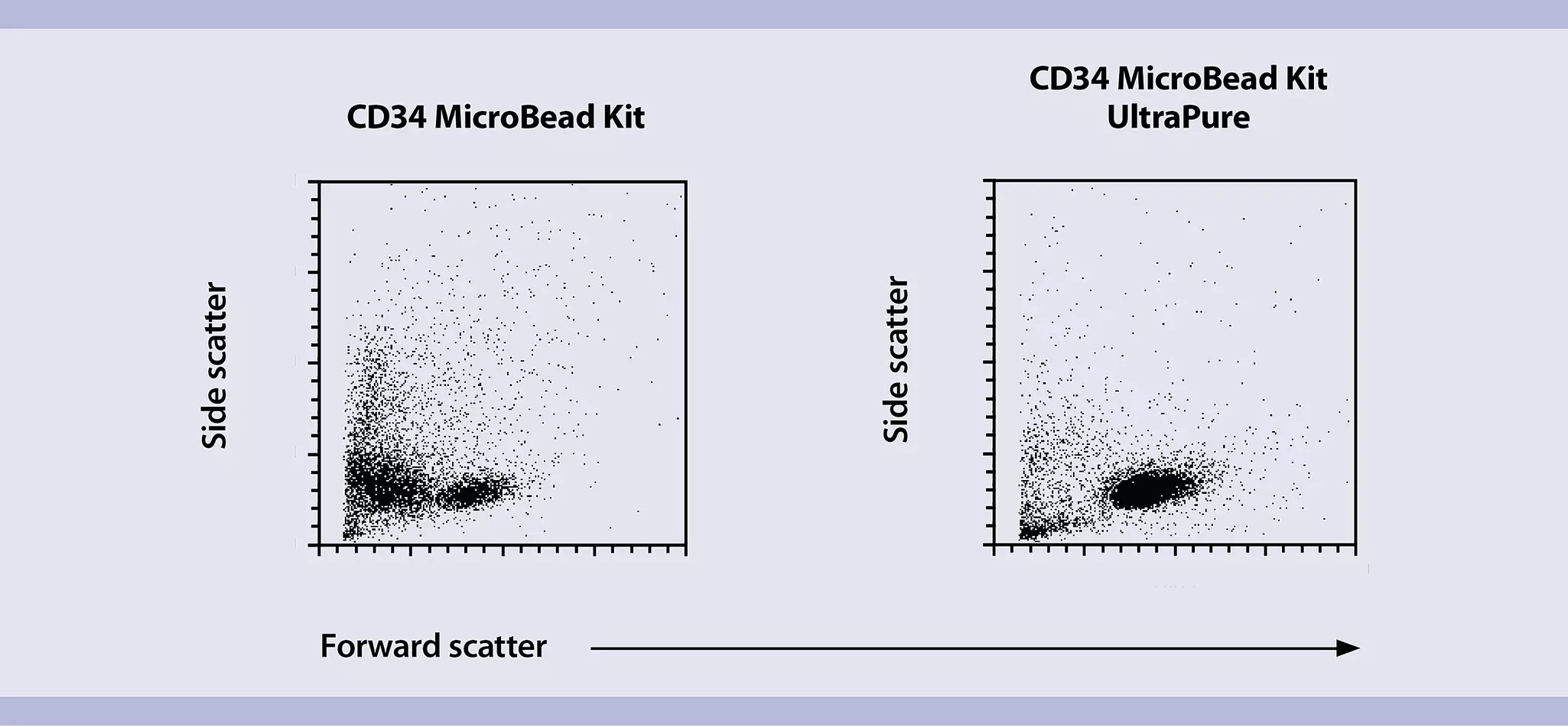
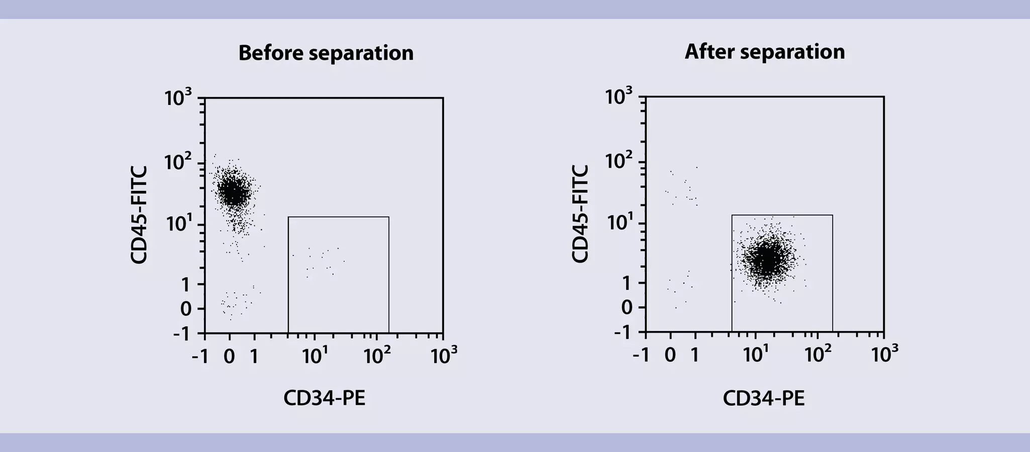
Isolate CD34+ cells among the mononuclear cells using the CD34 MicroBead Kit UltraPure, human and LS or MS columns. Follow the protocol of the kit data sheet.
It is recommended to filter the magnetically labeled cell suspension before separation on the column to guarantee a single-cell suspension.
- Place a Pre-Separation filter (30 µm) on the separation column.
- Rinse the column three times with PBE buffer, pre-wetting the filter.
- Apply the cell suspension and washing buffer to the filter on the column.
Download data sheet
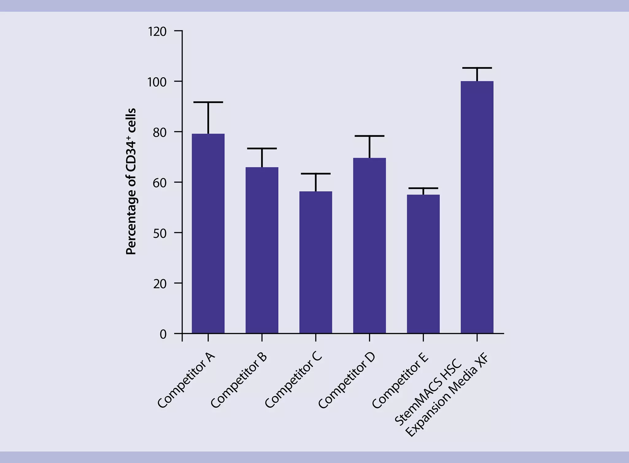
For expansion, culture the isolated CD34+ cells in StemMACS™ HSC Expansion Media XF supplemented with StemMACS HSC Expansion Cocktail. Follow the protocol of the media data sheet.
Download data sheet
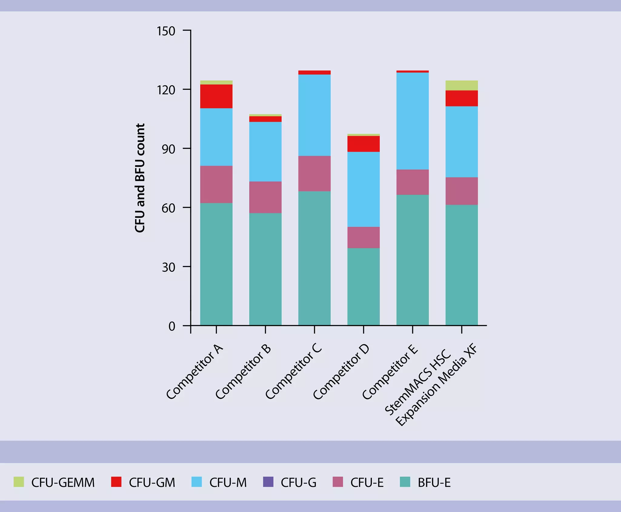
Analyze the expanded CD34+ cells for HSC-specific surface markers via flow cytometry using REAfinity™ antibodies, or perform CFU-GEMM analysis to assess their myeloid differentiation potential.
The following is a list of relevant resources that might be of interest:
- StemMACS™ HSC Expansion Media XF – A xeno-free culture system for hematopoietic stem cells
- CD34 MicroBead Kit UltraPure, human – Leading the way in magnetic HSC separation
- REAfinity Recombinant Antibodies – Flow cytometry is in their genes
- REAfinity Recombinant Antibodies – Frequently Asked Questions
- Nölle, V. et al., (2016) Recombinant antibodies for improved standardization in flow cytometry.
- StemMACS™ HSC-CFU Media – Need to estimate HSC differentiation potential? We have the media!
- Web-based training to perform the standard HSC-CFU assay with our StemMACS HSC-CFU Media
You have previously chosen International as your location.
What do you want to do?