Human CD4+CD25+CD127dim/– regulatory T cell isolation, in vitro expansion, and analysis
In vitro human regulatory T cell expansion
This application protocol describes the use of the Treg Expansion Kit, human, which is designed to efficiently expand Treg cells that maintain FoxP3 expression after isolation with the CD4+CD25+CD127dim/– Regulatory T Cell Isolation Kit II, human. It includes a complete protocol for Treg cell workflows, describing human Treg cell isolation from peripheral blood mononuclear cells (PBMCs), their in vitro expansion, and subsequent flow cytometric analysis.
Protocol
Human CD4+CD25+CD127dim/– regulatory T cell isolation, in vitro expansion, and analysis
3 h
Isolation of CD4+CD25+CD127dim/– Treg cells
2 h
Staining of Treg cells and flow cytometric analysis
30 min
Preparation and start of in vitro expansion
14 d
Cell harvest and analysis or further restimulation
21 d
Cell harvest
2 h
Flow cytometric analysis after expansion
Protocol

This protocol includes detailed instructions for preparing necessary reagents and materials and then carrying out each step.
Separation buffer
1× PBS (pH 7.2)
+ 0.5% BSA
+ 2 mM EDTA
Preparation of Treg cell staining and flow cytometric analysis
FoxP3 staining buffer (components provided in Treg Detection Kit)
- Always prepare reagents freshly on the same day that intracellular FoxP3 staining is performed.
- The required total buffer volumes should be calculated beforehand; volumes will depend on the number of cells to be analyzed as well as the number of tests to be performed.
Example:
For one sample with 1×10⁶ Treg cells: Prepare 1 mL of fixation/permeabilization solution and 4 mL of 1× Permeabilization Buffer.
- Fixation/permeabilization solution: Dilute Fixation/Permeabilization Solution 1 (e.g. 0.25 mL) 1:4 with
Fixation/Permeabilization Solution 2 (e.g. 0.75 mL). - 1× Permeabilization Buffer: Dilute 10× Permeabilization Buffer (e.g. 1 mL) 1:10 with deionized or
distilled water (e.g. 9 mL).
Preparation of Treg cell in vitro expansion
Expansion medium
The expansion medium can be stored for up to 10 days under sterile conditions at 4 °C.
500 mL TexMACS™ Medium
+ 500 IU/mL Human IL-2 IS, premium grade
+ 5% human AB Serum
+ 0.01 mM β-ME
+ 1% PenStrep
+ (Optional) 100 nmol/L rapamycin
▲ Note: Rapamycin has been shown to increase the frequency of FoxP3+ Treg cells during expansion (Battaglia, M. et al. (2005) Blood 105: 4743–4748).
- When working with anticoagulated peripheral blood or buffy coat, PBMCs should be isolated
by density gradient centrifugation, for example, using Ficoll-Paque™ according to the manufacturer’s instructions. - Collect a sample of PBMCs for subsequent flow cytometric analysis. See section "Staining of Treg cells and flow cytometric analysis".
Magnetic labeling of non-CD4+ and CD127high cells
- All steps described below for magnetic labeling and depletion of non-CD4+ and CD127high cells, as well as labeling and selection of CD25+ cells, are performed using the CD4+CD25+CD127dim/– Regulatory T Cell Isolation Kit II, human.
- Volumes for magnetic labeling given below are for up to 1×10⁷ total cells. When working with fewer than 1×10⁷ cells, use the same volumes as indicated. When working with higher cell numbers, scale up all reagent volumes and total volumes accordingly (e.g. for 2×10⁷ total cells, use twice the volume of all indicated reagent volumes and total volumes).
- Determine cell number.
- Centrifuge cell suspension at 300×g for 10 minutes. Discard supernatant.
- Resuspend cell pellet in 40 μL of separation buffer per 1×10⁷ total cells.
- Add 10 μL of CD4+CD25+CD127dim/– T Cell Biotin-Antibody Cocktail II per 1×10⁷ total cells.
- Mix well and incubate for 5 minutes in the refrigerator (2–8 °C).
- Add 30 μL of separation buffer and 20 μL of Anti-Biotin MicroBeads per 1×10⁷ total cells.
- Mix well and incubate for 10 minutes in the refrigerator (2–8 °C).
- Adjust the volume to a minimum of 500 μL of separation buffer.
▲ Note: Resuspend up to 1×10⁸ cells in 500 μL of separation buffer. For higher cell numbers, scale up buffer volume accordingly.
Magnetic depletion of non-CD4+ and CD127high cells
- The first step of Treg cell isolation is a depletion of non-CD4+ and CD127high cells. Here, an LD Column is used, which has a capacity of 1×10⁸ labeled cells and 5×10⁸ total cells.
- When using the CD4+CD25+CD127dim/– Regulatory T cell Isolation Kit II, human, it is not recommended to process more than 1.3×10⁸ total cells per LD Column. When exceeding this cell number, it is strongly recommended to split the sample and use additional LD Columns.
- Place LD Column(s) in the magnetic field of a MidiMACS™ Separator.
- Prepare column by rinsing with 2 mL of separation buffer. Always wait until the column reservoir is empty before proceeding to the next step.
- Apply the cell suspension onto the column.
- Collect unlabeled cells that pass through and wash column twice with 1 mL of separation buffer each. Collect total effluent; this is the unlabeled pre-enriched CD4+ cell fraction which is needed for further Treg cell isolation.
- Determine cell number.
Magnetic labeling of CD25+ cells
- Volumes for magnetic labeling given below are for an initial starting cell number of up to 1×10⁷ total cells. For higher initial cell numbers, scale up all volumes accordingly.
- Centrifuge cell suspension at 300×g for 10 minutes. Discard supernatant.
- Resuspend cell pellet in 90 μL of separation buffer per 1×10⁷ total cells.
- Add 10 μL of CD25 MicroBeads II per 1×10⁷ total cells.
- Mix well and incubate for 15 minutes in the refrigerator (2–8 °C).
- Wash cells by adding 1–2 mL of separation buffer and centrifuge at 300×g for 10 minutes. Discard supernatant.
- Resuspend up to 1×10⁸ cells in 500 μL of separation buffer.
▲ Note: For higher cell numbers, scale up buffer volume accordingly.
Magnetic separation of CD25+ cells
- The second step during Treg cell isolation is a positive selection of CD25+ cells. Here, two consecutive MS Columns are used, with a capacity of 1×10⁷ labeled cells. To not exceed the capacity, it is recommended to determine the frequency of CD25+ cells in the cell suspension by flow cytometry beforehand.
- Place an MS Column in the magnetic field of a MiniMACS™ Separator.
- Prepare the column by rinsing with 500 μL of separation buffer.
- Apply the cell suspension onto the column. Collect flow-through containing unlabeled cells.
- Wash column three times with 500 μL of separation buffer each. Collect unlabeled cells that pass through and combine with the effluent from step 3.
- Remove column from the separator and place it on a suitable collection tube.
- Pipette 1 mL of separation buffer onto the column. Immediately flush out magnetically labeled cells by firmly pushing the plunger into the column.
- (Optional) To increase purity of CD4+CD25+CD127dim/– cells, the eluted fraction can be enriched over a second MS Column. Repeat the magnetic separation procedure described in steps 1 to 6 using a new MS Column. The isolated Treg cells are now ready-to-use for in vitro expansion.
- Collect a sample of isolated CD4+CD25+CD127dim/– cells for subsequent flow cytometric analysis. See section "Staining of Treg cells and flow cytometric analysis".
General
- To assess the purity and to determine the frequency of FoxP3-expressing cells as start population for expansion, surface and intracellular flow cytometry staining of the isolated Treg cells must be performed. Purity is determined by surface staining of CD4, CD25, and CD127.
Furthermore, for the final analysis of Treg expansion, it is also important to determine:
a) The initial cell number.
b) The initial cell number and frequency of FoxP3-expressing CD4+ cells. - Volumes given below are for up to 1×10⁶ nucleated cells. When working with fewer than 1×10⁶ cells, use the same volumes as indicated except for the fixation step due to the impact on cell morphology. When working with higher cell numbers, scale up all reagent volumes and total volumes, accordingly (e.g. for 2×10⁶ nucleated cells, use twice the volume of all indicated reagent volumes and total volumes).
Surface staining of Treg cells with CD4 and CD25 antibodies (optional: CD127 antibody)
- Determine cell number.
- Centrifuge cell suspension at 300×g for 10 minutes. Discard supernatant.
- Resuspend up to 1×10⁶ nucleated cells per 90 μL of separation buffer.
- Add 10 μL of CD4-FITC and 20 μL of CD25 antibody (either PE- or APC-conjugated).
- (Optional) Add 10 µL of CD127 PE-Vio® 770 or CD127-PE-Vio 615.
- Mix well and incubate for 10 minutes in the dark in the refrigerator (2–8 °C).
- Wash cells by adding 1–2 mL of separation buffer per 1×10⁶ cells and centrifuge at 300×g for 5 minutes at 4 °C. Discard supernatant.
- Proceed immediately to "Intracellular staining with FoxP3 Antibody".
Intracellular staining with FoxP3 Antibody
- Resuspend 1×10⁶ nucleated cells in 1 mL cold, freshly prepared fixation/permeabilization solution. When working with fewer cells, scale down the buffer volume accordingly (e.g. for 5×10⁵ nucleated cells use 500 µL of fixation/permeabilization solution).
- Mix well and incubate for 30 minutes in the dark in the refrigerator (2–8 °C).
- Wash cells by adding 1–2 mL of cold separation buffer per 1×10⁶ cells and centrifuge at 300×g for 5 minutes at 4 °C. Discard supernatant.
- Wash cells by adding 1–2 mL of cold 1× Permeabilization Buffer per 1×10⁶ cells and centrifuge at 300×g for 5 minutes at 4 °C. Discard supernatant.
- Resuspend up to 1×10⁶ nucleated cells in 80 μL of cold 1× Permeabilization Buffer.
- Add 20 μL of FcR Blocking Reagent.
- Mix well and incubate for 5 minutes in the refrigerator (2–8 °C).
- Add 10 μL of FoxP3 Antibody (either APC- or PE-conjugated).
- Mix well and incubate for 30 minutes in the dark in the refrigerator (2–8 °C).
- Wash cells by adding 1–2 mL of cold 1× Permeabilization Buffer per 1×10⁶ cells and centrifuge at 300×g for 5 minutes at 4 °C. Discard supernatant.
- Resuspend cell pellet in a suitable amount of separation buffer for analysis by flow cytometry.
▲ Note: Due to fixation and permeabilization, cells can be smaller than viable cells. Thus, FSC/SSC settings of the flow cytometer might need to be adjusted.
Flow cytometric analysis of isolated Treg cells
- To assess the purity of the isolated Treg cells and the initial frequency of FoxP3+ CD4+ cells, the cells are analyzed by flow cytometry. The analysis should be performed with the cell sample taken in section "Isolation of PBMCs" and the cell sample taken in "Magnetic separation of CD25+ cells".
- Identify lymphocytes by FSC/SSC gating.
- Exclude dead cells from the analysis by using the live/dead cell exclusion marker propidium iodide (PI).
- Identify CD4+ cells according to CD4/FSC analysis among lymphocytes.
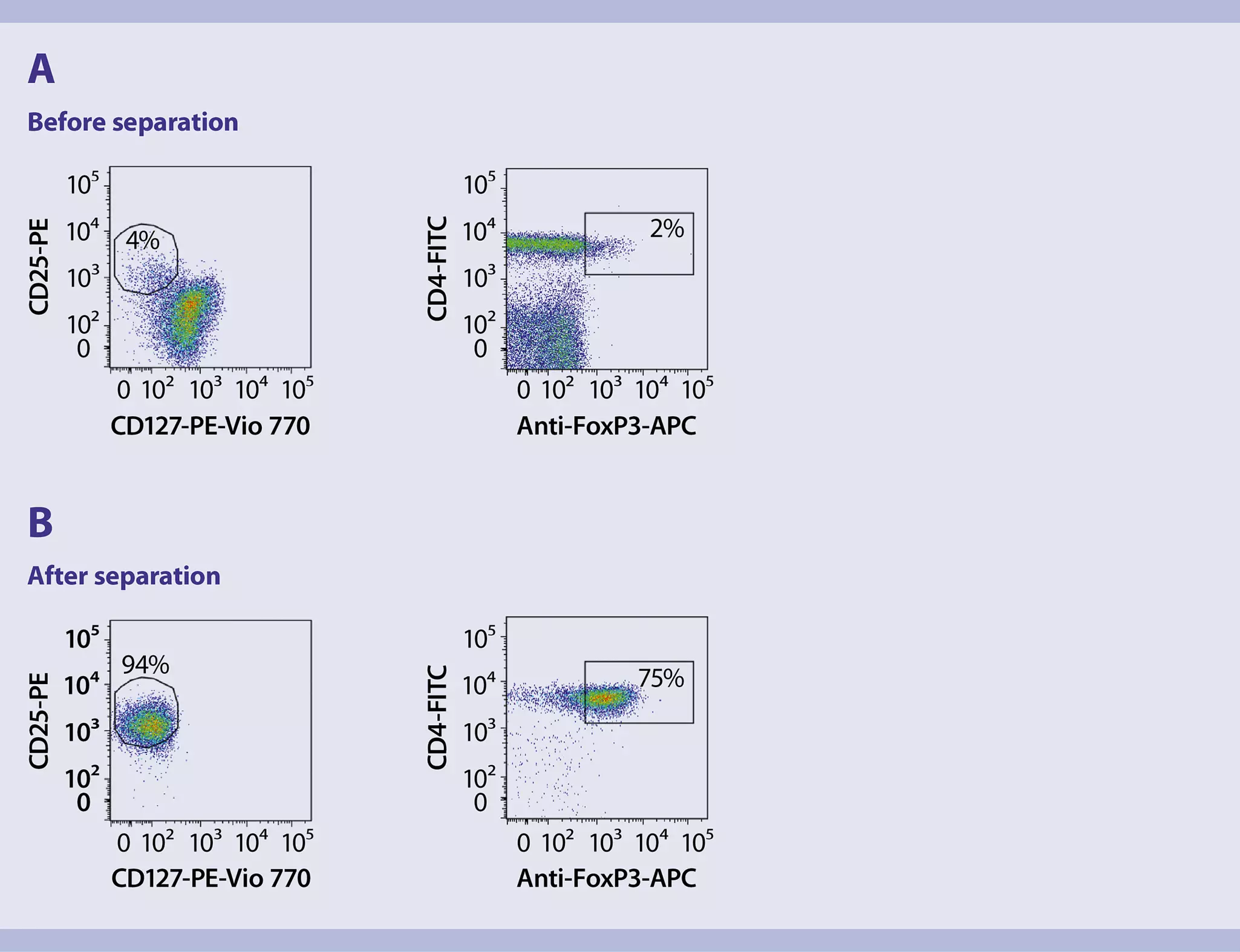
- Analyze CD4+ cells further for their expression of CD25 (y-axis) and absence of CD127 (x-axis) to assess the purity of CD25+CD127dim/– cells among CD4+ cells (figure 1, A and B, left plots).
- Analyze lymphocytes further for their expression of CD4 (y-axis) and FoxP3 (x-axis) to assess the initial frequency of FoxP3-expressing CD4+ cells among lymphocytes (figure 1, A and B, right plots).
 View details
General
- Prepare the expansion medium as described in "Things to prepare in advance”. The Treg Expansion Kit is based on MACSiBead™ Particles preloaded with CD3 and CD28 antibodies. Best expansion of Treg cells is accomplished by using MACSiBead Particles and Treg cells at a bead-to-cell ratio of 4:1.
Preparation of Treg cells
- Determine the total number of Treg cells.
- Wash Treg cells by adding 5–10 volumes expansion medium to the cells and centrifuge at 300×g for 10 minutes. Discard supernatant.
- Resuspend cells at a concentration of 1×10⁶ cells/mL in expansion medium. Add 100 μL (1×10⁵ Treg cells) to each well of a 96-well round-bottom plate.
Preparation of CD3/CD28 MACSiBead Particles
- The CD3/CD28 MACSiBead Particles have a concentration of 2×10⁷ beads/mL and should be used at a bead-to-cell ratio of 4:1.
- MACSiBead Particles are bigger in size than MACS® MicroBeads and sediment rapidly. It is therefore mandatory to bring the MACSiBead Particles into suspension by vortexing prior to use.
- Resuspend CD3/CD28 MACSiBead Particles thoroughly by vortexing and transfer four times the amount of Treg cells to a suitable tube (e.g. for expansion of 1×10⁶ Treg cells, transfer 4×10⁶ (= 200 µL) of MACSiBead Particles).
- Wash MACSiBead Particles by adding 300–600 μL of culture medium and centrifuge at 300×g for 5 minutes. Aspirate supernatant completely.
- Resuspend CD3/CD28 MACSiBead Particles in expansion medium to get a final concentration of 2×10⁷ beads/mL (e.g. when 4×10⁶ of MACSiBead Particles are used, resuspend in 200 µL of expansion medium)
Co-cultivation of Treg cells and MACSiBead Particles
- Add 20 μL of CD3/CD28 MACSiBead Particles to each well containing the Treg cells. As MACSiBead Particles sediment rapidly, the bead suspension should be vortexed from time to time prior addition to the well.
- The final volume of each well is now 120 µL. Let the co-culture of Treg cells and MACSiBead Particles rest for one day at 37 °C and 5–7% CO2.
Expansion process
- Day 1: Add 100 µL of expansion medium to each well.
- Day 3–5 (depending on medium color): Either split Treg cells (including MACSiBead Particles) at a ratio of 1:2 and resuspend cells in 200 µL of fresh expansion medium or carefully aspirate 100 µL of old medium and add 100 µL of fresh expansion medium.
- Day 5–8: Either transfer Treg cells (including MACSiBead Particles) to a 24-well plate in higher volumes, e.g., 5×10⁵ Treg cells in 500 µL, or split Treg cells (including MACSiBead Particles) at a ratio of 1:2 and resuspend cells in 200 µL of fresh expansion medium.
- Day 14: The Treg expansion process can be stopped here or proceeded by restimulation of Treg cells. To stop Treg expansion, determine the total cell number and the frequency of CD4+ FoxP3-expressing cells by performing a surface and intracellular flow cytometry staining to assess the result of expansion. For details refer to section "Flow cytometric analysis after expansion". To proceed with restimulation, CD3/CD28 MACSiBead Particles may be removed prior to restimulation. For details refer to section "Restimulation of Treg cells".
General
- Removal of MACSiBead™ Particles may be required before restimulation with MACSiBead Particles. In addition, bead removal is required before magnetic separation of cells with MACS® MicroBeads or before stimulation with different agents or antigens.
Removal of MACSiBead™ Particles
- Harvest cells, transfer to an appropriate tube and wash once with separation buffer.
- Determine cell number and resuspend cells in separation buffer at a density of up to 2×10⁷ cells/mL and vortex thoroughly.
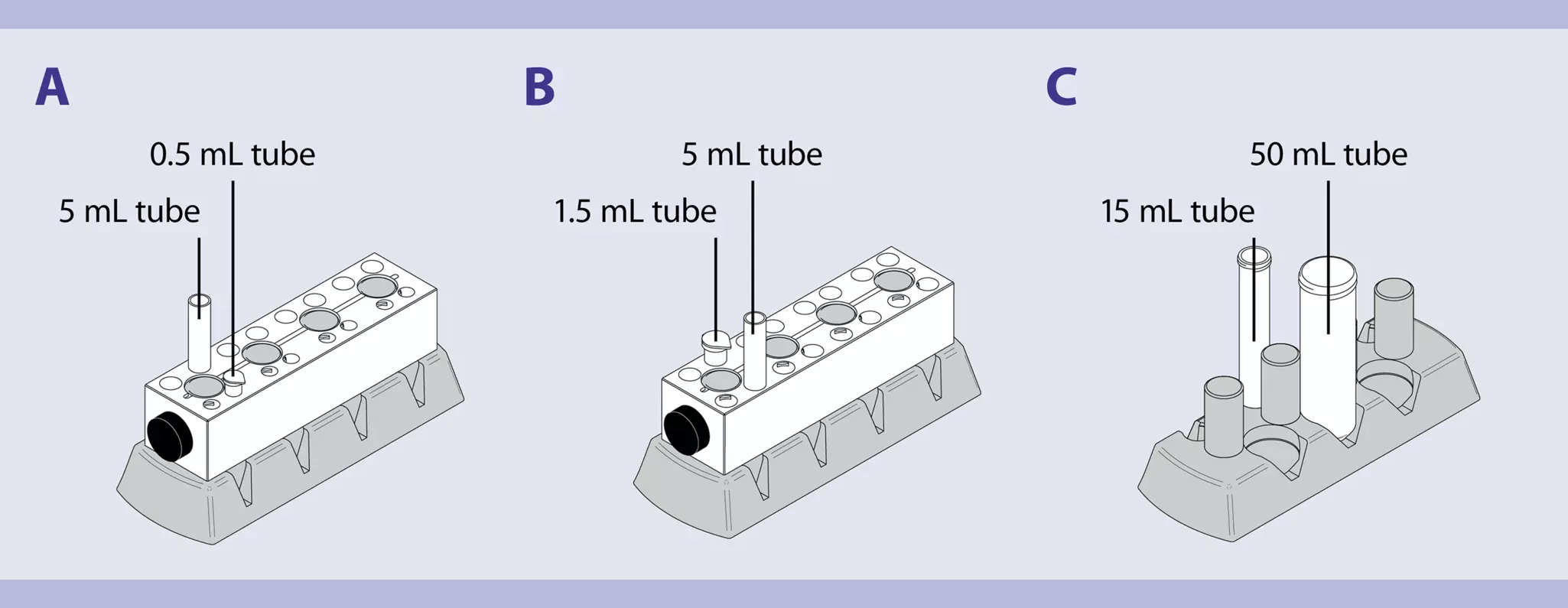
- Place the tube in the magnetic field of the MACSiMAG™ Separator (figure 2). Detailed instructions on how to use the MACSiMAG Separator can be found in the MACSiMAG Separator data sheet.
- Allow the MACSiBead Particles to adhere to the wall of the tube:
5 mL tubes: 2 minutes
15 mL or 50 mL tubes: 4 minutes - Retaining the tube in the magnet, carefully remove the supernatant containing the MACSiBead Particles-depleted cells and place in a new tube.
- Remove the tube from the separator and add separation buffer to reach the same volume as before.
- Vortex sample, replace tube in the MACSiMAG Separator, and repeat steps 4–5.
- Collected cells can now be further processed as required, e.g., for restimulation with MACSiBead Particles.
Restimulation of Treg cells
- Restimulation is performed with a bead-to-cell ratio of 4:1.
- Depending on Treg number and resulting volumes, restimulation can either be performed in a 96-well plate or in a 24-well plate. If restimulate of Treg cells is done in a 96-well-plate, refer to section "In vitro expansion of Treg cells". Alternatively, restimulation may also be performed in 24-well plates in higher volumes, for example, 5×10⁵ Treg cells in 500 μL of expansion medium and 100 μL of CD3/CD28 MACSiBead Particles (= 2×10⁶ particles).
- Day 15: Add 100 µL of expansion medium per well when restimulation is performed in a 96-well plate, or 500 µL of expansion medium per well when restimulation is performed in a 24-well plate.
- Day 17–19 (depending on medium color): Either split Treg cells again (including the MACSiBead Particles) at a ratio of 1:2 and fill the well to 1 mL with fresh expansion medium or carefully aspirate 500 µL old medium and add 500 µL fresh expansion medium.
- Day 21: The Treg expansion process should be stopped after 21 days. To assess the result of expansion, determine the total cell number and the frequency of FoxP3-expressing cells by performing a surface and intracellular flow cytometry staining. For details see "Staining of Treg cells and flow cytometry analysis".
▲ Note: Bead removal is required before certain downstream applications, i.e., magnetic separation of cells with MACS MicroBeads or before stimulation with different agents or antigens. For details refer to "Removal of MACSiBead Particles".
Flow cytometric analysis
- To determine the frequency of FoxP3+CD4+ cells after expansion analyze cells by flow cytometry.
- Identify lymphocytes according to FSC and SSC.
- Exclude dead cells from the analysis by using the live/dead cell exclusion marker PI.
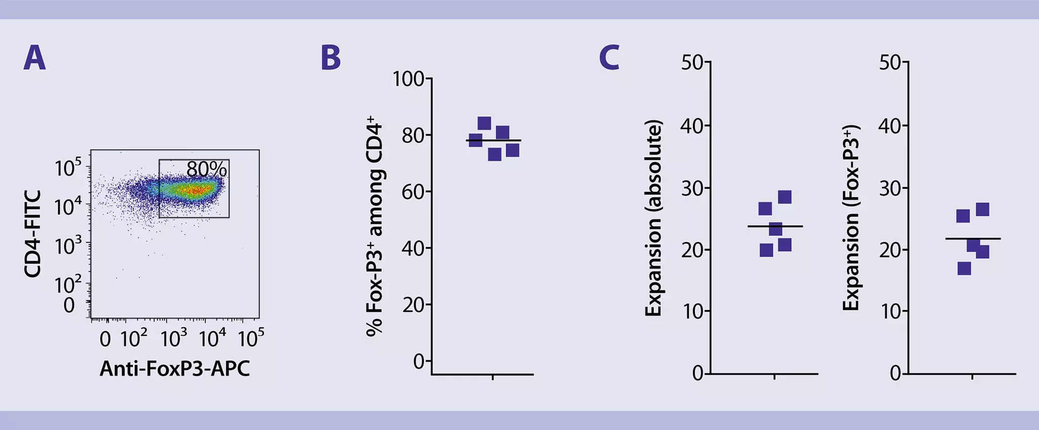
- Analyze lymphocytes further for their expression of CD4 (y-axis) and FoxP3 (x-axis) to assess the frequency of FoxP3-expressing CD4+ cells among lymphocytes (figure 3).
▲ Note: CD4 should be used here to identify Treg cells as CD25 is not an appropriate Treg marker after expansion as it might also be upregulated on activated T cells.
You have previously chosen International as your location.
What do you want to do?