
Synonyms: NRP1, BDCA-4, NP1, Nrp, VEGF165R, Neuropilin-1
RECOMBINANT EXTENDED VALIDATION
- Clone:
- REAL222
- Type of antibody:
- Releasable antibodies, Primary antibodies, Recombinant antibodies
- Applications:
- FC, MICS, IF, IHC Glossary
- Select a conjugate:
- Select a size:


Other sizes
Related products for CD304 (BDCA-4) Antibody, anti-human, REAlease®




Flow cytometry applications
APC
PE
Recommended dilution:
1:50
Data source:
Miltenyi Biotec
Conjugate:
APC
Used products:
130-115-428, 130-115-549, 130-122-669
Description:
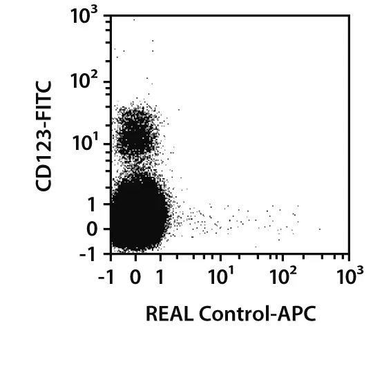
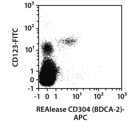
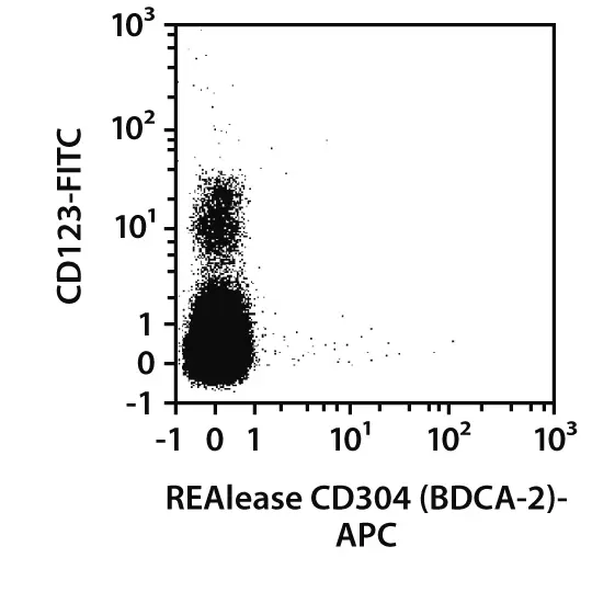
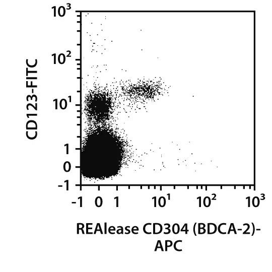
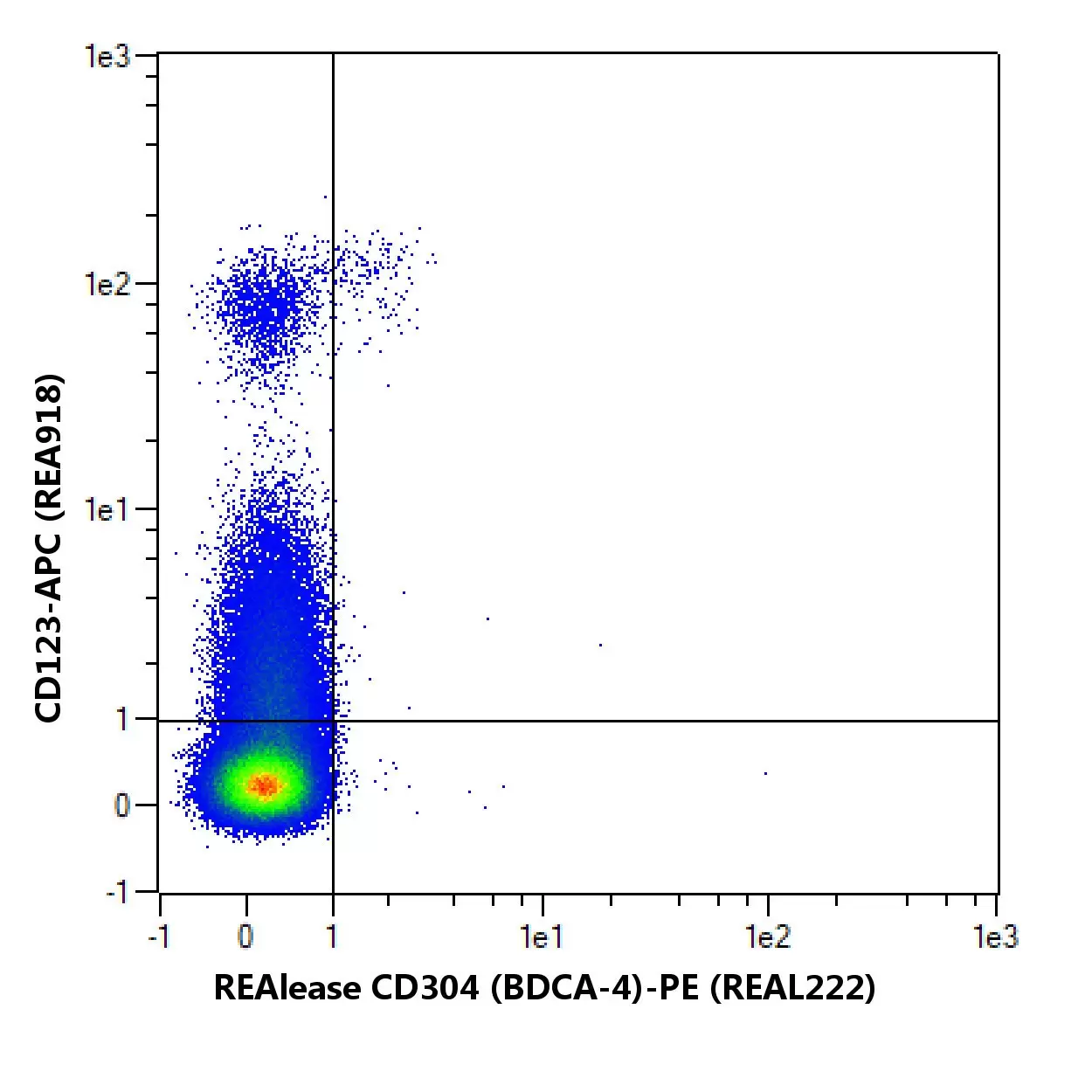
Human peripheral blood mononuclear cells (PBMCs) were stained with REAlease CD304 (BDCA-4) Complexes or with the corresponding REAL Control (left images) as well as with CD123 antibodies and analyzed by flow cytometry using the MACSQuant® Analyzer. Cell debris and dead cells were excluded from the analysis based on scatter signals and propidium iodide (PI) fluorescence.
 view details
 view details
 view details
 view details
MICS (MACSima Imaging Cyclic Staining) applications
APC
Fixation:
frozen – Acetone
Species tested:
human
Tissue used:
tonsil
Recommended dilution:
1:50
Data source:
Miltenyi Biotec
Compatible applications:
IF, IHC
Conjugate:
APC
Used products:
130-115-428, 130-115-549
Testing grade:
Description:
Immunofluorescence analysis of a human tonsil cryosection fixed with ice-cold acetone. The section was stained using CD304 (BDCA-4) Antibody, anti-human, APC (green) for 10 min at room temperature. Nuclei were visualized with Hoechst™ (blue).Scale bar: 30 µm.

Extended validation for CD304 (BDCA-4) Antibody, anti-human, REAlease®
Specificity
In order to compare the epitope specificity of an antibody, the clone being used is compared with other known clones recognizing the same antigen in a competition assay.
Other clones
Overlap in epitope recognition with REAL222
AD5-17F6
high
446921
high
U21-1283
high
12C2
high
REA774
high
Cells were incubated with an excess of purified unconjugated CD304 (BDCA-4) (REAL222) antibody followed by staining with fluorochrome-conjugated antibodies from other known clones targeting the same antigen. Fluorescence intensity was then used to assess epitope overlap: a minimal or absent signal indicated high epitope overlap, an intermediate signal suggested partial overlap, and a strong signal reflected low overlap.
Sensitivity
Selected fluorochrome conjugated antibodies from Miltenyi Biotec were compared to commercially available hybridoma clones in flow cytometry analysis.
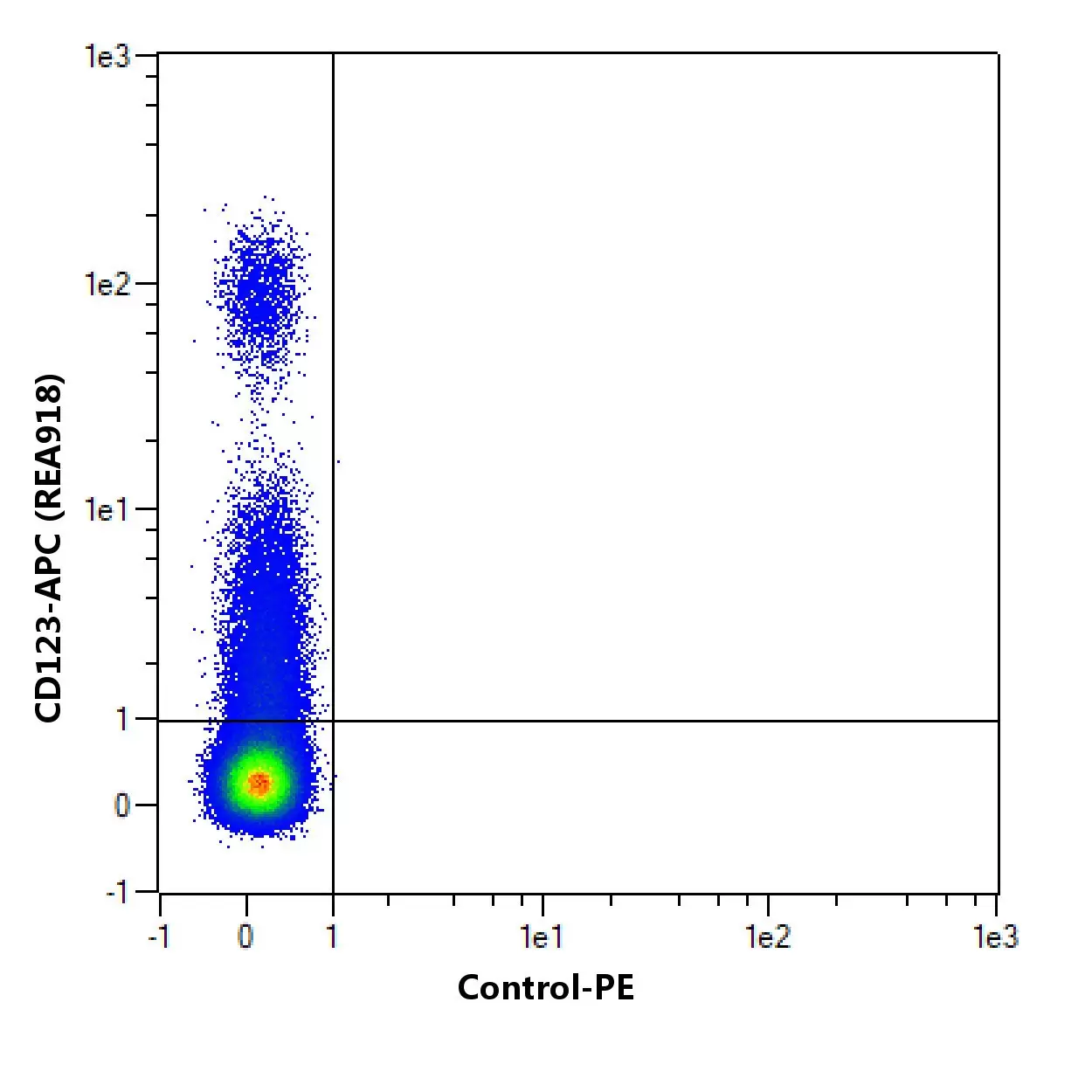
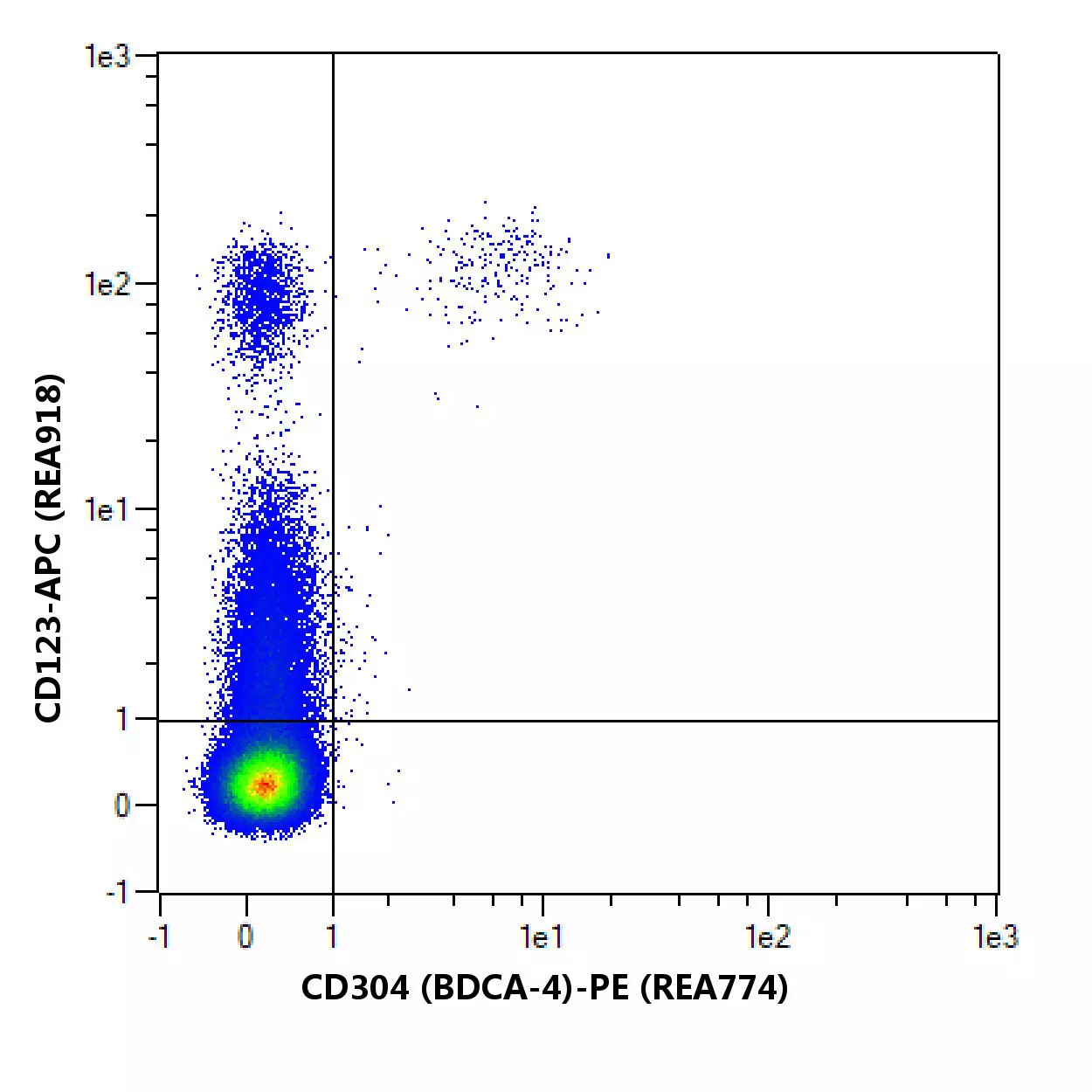

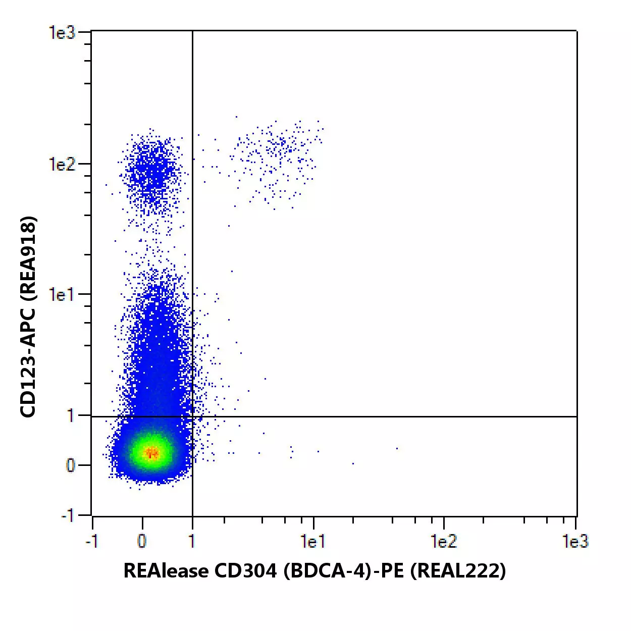

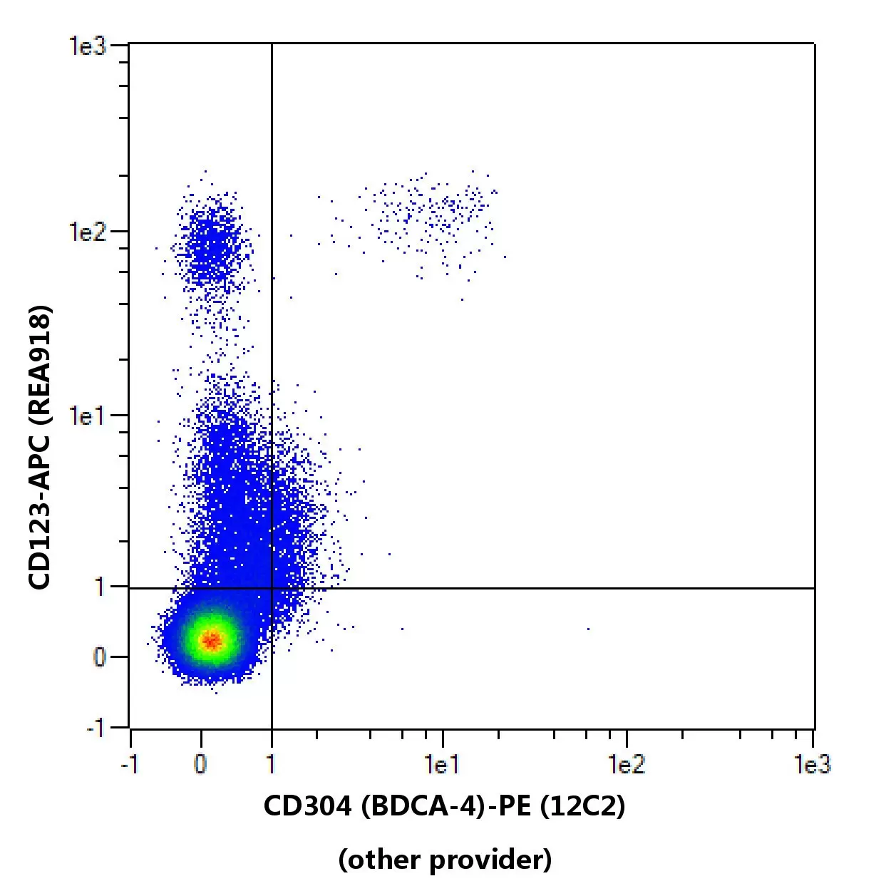
Flow cytometric comparison of different clones for CD304 (BDCA-4). Human peripheral blood mononuclear cells (PBMCs) were stained with CD304 (BDCA-4) antibodies and with a suitable counterstaining. As a control, CD304 (BDCA-4) antibody staining was omitted and cells were measured in the same channels. Flow cytometry was performed with the MACSQuant® Analyzer. Cell debris, dead cells, and cell doublets were excluded from the analysis based on scatter signals and 4',6-diamidino-2-phenylindole (DAPI) fluorescence. No FcR Blocking Reagent was used. The recommended titers of respective antibodies from different suppliers were used.
To provide an indication on how an antibody performs after fixation of cells, in-house data on staining results before and after fixation with 3.7% formaldehyde using Miltenyi Biotec antibodies are provided. Different experimental settings may lead to different results.
No fixation
Post fixation
Comparison of staining pattern on non-fixed and fixed cells using CD304 (BDCA-4) (REAL222). The performance of the antibody after fixation was tested by comparing the staining pattern on fresh (no fixation) versus fixed (post fixation) cells.
Specifications for CD304 (BDCA-4) Antibody, anti-human, REAlease®
Overview
Clone REAL222 is an antibody fragment derived from the full CD304 (BDCA-4) antibody molecule. It displays no binding to Fc receptors. The recombinantly engineered antibody fragments are multimerized to form the REAlease Complex to bind markers with high avidity.
Clone REAL222 recognizes the human CD304 (BDCA-4) antigen. Like CD303 (BDCA-2), the CD304 (BDCA-4) antigen is specifically expressed on human plasmacytoid dendritic cells in blood and bone marrow. Unlike CD303 (BDCA-2), expression of CD304 (BDCA-4) is up-regulated on plasmacytoid dendritic cells and induced on myeloid cells upon culturing. In inflamed tonsils, CD304 expression, apart from plasmacytoid dendritic cells, is also detected primarily on follicular B helper memory T cells. The CD304 (BDCA-4) is identical to neuropilin-1 (NP-1), a neuronal receptor, which is known to be expressed on numerous nonhematopoietic cell types, including neurons, endothelial, and tumor cells. CD1a+ dendritic cells generated ex vivo from monocytes or hematopoietic precursor cells are CD304+ but CD303 (BDCA-2).
The REAlease Kits consist of the respective fluorochrome-conjugated REAlease Complexes and the REAlease Support Kit for removal of the REAlease Complexes and optional relabeling with different fluorochrome-conjugated REAlease Complexes.
NRP1, BDCA-4, NP1, Nrp, VEGF165R, Neuropilin-1
Detailed product information
Technical specifications
Clone
REAL222
Clonality
monoclonal
Isotype control
Control Antibody
Host
cell line
Type of antibody
Releasable antibodies, Primary antibodies, Recombinant antibodies
Species reactivity
human
Antigen
CD304 (BDCA-4)
Distribution of antigen
dendritic cells, B cells, T cells, bone marrow
RRID
AB_2784121, AB_2751264, AB_2751420, AB_2751271, AB_2784120, AB_2751438
Resources for CD304 (BDCA-4) Antibody, anti-human, REAlease®
Certificates
To access a Certificate of Analysis (CoA) follow the link below and enter the specific lot of your product.
Should you wish to request additional documentation, such as a Safety Data Sheet (SDS) in other languages than English or Swedish, an SDS-replacement document, or a Certificate of Origin (CoO), please direct your inquiry to your local technical support team.
Citations for CD304 (BDCA-4) Antibody, anti-human, REAlease®
Publications
- Grabbe, S. et al. (2000) Dendritic cells: multi-lineal and multi-functional. Immunol. Today 21: 431-433
- Jardine, L. et al. (2013) Rapid detection of dendritic cell and monocyte disorders using CD4 as a lineage marker of the human peripheral blood antigen-presenting cell compartment. Front Immunol 4: 495
- Dzionek, A. et al. (2001) BDCA-2, a novel plasmacytoid dendritic cell–specific type II C-type lectin, mediates antigen capture and is a potent inhibitor of interferon α/β induction. J. Exp. Med. 194: 1823-1834
- Jongbloed, S. L. et al. (2010) Human CD141+ (BDCA-3)+ dendritic cells (DCs) represent a unique myeloid DC subset that cross-presents necrotic cell antigens. J. Exp. Med. 207 (6): 1247-1260
- Dzionek, A. et al. (2000) BDCA-2, BDCA-3, BDCA-4: Three markers for distinct subsets of dendritic cells in human peripheral blood. J Immunol 165: 6037-6046
- Dzionek, A. et al. (2002) Plasmacytoid dendritic cells: from specific surface markers to specific cellular functions. Hum Immunol 63: 1133-1148
- Mittag, D. et al. (2011) Human dendritic cell subsets from spleen and blood are similar in phenotype and function but modified by donor health status. J Immunol 186 (11): 6207-6217