Recombinant SARS-CoV-2 Spike-Prot (HEK)-Biotin, Streptavidin, PE, Streptavidin, PE-Vio® 770, and fluorochrome-conjugated antibodies as well as the 7-AAD Staining Solution are available as single products or all-in-one in the SARS-CoV-2 Spike B Cell Analysis Kit, anti-human (# 130-128-022).
Microchip-based cell sorting and expansion of SARS-CoV-2–specific B cells using recombinant SARS-CoV-2 proteins and the MACSQuant® Tyto® Cell Sorter
This application protocol describes the preparation of tetramer solutions, staining, and sorting of SARS-CoV-2–specific B cells using recombinant SARS-CoV-2 spike proteins and the MACSQuant Tyto Cell Sorter.
Protocol
Preparation of spike-tetramer solutions
The following protocol is an example when using Streptavidin, PE and Streptavidin, PE-Vio 770. Other streptavidin conjugates can be used as well. Always check the concentration of the stock beforehand and adapt the protocol accordingly.
For preparation of spike-tetramer solutions, Recombinant SARS-CoV-2 Spike-Prot (HEK)-Biotin was conjugated to Streptavidin, PE and Strepdavidin, PE-Vio 770 with a molar ratio of 4:1, respectively. For ease of use calculations were performed. Volumetric measurements are shown in table 1.
▲ Note: Always freshly prepare spike-tetramer solutions before the use. Solutions can be stored for up to 24 hours at 2–8 °C protected from light.
- Resuspend Recombinant SARS-CoV-2 Spike-Prot (HEK)-Biotin in deionized, sterile-filtered water to a final concentration of 0.1 mg/mL (e.g. use 250 µL water for 25 µg protein).
▲ Note: To avoid repeated freeze-thaw cycles prepare working aliquots from the stock solution of Recombinant SARS-CoV-2 Spike-Prot (HEK)-Biotin. Store the working aliquots at –70 °C. Once thawed the protein can be stored for up to three days at 2–8 °C. - Dilute Streptavidin, PE as well as Streptavidin, PE-Vio 770 to a concentration of 50 µg/mL in MACSQuant Tyto Running Buffer.
▲ Note: Check the concentration of the streptavidin conjugates and dilute if needed. - Mix Recombinant SARS-CoV-2 Spike-Prot (HEK)-Biotin, Streptavidin, PE or Streptavidin, PE-Vio 770, and MACSQuant Tyto Running Buffer carefully as indicated in table 1 and incubate for 15 minutes at room temperature.
Each test indicated in table 1 corresponds to 5×10⁶ B cells (or 107 PBMCs). When working with less than 5×10⁶ B cells (or 107 PBMCs), use the same volumes as indicated for one test. When working with more than 5×10⁶ B cells (or 107 PBMCs), scale up all reagent volumes accordingly.
Table 1: Volumes of spike-tetramer solutions
| Spike-tetramer-PE | |||
| No. of tests | Recombinant SARS-CoV-2 Spike-Prot (HEK)-Biotin (0.1 mg/mL) | Streptavidin, PE (50 µg/mL) | MACSQuant Tyto Running Buffer |
| 1 | 3 µL | 0.6 µL | 1.4 µL |
| 5 | 15 µL | 3 µL | 7 µL |
| 10 | 30 µL | 6 µL | 14 µL |
| 20 | 60 µL | 12 µL | 28 µL |
| Spike-tetramer-PE-Vio 770 | |||
| No. of tests | Recombinant SARS-CoV-2 Spike-Prot (HEK)-Biotin (0.1 mg/mL) | Streptavidin, PE-Vio 770 (50 µg/mL) | MACSQuant Tyto Running Buffer |
| 1 | 6 µL | 1.2 µL | 2.8 µL |
| 5 | 30 µL | 6 µL | 14 µL |
| 10 | 60 µL | 12 µL | 28 µL |
| 20 | 120 µL | 24 µL | 56 µL |
▲ Note: If using an IgG antibody, the other antibodies should have another isotype. Therefore, the use of REAfinity™ Antibodies for this panel design is not recommended, as REAfinity Antibodies are all IgG1 isotype.
Preparation of the antibody staining cocktail
For each test (5×10⁶ B cells or 107 PBMCs) mix 2 μL of each fluorochrome-conjugated antibody, 5 μL of 7-AAD Staining Solution, 5 μL of spike‑tetramer-PE, and 10 μL of spike-tetramer-PE-Vio 770.
Fill up to 100 μL with MACSQuant Tyto Running Buffer.
▲ Note: Try different volumes of tetramer conjugates in order to achieve the best double staining results (refer to section flow cytometry analysis).
Table 2: Suggested antibody panel
| Channel | Fluorochrome | Clone |
| V1 | IgG Antibody, anti-human, VioBlue® | IS11-3B2.2.3 |
| V2 | IgA Antibody, anti-human, VioGreen™ | IS11-8E10 |
| B1 | CD27 Antibody, anti-human, Vio Bright FITC | M-T271 |
| B2 | Spike-tetramer-PE | |
| B3 | 7-AAD Staining Solution | |
| R1 | IgM Antibody, anti-human, APC | PJ2-22H3 |
| R2 | CD19 Antibody, anti-human, APC-Vio 770 | LT19 |
| B4 | Spike-tetramer-PE-Vio 770 |
(Optional) Enrichment of CD19+ cells
B cells can be enriched from single-cell suspension of peripheral blood mononuclear cells (PBMCs) using the REAlease® CD19 MicroBead Kit (# 130-117-034) accordingly to the data sheet including removal of magnetic labeling.
- Resuspend up to 5x10⁶ B cells in 100 μL of antibody staining cocktail. Incubate for 30 minutes at 2–8 °C.
- Wash cells by adding 1–2 mL of MACSQuant Tyto Running Buffer and centrifuge at 300×g for 10 minutes. Aspirate supernatant completely.
- Resuspend cell pellet in a suitable amount of MACSQuant Tyto Running Buffer and proceed to sorting.
▲ Note: Resuspend cautiously as B cells are very sensitive. Shear forces induce stress, what could lead to lower viability of the cells and unspecific staining.
▲ Note: The MACSQuant Tyto Cell Sorter can sort between 100 µL and 10 mL. It is recommended to use 2–10 mL volumes for optimal performance.
- B cells have been pre-enriched using the REAlease CD19 MicroBead Kit (# 130-117-034).
- Refer to the MACSQuant Tyto Cell Sorter user manual and MACSQuant Tyto Cartridge data sheet for instructions on how to use the instrument and cartridges.
- Load B cells into the input chamber of a MACSQuant Tyto Cartridge according to the MACSQuant Tyto Cartridge data sheet.
▲ Note: Take an aliquot of the original fraction from the input chamber for further cell analysis after sorting. - Start the MACSQuant Tyto Cell Sorter and software.
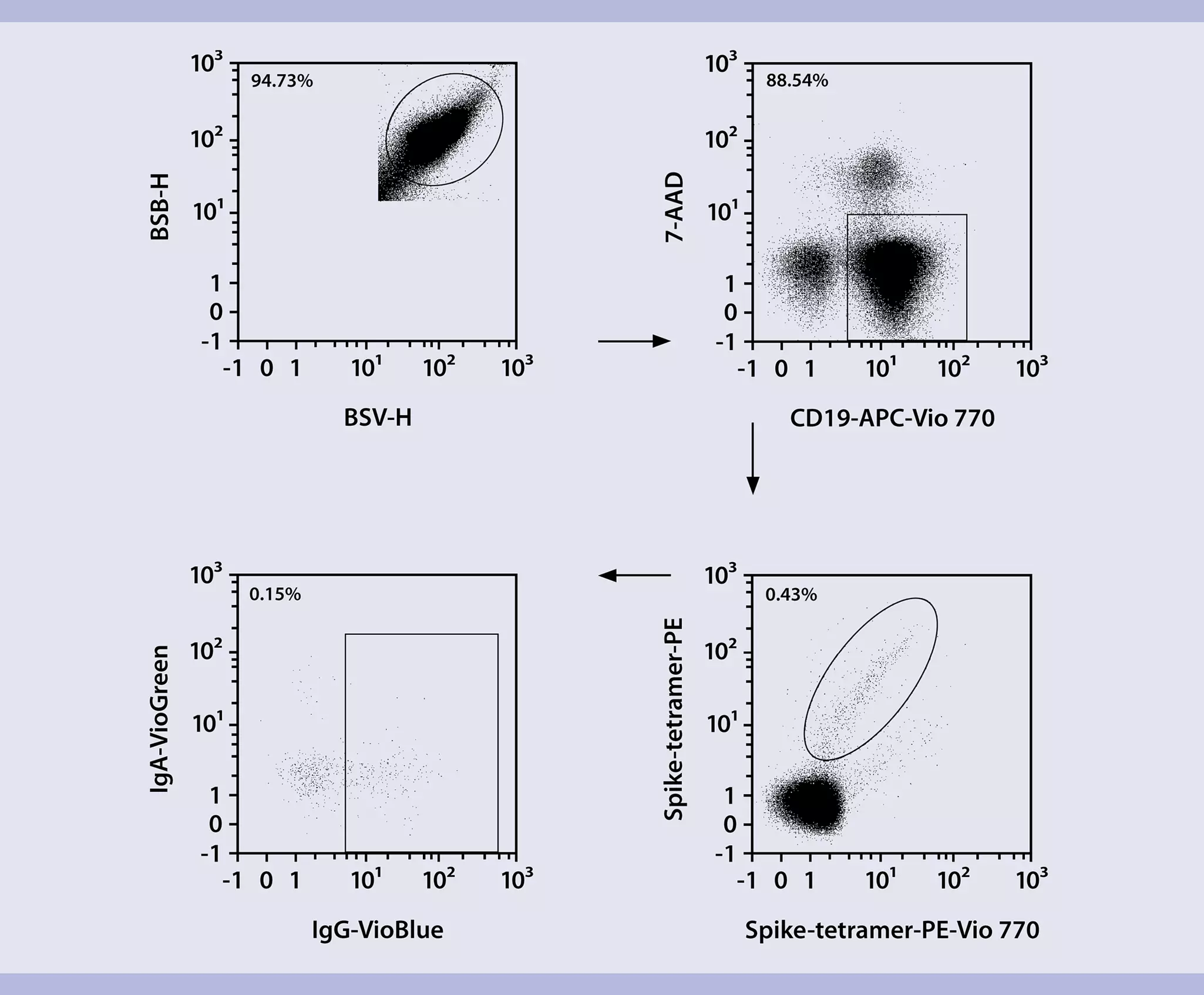
- Load a workspace (gating strategy is shown in figure 1).
▲ Note: Compensate panel if needed. - (Optional) Adjust sample volume in the Experiment tab according to the particular sample.
- Load the MACSQuant Tyto Cartridge into the MACSQuant Tyto Cell Sorter following the instructions in the cartridge data sheet.
- Click the Start measurement button in the status bar to initialize data acquisition.
- Adjust PMTs, trigger, and gating settings.
▲ Note: Adjust compensation if needed. - Click the Sort button in the status bar to start the sort process.
- Click the Stop measurement button in the status bar and take out the cartridge.
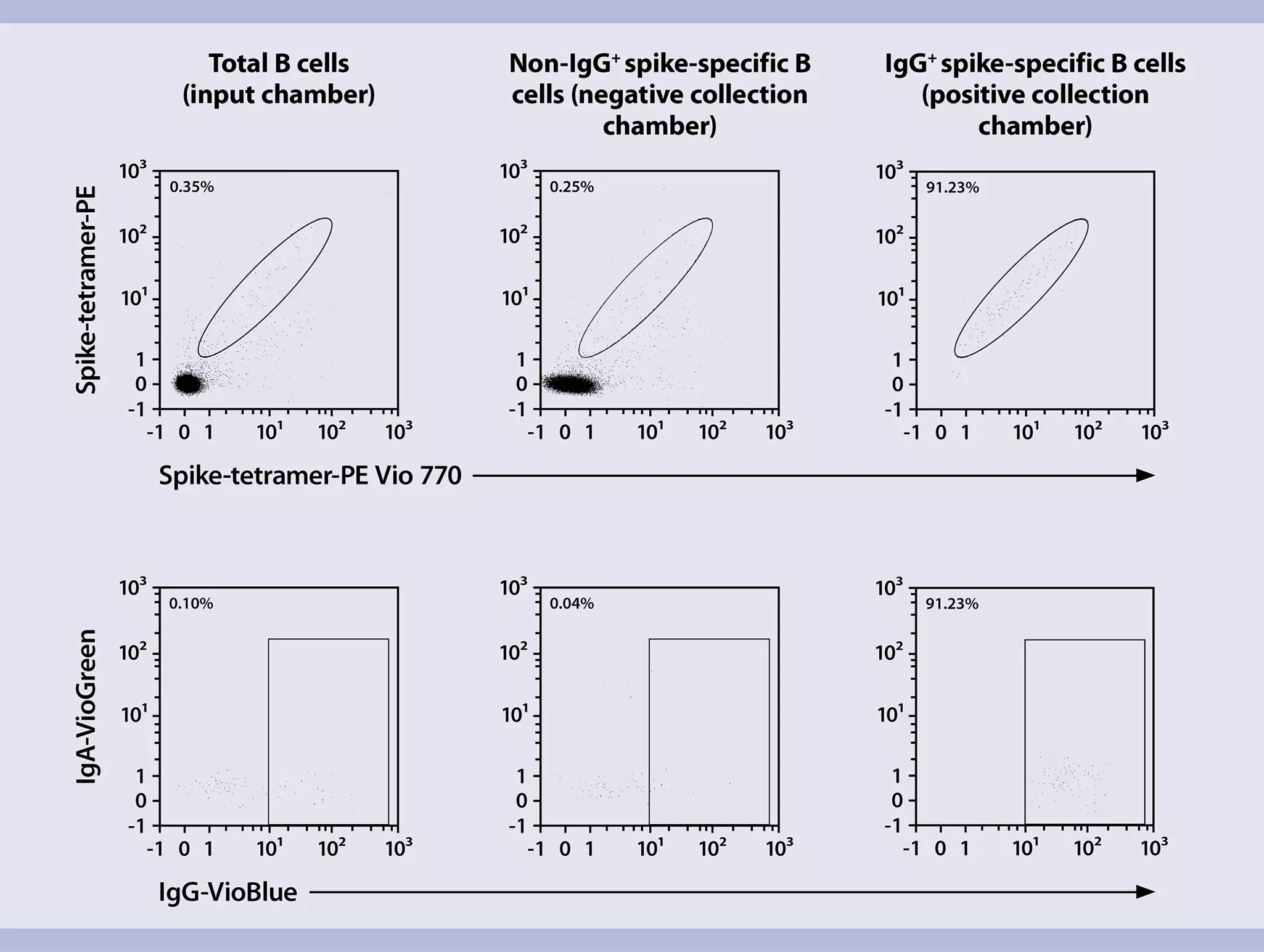
▲ Note: After the input chamber is empty, the sort will automatically stop. - Collect the sorted cells from the positive collection chamber and the non-target cells from the negative collection chamber.
▲ Note: To increase the purity of target cells, the sorted cells can be sorted again. - Take an aliquot from both fractions and proceed to flow cytometry analysis using a MACSQuant Analyzer 10.
▲ Note: Sorted B cells are already stained and just need to be applied to the flow cytometer for analysis.
4.1 Reconstitution of lyophilized reagents
- Reconstitute lyophilized Human CD40-Ligand (component of the B Cell Expansion Kit, human) with deionized sterile-filtered water to a final concentration of 0.5 mg/mL in a volume of 200 μL (for 100 μg size) or 1000 μL (for 500 μg size) and store at –20 °C.
- Reconstitute human IL-4 with deionized sterile-filtered water to a final concentration of 2.5×10⁵ U/mL, e.g., if the lot has a biological activity of 8×10⁶ U/mg, 160 μL deionized sterile-filtered water should be used for reconstitution of 5 μg.
- Mix components by pipetting up and down until resuspended. To avoid repeated freeze-thaw cycles, reconstituted reagents should be aliquoted and stored at –20 °C or below until use.
4.2 B cell expansion
This protocol is optimized for the activation and expansion of B cells, using the Human CD40-Ligand Multimer Kit (component of the B Cell Expansion Kit, human).
▲ Note: Volumes given below are for 10 mL of final expansion medium. When working with larger volumes please scale up accordingly.
▲ Note: The final expansion medium should be prepared directly before the stimulation. Long-term storage is not recommended.
- To increase the biological activity, Human CD40-Ligand and Cross-Linking Antibody (component of the B Cell Expansion Kit, human) must be pre-incubated. Mix 40 μL reconstituted Human CD40-Ligand with 40 μL Cross-Linking Antibody. Incubate for 30 minutes at room temperature (19–25 °C).
- Prepare 10 mL StemMACS™ HSC Expansion Media XF (component of the B Cell Expansion Kit, human) with 2 µL reconstituted Human IL-4, and 5% AB serum. Add the multimerized Human CD40-Ligand (80 μL) to prepared media to get the final expansion medium.
- Remove cells from the sort chamber using HSC Expansion Media with all additives in three sequential steps using 200 µL per step to wash out the sorting chamber. Count cells to determine the total cell number.
- Spin cells down at 200×g for 10 min and resuspend them in appropriate amount of Expansion medium with all indicated additives. Plate at least 3000 cells/mL and up to 7500 cells/mL in 200 µL of the final expansion medium in a 96-U bottom cell culture plate. The same number of cells should be plated for all control fractions within one experiment.
▲ Note: For higher cell numbers plate cells as indicated in the data sheet of the B cell Expansion Kit. - Incubate at 37 °C, 5% CO₂.
- On days 7 and 10 exchange half of the medium with fresh medium supplemented with 5% AB serum and 50 units IL-4/mL (0.2 μL human IL-4 per mL medium from step 2). Harvested supernatants can be stored at –20°C and used for downstream applications, i.e., tested for the presence of spike-specific Ig.
▲ Note: Pipette gently to avoid cell loss as B cell grow in non-adherend cell clusters. - On day 14 harvest B cells by pipetting gently and optionally use them for further downstream applications.
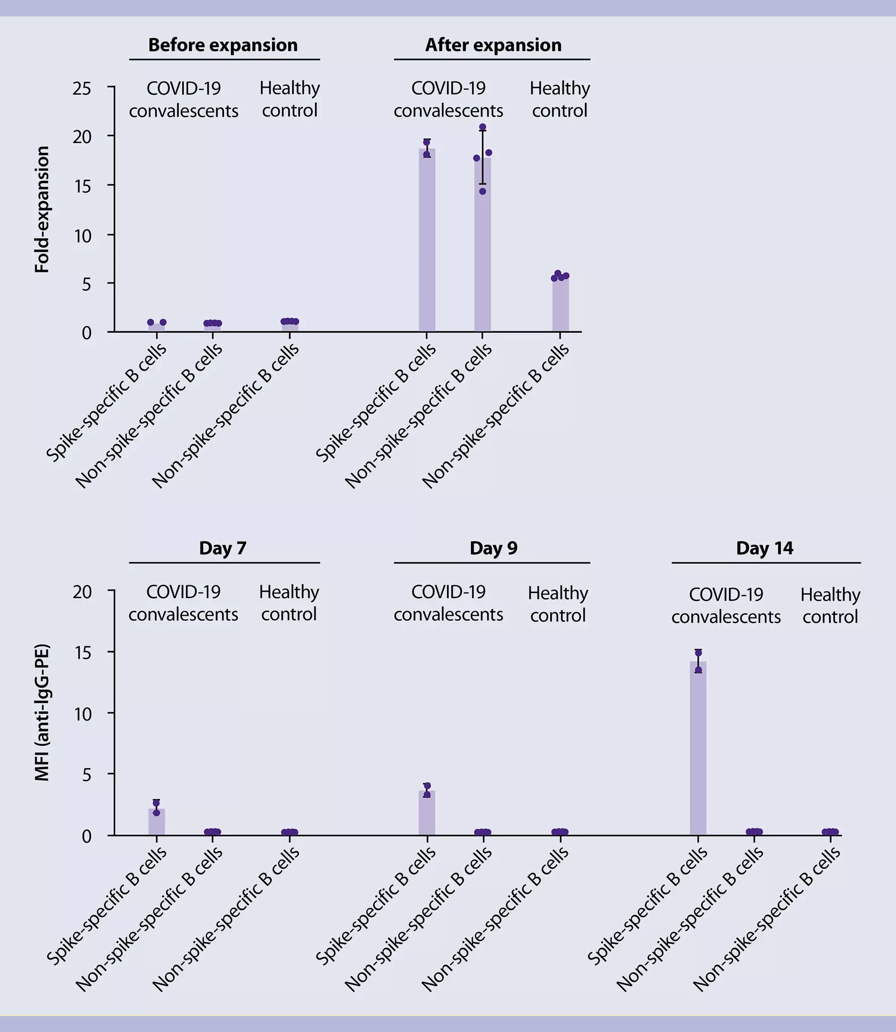
▲ Note: Expansion rates calculated from cell counts are shown in figure 3.
4.3 Analysis of spike-specific antibody production in cell culture supernatants
For the analysis of spike-specific antibody production of expanded B cell cultures refer to the protocol “Bead-based immunoassay for detection of patient-derived SARS-CoV-2 antibodies”. B cell culture supernatants were processed as described for the plasma or serum samples of COVID-19 convalescents in this protocol. Analysis data is shown in figure 3.