TNF-α stands for tumor necrosis factor α, also termed TNFSF2 (TNF ligand superfamily member 2). Human TNF-α is a recombinant protein optimized for use in cell culture, differentiation studies, and functional assays.


Related products for Human TNF-α
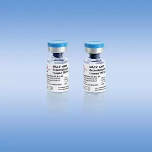
TNF-α stands for tumor necrosis factor α, also termed TNFSF2 (TNF ligand superfamily member 2). Human TNF-α is a recombinant protein optimized for use in cell culture, differentiation studies, and functional assays.
TNF-α can be used for a variety of applications, including:
- Induction of Mo-DC maturation.
- Cytotoxicity and cell proliferation assays.
- Assessment of apoptosis and viral protection.
- Investigation of TNF-α–induced signaling pathways.
TNFSF2
Biological activity
Tumor necrosis factor α (TNF-α) is a proinflammatory cytokine mainly produced by activated monocytes and macrophages in response to infection, injury, and tumor burden. TNF-α production has also been reported for a variety of other cell types involved in inflammatory responses, including T cells, NK cells, and neutrophils as well as a number of non-immune cells, such as keratinocytes and astrocytes. TNF-α has a broad spectrum of biological activities. In addition to its central role in inflammation, TNF-α is noted for its cytotoxic and tumoricidal abilities either by necrosis or induction of apoptosis. Further functions include antiviral activity, growth modulation, and induction of cellular differentiation. Despite its various beneficial actions, TNF-α also plays a detrimental role in, for example, septic shock syndrome, tissue injury, inflammation, cachexia, and diabetes.
Research-grade cytokines are suitable for a wide variety of cell culture applications. They are sterile-filtered prior to lyophilization. Generally, endotoxin levels are <0.1 ng/μg (<1 EU/μg), and purities are >95%. The biological activity is tested in appropriate bioassays.
Premium-grade cytokines offer the convenience of high and well-defined biological activities and allow exact unit dosing for demanding applications. The biological activity is determined after lyophilization and reconstitution, and normalized to WHO/NIBSC standards whenever available. In general, endotoxin levels are <0.01 ng/μg (<0.1 EU/μg), and purities are >97%. Lot-specific activities are stated in the Certificate of Analysis (www. miltenyibiotec.com/certificates).
Human TNF-α activity assay. The biological activity of Human TNF-α, premium grade was determined by inhibition assay using L-929 cells.
SDS-PAGE of Human TNF-alpha, premium grade under reducing (R) and non reducing (NR) conditions.
Mass spectrometry analysis (ESI-MS) of Human TNF-α, premium grade. The peak corresponds to the calculated molecular mass of 17353 Da.
To access a Certificate of Analysis (CoA) follow the link below and enter the specific lot of your product.
Should you wish to request additional documentation, such as a Safety Data Sheet (SDS) in other languages than English or Swedish, an SDS-replacement document, or a Certificate of Origin (CoO), please direct your inquiry to your local technical support team.
- Clement, M. et al. (2016) Targeted suppression of autoreactive CD8 + T-cell activation using blocking anti-CD8 antibodies. Sci Rep 6: 35332
- Baarsch, M. J. et al. (1991) Detection of tumor necrosis factor alpha from porcine alveolar macrophages using an L929 fibroblast bioassay. J. Immunol. Methods 140: 15-22
- Boink, M. A. et al. (2017) Saliva-Derived Host Defense Peptides Histatin1 and LL-37 Increase Secretion of Antimicrobial Skin and Oral Mucosa Chemokine CCL20 in an IL-1 α-Independent Manner. J Immunol Res 2017: 3078194
- Barbara, J. A. et al. (1996) Tumour necrosis factor-alpha (TNF-alpha): the good, the bad and potentially very effective. Immunol. Cell Biol. 74: 434-443
- Bacher, P. et al. (2014) Antigen-specific expansion of human regulatory T cells as a major tolerance mechanism against mucosal fungi. Mucosal Immunol 7 (4): 916-928
- Ferroni, L. et al. (2020) Fluorescent Light Energy (FLE) Acts on Mitochondrial Physiology Improving Wound Healing. J Clin Med 9 (2): 559
- Voskamp, A. et al. (2013) MHC class II expression in human basophils: induction and lack of functional significance. PLoS One 8 (12): e81777
- Schweikert, E. M. et al. (2012) PON3 is upregulated in cancer tissues and protects against mitochondrial superoxide-mediated cell death. Cell Death Differ. 19 (9): 1549-1560
- Mayordomo, A. C. et al. (2018) IL-12/23p40 overproduction by dendritic cells leads to an increased Th1 and Th17 polarization in a model of Yersinia enterocolitica-induced reactive arthritis in TNFRp55-/- mice. PLoS One 13 (3): e0193573
- Islam, S. A. et al. (2013) Identification of human CCR8 as a CCL18 receptor. J. Exp. Med. 210 (10): 1889-1898
- Meurer, T. et al. (2018) Dissecting Genetic Control of HLA-DPB1 Expression and Its Relation to Structural Mismatch Models in Hematopoietic Stem Cell Transplantation. Front Immunol 9: 2236
- Kronenberg, D. et al. (2012) Circulating preproinsulin signal peptide-specific CD8 T cells restricted by the susceptibility molecule HLA-A24 are expanded at onset of type 1 diabetes and kill β-cells. Diabetes 61 (7): 1752-1759
- Garufi, A. et al. (2012) Targeting COX-2/PGE(2) pathway in HIPK2 knockdown cancer cells: impact on dendritic cell maturation. PLoS One 7 (11): e48342
- Black, R. A. et al. (1997) A metalloproteinase disintegrin that releases tumour-necrosis factor-alpha from cells. Nature 385: 729-733
- Paravati, R. et al. (2020) Differential regulation of osteopontin and CD44 correlates with infertility status in PCOS patients. J. Mol. Med. 98 (12): 1713-1725
- Schipper, H. S. et al. (2010) A multiplex immunoassay for human adipokine profiling. Clin. Chem. 56 (8): 1320-1328
- Nicoli, F. et al. (2021) Use of a Novel Peptide Welding Technology Platform for the Development of B- and T-Cell Epitope-Based Vaccines. Vaccines (Basel) 9 (5): 526
- Zhou, R. et al. (2020) Acute SARS-CoV-2 Infection Impairs Dendritic Cell and T Cell Responses. Immunity 53 (4): 864-877
- Cantoni, C. et al. (2021) Stromal-like Wilms tumor cells induce human Natural Killer cell degranulation and display immunomodulatory properties towards NK cells. Oncoimmunology 10 (1): 1879530
- Yeung, M. C. et al. (1996) An essential role for the interferon-inducible, double-stranded RNA-activated protein kinase PKR in the tumor necrosis factor-induced apoptosis in U937 cells. Proc. Natl. Acad. Sci. U.S.A. 93: 12451-12455
- Simo, R. et al. (2012) Potential role of tumor necrosis factor-α in downregulating sex hormone-binding globulin. Diabetes 61 (2): 372-382
You have previously chosen International as your location.
What do you want to do?