- Volumes given below are for up to 106 total cells (e.g. PBMCs) per staining reaction. Depending on sample composition (e.g. whole blood, isolated PBMCs, enriched T cells) and the individual application field, the optimum amount of peptide-loaded MHC MACSimers for T cell staining can greatly vary. It is recommended to perform an application-specific titration assay prior to final experiment setup.
- Determine cell number.
- Centrifuge peptide-loaded MHC MACSimers at 3300×g for 5 minutes.
▲ Note: Potential aggregates will be pelleted at the bottom of the tube and should not be used for staining. - Carefully add 5 µL peptide-loaded MHC MACSimers from the supernatant to 1×10⁶ cells in 50 µL staining buffer and mix well.
▲ Note: When using allele-matched MHC MACSimer Negative Control Reagents, 3 µL Control Reagent per 1×10⁶ cells in 50 µL staining buffer are recommended to precisely match titers of reagents. - Incubate at +4 °C in the dark for 10 minutes.
▲ Note: Alternatively, incubation can be performed at room temperature, which in some cases may optimize brightness of the staining. However, it is not recommended to perform incubation at room temperature if subsequent release of the MHC MACSimer Reagent from the stained T cells is planned (e.g. for sequential multi-sorting or downstream analyses). Release of the constructs using the REAlease® Support Kit can be significantly impaired by MHC MACSimer incubation at room temperature due to accelerated receptor internalization. - Wash by adding 1 mL staining buffer and centrifuge at 300×g for 10 minutes. Aspirate supernatant completely.
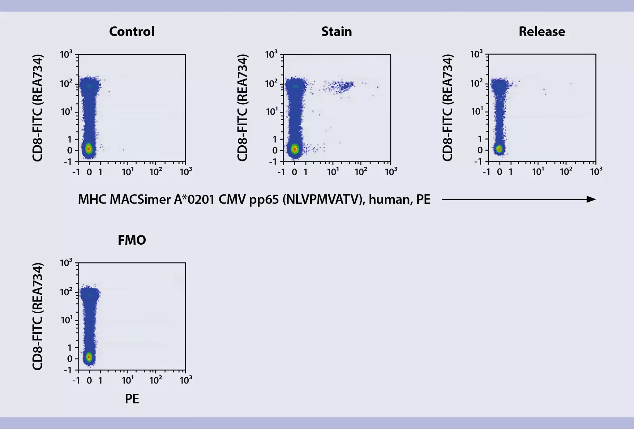
- Proceed to secondary fluorescent staining for immune cell phenotyping, e.g., using CD8 antibodies for identification of cytotoxic T cells, CD19 or CD20 for exclusion of B cells, CD14 for exclusion of monocytes, markers for exclusion of dead cells, and additional markers of interest. For more information on antibodies refer to Recombinant antibodies for flow cytometry.
▲ Note: Note that certain CD8 antibody clones interfere with the peptide-MHC-TCR interaction and thus might impair MHC MACSimer staining efficiency. Refer to table 1 for recommended antibodies. - (Optional) For negative staining control, use allele-matched MHC MACSimer Control Reagents diluted in staining buffer.
Table 1: Recommended antibodies for CD8 co-staining. Antibodies have been tested and approved via flow cytometry for their compatibility with MHC I MACSimers.
| Antibody | Clone |
| CD8 Antibody, anti-human, REAfinity® | REA734 |
| CD8a Antibody, anti-mouse, REAfinity | REA601 |
| CD8a Antibody, anti-mouse | 53-6.7 |
 View details

