CD14 MicroBeads can be used for the positive selection or depletion of human monocytes and macrophages from cord blood or PBMCs, as well as pleural, peritoneal, or synovial fluids or from various tissues, such as spleen and lymph node.


Related products for CD14 MicroBeads, human

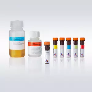
Separation of CD14+ cells from PBMCs using CD14 MicroBeads, an MS Column, and a MiniMACS™ Separator.
Monocytes were isolated from PBMCs using CD14 MicroBeads and subsequently cultured for 7 days with IL-4 and GM-CSF to generate immature Mo-DCs and were, finally, cultured for additional 3 days with TNF-α for maturation. Mature Mo-DCs were stained for expression of CD1a, costimulatory molecules (CD80, CD86), CD83 and HLA-DR.
CD14 MicroBeads can be used for the positive selection or depletion of human monocytes and macrophages from cord blood or PBMCs, as well as pleural, peritoneal, or synovial fluids or from various tissues, such as spleen and lymph node.
The CD14 antigen belongs to the LPS receptor complex. Binding of antibody to CD14 does not trigger signal transduction since CD14 lacks a cytoplasmatic domain. CD14 is strongly expressed on most monocytes and macrophages and weakly on neutrophils and some myeloid dendritic cells.
The most eminent use of monocytes isolated by MACS® Technology is their ex vivo differentiation into monocyte-derived dendritic cells (Mo-DCs). The excellent purity and recovery obtained by using CD14 MicroBeads provide a pure and consistent cell source (fig. 1). Contamination with unwanted cells (e.g. platelets) which may interfere with a controlled differentiation process is markedly reduced, and the isolated monocytes can be induced to differentiate into a homogenous population of immature as well as mature Mo-DCs. Dendritic cells generated from monocytes isolated by MACS® Technology have been used in many different fields of application.1-6
Apart from the generation of dendritic cells, monocytes are isolated, for example, for ex vivo generation of macrophages7,8 or for studies on cytotoxicity9 or migration10.
For positive selection: MS, LS, XS, or autoMACS® Columns. For depletion: LD, D, or autoMACS Columns.
- Ebner, S. et al. (2002) A novel role for IL-13: Human monocytes cultured in the presence of IL-3 and IL-4 differentiate into dendritic cells that produce less IL-12 and shift the cell responses toward a Tʜ2 cytokine pattern. J Immunol 168: 6199-6207
- Matsumoto, M. et al. (2003) Subcellular localization of Toll-like receptor 3 in human dendritic cells. J Immunol 171: 3154-3162
- de Baey, A. and Lanzaveccia, A. (2000) The role of aquaporins in dendritic cell macropinocytosis. J. Exp. Med. 191: 743-747
- Pickl, W. F. et al. (1996) Molecular and functional characteristics of dendritic cells generated from highly purified CD14+ peripheral blood monocytes. J Immunol 157: 3850-3859
- Verreck, F. A. et al. (2004) Human IL-23-producing type 1 macrophages promote but IL-10-producing type 2 macrophages subvert immunity to (myco)bacteria. Proc. Natl. Acad. Sci. U.S.A. 101: 4560-4565
- Ryan, E. J. et al. (2002) Dendritic cell-associated lectin-1: a novel dendritic cell-associated, C-type lectin-like molecule enhances T cell secretion of IL-4. J Immunol 169: 5638-5648
- Salio, M. et al. (2000) Dendritic cell maturation is induced by mycoplasma infection but not by necrotic cells. Eur. J. Immunol. 30: 705-708
- Hanley, P. J. et al. (2004) Extracellular ATP induces oscillations of intracellular Ca2+ and membrane potential and promotes transcription of IL-6 in macrophages. Proc. Natl. Acad. Sci. U.S.A. 101: 9479-9484
- Vitale, S. et al. (2004) Soluble fractalkine prevents monocyte chemoattractant protein-1-induced monocyte migration via inhibition of stress-activated protein kinase 2/p38 and matrix metalloproteinase activities. J Immunol 172: 585-592
- Jefford, M. et al. (2003) Functional comparison of DCs generated in vivo with Flt3 ligand or in vitro from blood monocytes: differential regulation of function by specific classes of physiologic stimuli. Blood 102: 1753-1763
You have previously chosen International as your location.
What do you want to do?