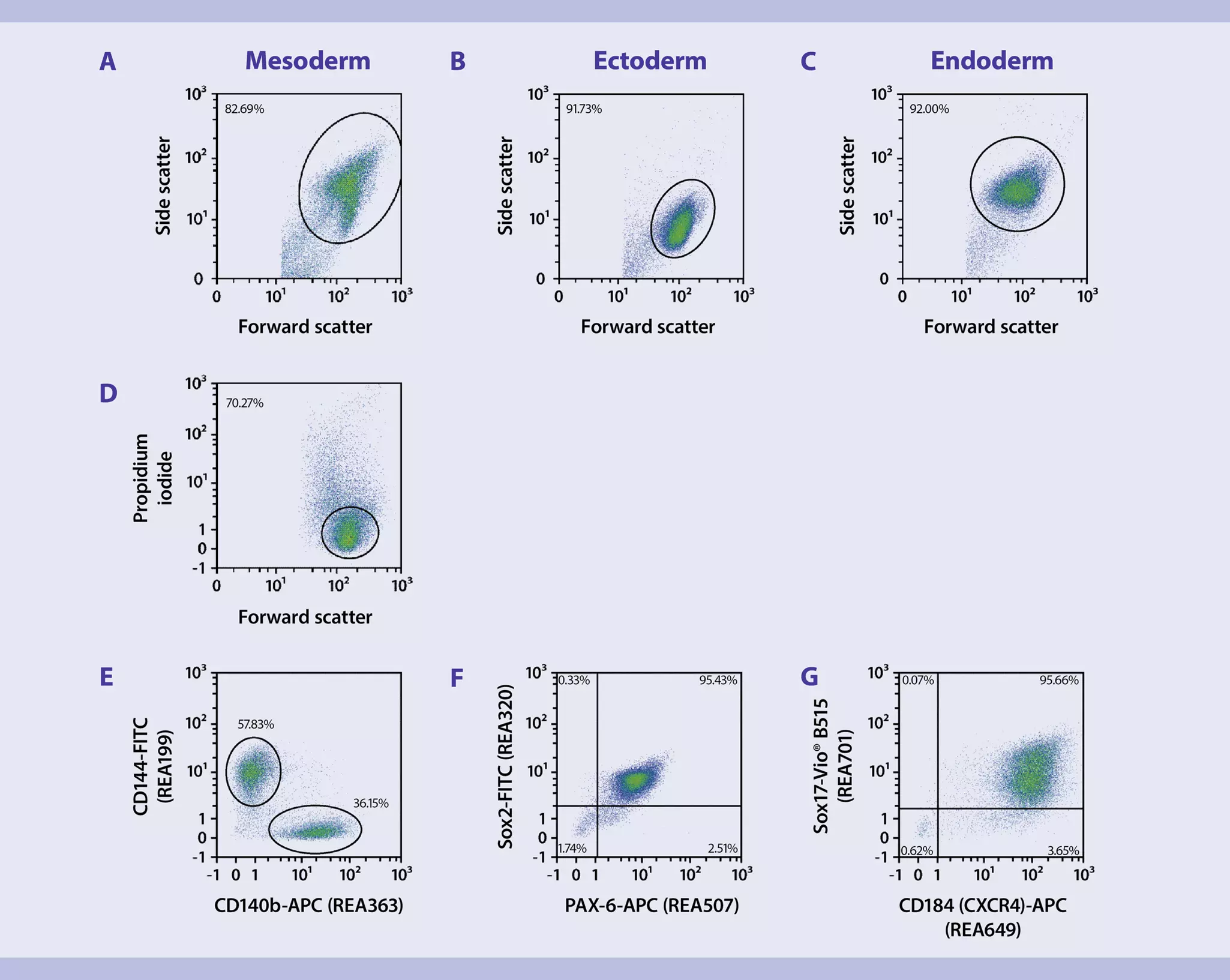
The staining panel for pluripotent stem cell differentiation is based on a set of REAfinity™ Antibodies to ensure low background and high staining specificity. The panel consists of the three marker combinations (combination 1: CD144 and CD140b for mesoderm analysis; combination 2: CD184 and Sox17 for endoderm analysis; combination 3: PAX-6 and Sox2 for ectoderm analysis) and includes both surface and intracellular markers. Surface markers are stained first. Subsequently, cells are fixed and permeabilized for intracellular staining. Since the marker combination for ectoderm detection only includes intracellular markers, no surface staining is needed for this sample.
Analysis of the trilineage differentiation potential of human pluripotent stem cells (PSCs) using flow cytometry
MBTP 22 = Miltenyi Biotec-tested panel 22
This application protocol describes the quantitative analysis of the trilineage differentiation potential of human pluripotent stem cells (PSCs) using flow cytometry. To demonstrate this state-of-the-art panel for pluripotent stem cell differentiation, PSCs were differentiated using the StemMACS™ Trilineage Differentiation Kit. The StemMACS™ Trilineage Differentiation Kit enables the directed and reproducible differentiation of stem cells into ectodermal, mesodermal, and endodermal lineages ready for downstream applications, such as flow cytometric analysis.
Surface staining
- Volumes given below were for up to 106 nucleated cells. When working with fewer than 106 cells, use the same volumes as indicated. When working with higher cell numbers, scale up all reagent volumes and total volumes accordingly (e.g. for 2 × 10⁶ nucleated cells, use twice the volume of all indicated reagent volumes and total volumes).
- Cell number was determined.
- Cells were centrifuged at 300×g for 10 minutes. Supernatant was aspirated completely.
- Master mixes for the respective antibody combinations were prepared according to the dilutions mentioned in the respective antibody data sheets.
▲Note: Sox17, PAX-6, and Sox2 are intracellular markers and will be detected after fixation and permeabilization of cells.
▲Note: CD144 and CD140b were used for staining mesoderm samples, PAX-6 and Sox2 for endoderm samples, and CD184 and Sox17 for ectoderm samples. - 100 µL antibody master mix was added to the cells.
- Cells were resuspended and incubated for 10 minutes in the dark at +4 °C.
- Cells were washed by addition of 1 mL of PEB buffer and centrifuged at 300 × g for 5 minutes at +4 °C. Supernatant was aspirated completely.
- Mesoderm samples were resuspended in 100 µL of PEB buffer and 1 µL of propidium iodide was added right before flow cytometric analysis using the MACSQuant® Analyzer.
- Intracellular staining of endoderm and ectoderm samples was performed.
Intracellular staining
- Volumes given below were for up to 106 nucleated cells. When working with fewer than 106 cells, use the same volumes as indicated. When working with higher cell numbers, scale up all reagent volumes and total volumes accordingly (e.g. for 2 × 10⁶ nucleated cells, use twice the volume of all indicated reagent volumes and total volumes).
- Reagents were prepared freshly. Failure to do so may lead to sub-optimal results.
- The required total buffer volumes were calculated beforehand. Volumes depend on the number of cells to be analyzed as well as the number of tests to be performed.
- Fixation and Permeabilization solution was prepared by diluting 1:3 the Fixation/Permeabilization Solution 1 with the Fixation/Permeabilization Solution 2.
- 1× solution of Permeabilization Buffer was prepared by diluting the 10× Permeabilization buffer with deionized or distilled water.
▲Note: Before preparing the dilution make sure the buffer does not contain any precipitates. - Cells previously stained for surface markers were resuspended in 1 mL of cold, freshly prepared Fixation and Permeabilization Solution.
- Cells were mixed well and incubated for 30 minutes in the dark at +4 °C.
- Cells were washed by adding 1 mL of cold PEB buffer per 106 cells and centrifuged at 300 × g for 5 minutes at +4 °C. Supernatant was aspirated completely.
- Cells were washed by adding 1 mL of cold 1× Permeabilization Buffer and centrifuged at 300 × g for 5 minutes at +4 °C. Supernatant was aspirated completely.
- Cells were resuspended in 1× Permeabilization Buffer and the corresponding antibody volume according to the dilution mentioned in the respective data sheet was added.
- Cells were mixed well and incubated for 30 minutes in the dark at +4 °C.
- Cells were washed by adding 1 mL of cold 1× Permeabilization Buffer and centrifuged at 300×g for 5 minutes at +4 °C. Supernatant was aspirated completely.
- Cells were resuspended in 100 µL PEB buffer and flow cytometric analysis was performed using the MACSQuant® Analyzer.
Antibody panel
| Specificity | Clone | Purpose | Fluorochrome | Detection filter (nm) (laser) |
| CD144 (VE-Cadherin) | REA199 | Mesoderm | FITC | 525/50 (blue) |
| CD140b | REA363 | Mesoderm | APC | 655−730 (red) |
| CD184 (CXCR4) | REA649 | Endoderm | APC | 655−730 (red) |
| Sox17 | REA701 | Endoderm | Vio® B515 | 525/50 (blue) |
| PAX-6 | REA507 | Ectoderm | APC | 655−730 (red) |
| Sox2 | REA320 | Ectoderm | FITC | 525/50 (blue) |
| Propidium Iodide Solution | - | Viability | - | 655−730 (blue) |
Gating strategy
7 products found
|
For research use only
2 mL
For research use only
ICFC
100 tests in 200 µL
For research use only
FC, MICS, IF, IHC
30 tests in 60 µL
For research use only
FC
100 tests in 200 µL
For research use only
ICFC, MICS, IF, IHC
30 tests in 60 µL
For research use only
ICFC
30 tests in 60 µL
For research use only
FC
100 tests in 200 µL
You have previously chosen International as your location.
What do you want to do?